_____________________________________________________________________________________________________ *Corresponding author: E-mail: [email protected];
ISSN: 2231-0614, NLM ID: 101570965
SCIENCEDOMAIN international
www.sciencedomain.org
A Novel Decrease of Mrp3 Protein in Liver of
β-Thalassemic Mouse
Mohammed Qaisiya
1*, Lucia de Franceschi
2, Achille Iolascon
3,
Claudio Tiribelli
1,4and Cristina Bellarosa
11Fondazione Italiana Fegato ONLUS, Italian Liver Foundation ONLUS, Bldg Q AREA Science
Park -Basovizza Campus 34149 Trieste, Italy. 2
Department of Medicine, Section of Internal Medicine, University of Verona-AOUI-Verona, Verona, Italy. 3Department of Molecular Medicine and Medical Biotechnology, University of Naples Federico II,
CEINGE- Advanced Biotechnologies, 80145 Naples, Italy. 4
Department of Medical Sciences, University of Trieste, 34149 Trieste, Italy.
Authors’ contributions
This work was carried out in collaboration between all authors. Author MQ performed experiments, analyzed results and wrote the first draft of the manuscript. Authors LF and AI performed treatments and provide liver samples. Author CB designed the study, analyzed the data and revised the first draft of the manuscript. Author CT designed the study and analyzed the results. All authors read and approved the final manuscript.
Article Information
DOI: 10.9734/BJMMR/2016/25139 Editor(s): (1) Sinan INCE, Department of Pharmacology and Toxicology, University of Afyon Kocatepe, Turkey. Reviewers: (1) Wilairat Leeanansaksiri, Suranaree University of Technology, Thailand. (2)S. Vetriselvan, OPJS University, Churu, Rajasthan, India. (3)Sulaiman Shams, AbdulWali Khan University, Mardan, Pakistan. Complete Peer review History:http://sciencedomain.org/review-history/13957
Received 19th February 2016 Accepted 22nd March 2016 Published 31st March 2016
ABSTRACT
Background: Hepatocytes have a fundamental system of efflux proteins that protect cells from toxic insults. Unconjugated bilirubin at higher concentration is toxic to cells and its intracellular accumulation is limited by the induction of efflux proteins such as Mrp3. In vivo studies showed an induction of hepatic Mrp3 expression in response to non-hemolytic hyperbilirubinemia as a compensatory mechanism to reduce UCB toxicity.
hemolytic hyperbilirubinemia. We used β-thalassemic mouse and WT rodents treated with phenylhydrazine as an animal model of chronic and acute hemolysis, respectively.
Results: Unexpectedly, Mrp3 protein was 75% down-regulated in β-thalassemic mouse although Mrp3 mRNA was normal. Mrp3 mRNA was significantly induced in PHZ treated animals while again; Mrp3 protein was 60% down-regulated.
Conclusion: For the first time we observed a clear down-regulation for hepatic Mrp3 protein that linked to hemolysis, not to bilirubin. We hypothesize that a similar decrease for hepatic Mrp3 proteins is occur in hemolytic patients such as β-thalassemia.
Keywords:Hyperbilirubinemia; Mrp3, β-thalassemia; phenylhydrazine.
1. INTRODUCTION
The hepatocytes have a fundamental system of efflux proteins to protect them from toxic insults [1]. Unconugated bilirubin (UCB) is the metabolite that is produced mostly in spleen, and in hepatocytes it is conjugated with glucuronic acid by the activity of UGT1A1 enzyme and exported to the bile [2]. In cases of hyperbilirubinemia, the high serum level of UCB is toxic to cells and its accumulation in tissues was suggested to be limited by active transport through the basolateral membrane, backing UCB to blood serum [3]. The multidrug resistance proteins (Mrps) can transport a large number of xeno- and endo-biotic compounds [4]. MRP3 transporter is expressed in the liver, at the basolateral membrane of both intrahepatic bile duct epithelial cells (cholangiocytes) and hepatocytes [5] indicating a physiological function of this transporter. As described by others, Mrp3 expression is induced in vivo under non-hemolytic hyperbilirubinemia, during bile duct ligations [6–9] or in the absence of UGT1A1 activity [10]. This up-regulation was suggested as a compensatory mechanism in which endogenously conjugated and unconjugated bilirubin is exported from hepatocytes back to bloodstream for renal excretion [6–10]. The aim of the present study was to characterize the expression profile of Mrp3 in vivo in response to hemolytic hyperbilirubinemia.
2. MATERIALS AND METHODS
2.1 Animals and Phenylhydrazine
Treatment
The study plan was approved by the Local Committee for Care and Use of Laboratory. Animals were received human care. We choose a model of chronic hemolytic hyperbilirubinemia (β-Thalassemic mouse) [11] and two models of induced acute hemolytic hyperbilirubinemia, in which wild type mouse and wild type rat
were treated with phenylhydrazine (PHZ, 20 mg/kg/day for two days) as we described
previously (PHZ-mouse and PHZ-rat,
respectively) [12] (n = 6 / each group).
2.2 RNA Extraction, Reverse
Trans-criptase and Real-time PCR
Total RNA from liver tissues of different models was isolated using the TRI Reagent (Sigma, USA) following the manufacturer’s instructions. The cDNA was obtained form 1µg of purified MMLV-derived reverse transcriptase system kit (iScript ™cDNA Synthesis Kit, Bio Rad
laboratories, Hercules, CA, USA) following the manufacturer’s instructions. Reverse
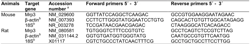
transcriptase was performed in a thermal cycler at 25°C for 5 min, 45°C for 30 min, and 85°C for 5 min. Real Time quantitative PCR (qRT-PCA) was performed according to the iQ SYBR Green Supermix (Bio-Rad Laboratories, Hercules, CA, USA) protocol. PCR amplification was carried out in 25 µL reaction volume containing 25 ng of cDNA, 1× iQ SYBR Green Supermix (100mM KCl; 40mM Tris-HCl; pH: 8.4; 0.4mM each dNTP; 40 U/mL iTaq DNA polymerase; 6mM MgCl2; SYBR Green I; 20 mM fluorescein; and stabilizers) (Bio-Rad) and 250 nM of gene specific primers. Primer sequences designed using Beacon Designer 4.02 software (PREMIER Biosoft International, Palo Alto, CA, USA) and listed in (Table 1). Cycling parameters were determined and the results were analyzed by using the comparative Ct method as the means of relative quantification, normalized to two references genes and expressed as 2−ΔΔCT. Melting curve analysis was performed to assess product specificity.
2.3 Western Blot Analysis
Qaisiya et al.; BJMMR, 14(10): 1-6, 2016; Article no.BJMMR.25139
Table 1.List of primers sequence
Animals Target gene
Accession number a
Forward primers 5´ - 3´ Reverse primers 5´ - 3´
Mouse Mrp3 NM_029600 GGTTATCCAGGCTCAAGAC GCCGTGGAAGGAATAGAAC
β-actinb NM_007393 CCTTCTTGGGTATGGAATCCTGTG CAGCACTGTGTTGGCATAGAGG
18Sb NR_003278 TCCGATAACGAACGAGAC CTAAGGGCATCACAGACC
Rat Mrp3 NM_080581 TGTGGGTCTTTCCGTGTC GCCTCAGTCTCCGTCTTAG
β-actinb NM_031144.2 GGTGTGATGGTGGGTATG CAATGCCGTGTTCAATGG
18Sb X01117 CGTCTGCCCTATCAACTTTCG GCCTGCTGCCTTCCTTGG
a
Accession numbers of target genes obtained from NCBI database
b
Housekeeping genes used for normalizing the expression of target genes
concentration were assessed using the bicinchoninic acid reaction [13]. Proteins were separated by SDS–PAGE in 10% acrylamide gel. At the end of electrophoresis the gel was obtained and blotted onto a nitrocellulose membrane (PVDF) with a wet blotting system (Sigma, St. Louis, MO, USA). The membrane was washed three times by T-TBS (0.2% Tween 20; 20mM Tris; and 500mM NaCl; pH 7.5), and blocked with the 4% milk-T-TBS solution for 1 hour at room temperature. Membranes were incubated with first antibodies anti-Mrp3 (C-18, santa cruz biotechnology, USA) and anti-actin (A2066, Sigma Aldrich). Next day, the membrane was washed and incubated with anti-rabbit HRP-conjugated (Dacko laboratories, Italy) for 1 hour at room temperature. The peroxidase reaction was obtained by exposure of the membrane in the ECL-Plus Western Blotting detection system solutions (Amersham–PharmaciaBiotech, UK). The optical density of the target proteins were analyzed by Scion Image program, normalized to α-actin and represented as protein relative expression.
2.4 Statistical Analysis
Data are reported as mean ± SD of 6 animals / each group. Differences between means were assessed by the two-tailed Student “t test”. * P < 0.05, ** P < 0.01 and *** P < 0.001.
3. RESULTS
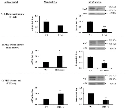
Results showed a clear decrease of hepatic
Mrp3 proteins (about 60%, P = 0.001) in β-thalassemic mouse compared to WT controls,
while no significant change was observed at mRNA level (Fig. 1A). The antibodies allowed the identification of two bands of Mrp3 protein that were described by others [5,14]. To determine whether other members of Mrps family maybe modulated to compensate for Mrp3 protein down-regulation in β-thalassemic mouse, we examined the level of expression of selected Mrp1 and Mrp2 that are located at basolateral and apical
membrane of hepatocytes, respectively. Both transporters have the same predicted topological structure of Mrp3 and shared a wide spectrum of related substrates with Mrp3 [4,15]. Mrp1 and Mrp2 mRNA and proteins were not changed in β-thalassemic mouse comparted to WT controls (data not shown). To confirm Mrp3 protein down-regulation we induced hemolysis in mice and rats by PHZ. Hepatic Mrp3 mRNA expression was 2 folds higher in PHZ-mouse (P = 0.05) and PHZ-rat (P = 0.01) compared to controls, while Mrp3 protein levels showed a decrease of 70% and 60% in PHZ-mouse and PHZ-rat (P = 0.01), respectively (Figs. 1B and 1C).
4. DISCUSSION
Impairment of the bilirubin conjugation or exportation via canalicular route leads to the higher induction of Mrp3, indicating that it functions in the export of bilirubin from hepatocytes to blood stream for renal excretion [6-10]. In the present study, we asked whether Mrp3 will also be up-regulated in vivo in response to higher level of bilirubin induced by hemolysis. Total serum bilirubin was 6 folds and 4 folds higher in β-thalassemic mouse and PHZ-treated rodents, respectively (data not shown).
generation of oxidative stress that leads to protein damage [16,17]. However, Mrp3 protein decrease appears to be Mrp3 specific since the protein expression of the closely related transporters (Mrp1 and Mrp2) was not affected by hemolysis (data not shown). Further studies are needed to identify the cause(s) of specific Mrp3 protein downregulation under hemolysis.
Using the same β-thalassemic mouse model, another study showed a down-regulation of Mrp6 at the transcriptional level [18]. As a results, authors suggested complicated syndromes
Fig. 1. Hepatic Mrp3 mRNA and protein expression. (A) β-thalassemic mouse (β-Thal) vs wild type mouse (WT). mouse) vs wild type mouse (WT).
panel: Mrp3 mRNA expression was analyzed by qRT
to housekeeping genes (β-actin and 18S) and represented as relative to WT controls. Right panel: Representative Western blot analysis for Mrp3 and α
density of Mrp3 proteins were normalized to actin and rep
generation of oxidative stress that leads to ]. However, Mrp3 protein decrease appears to be Mrp3 specific since the protein expression of the closely related transporters (Mrp1 and Mrp2) was not affected by hemolysis (data not shown). Further studies are needed to identify the cause(s) of specific
rp3 protein downregulation under hemolysis.
thalassemic mouse model, regulation of Mrp6 As a results, suggested complicated syndromes
(Pseudoxanthoma-elasticum-like syndrome implies the presence of abnormal circu molecules due to alteration in Mrp6 expression profile that will affects the transporter activity toward its biological substrates. Mrp3
include endogenous compounds (estradiol glucuronide, leukotriene C4, monovalent salts such as cholate and glycocholate), and several drugs, such as acetaminophen glucuronide and fexofenadine [19].
hepatic Mrp3 protein down-regul
hemolysis affects the transporter activity toward its substrates is still unknown.
Hepatic Mrp3 mRNA and protein expression. Left panel: Different animal models Thal) vs wild type mouse (WT). (B) PHZ treated mouse (PHZ mouse) vs wild type mouse (WT). (C) PHZ treated rat (PHZ-rat) vs wild type rat (WT).
Mrp3 mRNA expression was analyzed by qRT-PCR. mRNA expression was normalized actin and 18S) and represented as relative to WT controls. Representative Western blot analysis for Mrp3 and α-actin proteins. The optical density of Mrp3 proteins were normalized to actin and represented as relative expression
like syndrome) that implies the presence of abnormal circulating molecules due to alteration in Mrp6 expression profile that will affects the transporter activity toward its biological substrates. Mrp3 Substrates include endogenous compounds (estradiol-17β-glucuronide, leukotriene C4, monovalent bile salts such as cholate and glycocholate), and several drugs, such as acetaminophen ]. Weather the regulation under hemolysis affects the transporter activity toward
animal models. PHZ treated mouse (PHZ-rat) vs wild type rat (WT). Middle PCR. mRNA expression was normalized actin and 18S) and represented as relative to WT controls.
Qaisiya et al.; BJMMR, 14(10): 1-6, 2016; Article no.BJMMR.25139
5. CONCLUSION
Our present data showed a novel observation of down-regulation of hepatic Mrp3 protein during hemolysis. We hypothesize that a similar decrease might occur in patients of different hemolytic disorders and further studies are needed to discover the effects of this decrease on liver physiology under hemolysis.
CONSENT
It is not applicable.
ETHECAL APPROVAL
All authors hereby declare that "Principles of laboratory animal care" (NIH publication No. 85-23, revised 1985) were followed, as well as specific national laws where applicable. All experiments have been examined and approved by the appropriate ethics committee.
COMPETING INTERESTS
Authors have declared that no competing interests exist.
REFERENCES
1. Moscovitz JE, Aleksunes LM.
Establishment of metabolism and transport pathways in the rodent and human fetal liver. Int. J. Mol. Sci. 2013;14:23801– 23827.
2. Erlinger S, Arias IM, Dhumeaux D. Inherited disorders of bilirubin transport and conjugation: New insights into
molecular mechanisms and
consequences. Gastroenterology. 2014; 146:1625–1638.
3. Bellarosa C, Bortolussi G, Tiribelli C. The role of ABC transporters in protecting cells from bilirubin toxicity. Curr. Pharm. Des. 2009;15:2884–2892.
4. Deeley RG, Westlake C, Cole SPC. Transmembrane transport of endo- and xenobiotics by mammalian ATP-binding cassette multidrug resistance proteins. Physiol. Rev. 2006;86:849–899.
5. Scheffer GL, et al. Tissue distribution and induction of human multidrug resistant protein 3. Lab. Investig. J. Tech. Methods Pathol. 2002;82:193–201.
6. Keppler D. The roles of MRP2, MRP3, OATP1B1, and OATP1B3 in conjugated hyperbilirubinemia. Drug Metab. Dispos. 2014;42:561–565.
7. Nishiya T, et al. Tienilic acid enhances hyperbilirubinemia in Eisai hyperbilirubinuria rats through hepatic multidrug resistance-associated protein 3 and heme oxygenase-1 induction. Toxicol. Sci. Off. J. Soc. Toxicol. 2006;91:651–659. 8. Chai J, et al. Elevated hepatic
MRP3/ABCC3 expression in human
obstructive cholestasis is mediated through TNFα and JNK/SAPK signaling
pathway. Hepatol. Baltim. Md.
2012;55:1485–1494.
9. Belinsky MG, et al. Analysis of the in vivo functions of Mrp3. Mol. Pharmacol. 2005; 68:160–168.
10. Higuchi K, et al. Modulation of organic anion transporting polypeptide 1 and multidrug resistance protein 3 expression in the liver and kidney of Gunn rats. Hepatol. Res. Off. J. Jpn. Soc. Hepatol. 2004;29:60–66.
11. De Franceschi L, et al. Oxidative stress and β-thalassemic erythroid cells behind the molecular defect. Oxid. Med. Cell. Longev; 2013.
12. Cekic D, et al. Upregulation in the expression of multidrug resistance protein Mrp1 mRNA and protein by increased bilirubin production in rat. Biochem. Biophys. Res. Commun. 2003;311:891– 896.
13. Smith PK, et al. Measurement of protein using bicinchoninic acid. Anal. Biochem. 1985;150:76–85.
14. Hitzl M, et al. Influence of omeprazole on multidrug resistance protein 3 expression in human liver. J. Pharmacol. Exp. Ther. 2003;304:524–530.
15. Haimeur A, Conseil G, Deeley RG, Cole SPC. The MRP-related and BCRP/ABCG2 multidrug resistance proteins: Biology, substrate specificity and regulation. Curr. Drug Metab. 2004;5:21–53.
16. Mariani R, Trombini P, Pozzi M, Piperno A. Iron metabolism in thalassemia and sickle cell disease. Mediterr. J. Hematol. Infect. Dis. 2009;1.
17. Nienhuis AW, Nathan DG.
manifestations of the β-thalassemias. Cold Spring Harb. Perspect. Med. 2012;2. 18. Martin L, Douet V, VanWart CM, Heller
MB, Le Saux O. A mouse model of β-thalassemia shows a liver-specific down-regulation of Abcc6 expression. Am. J. Pathol. 2011;178:774–783.
19. Zhou SF, et al. Substrates and inhibitors of human multidrug resistance associated proteins and the implications in drug development. Curr. Med. Chem. 2008;15: 1981–2039.
_________________________________________________________________________________
© 2016 Qaisiya et al.; This is an Open Access article distributed under the terms of the Creative Commons Attribution License (http://creativecommons.org/licenses/by/4.0), which permits unrestricted use, distribution, and reproduction in any medium, provided the original work is properly cited.
Peer-review history: