_____________________________________________________________________________________________________ *Corresponding author: E-mail: [email protected];
SCIENCEDOMAIN international
www.sciencedomain.org
Novel Therapeutic Strategy in the Treatment of
Diabetes Type 2, the Use of Autologous Peripheral
Blood Stem Cells in 15 Patients: Is There Any
Relation with the Incretin-GLP-1/GIP Axis?
Ciro Gargiulo
1*, Van H. Pham
2, Tao D. Huynh
3, Vo L. H. Trieu
1,
Nguyen C. D. Kieu
1, Melvin Shiffman
4, Mark J. Holterman
5, Sergey K. Aityan
6and Luis Filgueira
71
Faculty of Medicine, HMIC-Tan Tao University, Ho Chi Minh City, Vietnam. 2
Faculty of Medicine, Nam Khoa Bioteck-Tan Tao University, Ho Chi Minh City, Vietnam. 3
Department of Stem Cell Research, Pham Ngoc Thach University of Medicine, Ho Chi Minh City, Vietnam. 4
Plastic Surgeon and Stem Cell Specialist Private Consultant, Tustin, CA, USA. 5
Faculty of Surgery, UICOMP, Chicago, IL, USA. 6
Multidisciplinary Research Center, Lincoln University, Oakland, CA, USA. 7
Department of Medicine, Faculty of Anatomy University of Fribourg, Fribourg, Switzerland.
Authors’ contributions
This work was carried out in collaboration between all authors. Authors CG and MJH designed the project, wrote and conducted the project. Authors VHP and LF conducted RT-PCR and cell analysis and organized data. Authors VLHT and TDH supervised the work procedure in the lab and performed culture of stem cells. Author NCDK researched data and organized funds. Author MS checked and revisited the manuscript. Author SKA performed statistical analysis. All authors read and approved the final manuscript.
Article Information
DOI: 10.9734/BJMMR/2017/32206 Editor(s): (1) Domenico Lapenna, Associate Professor of Internal Medicine, Department of Medicine and Aging Sciences, University “G. d’Annunzio” Chieti-Pescara, Chieti, Italy. Reviewers: (1) Agbo John, Abubakar Tafawa Balewa University, Bauchi, Nigeria. (2)Jessica Elena Mendieta-Wejebe, Escuela Superior de Medicina del Instituto Politécnico Nacional, México. Complete Peer review History:http://www.sciencedomain.org/review-history/18350
ABSTRACT
Peripheral blood stem cells (PB-SCs) are probably the most common and the most “slighted” stem cells utilized in medicine, their clinical application is back to year 1986 with the intent of replacing BM as a stem-cell source. This brief manuscript provides a general view into the amazing world of PB-SCs. Since then PB-SCs have been widely studied and the outcomes revealed a very particular biological character that lead to their clinical use in degenerative metabolic diseases as diabetes type 2 (DM2). Based on published data, we have proposed that a combination of both low glycemic index diet (LGI diet) and PB-SCs would generate major improvements in glucose metabolism via positive modification on GLP-1/GIP-Insulin axis. We have elucidated the beneficial effects of the LGI diet combined with PB-SCs on glucose tolerance in 15 individuals. We examined physiologic changes in whole-body insulin sensitivity and insulin and lipid profile after autologous PB-SCs, followed by a LGI diet regimen, which is a central tool in glucose clearance in the post-treatment period. Thus, it was discussed the modulating and regenerative activity of PB-SCs and LGI diet on the insulin, incretin/GLP-1 axis in response to sugar drive typical of DM2 condition.
Keywords: HPB-SCs; LGI diet; type 2DM; HbA1c; MSCs; NSCs; ESCs; HSCs; GLP-1; incretin; insulin.
1. INTRODUCTION
1.1 Brief Story on Peripheral Blood Stem
Cells
It has been already 3 decades since PB-SCs were used in clinical procedures in human transplantation. This marks the first clinical attempt evidencing the enormous potential of circulating blood stem cells in regenerating permanently the lympho-hematopoietic system after a myeloablative treatment. Remarkably, the outcomes have revealed the important homeostatic potential of hematopoietic blood stem cells and their capacity to rebuild and preserve an identical cellular concentration within marrow spaces and niches [1]. The existence of circulating progenitor cells with multipotential and pluripotential features was elucidated during the 50s by the presence of multiplying, developing non-leukemic DNA-synthesizing cells in peripheral blood, an event that clearly show a migratory event of stem cells from bone marrow (BM) able to repopulate a previously irradiated BM [2,3]. A cornerstone of blood stem-cell transplantation which revealed close similarities with embryonic hematopoietic development, where circulating multipotent hematopoietic progenitor cells locate and colonize the vascularized cellular matrix [4].
In the early ‘60s, it was introduced the term “Blood Stem Cells”. However, the initial clinical attempts performed in New York and Seattle faced few difficulties mainly due to the lower amount of viable stem cells compared with the total viability obtained from BM [1,5-7]. Scientists
concluded that the viable quantity was an important factor and thus, the real step forward was reached when the continuous-flow apheresis technology got in the scene. With this new equipment scientists from University of Texas MD Anderson Cancer Center were able to process 2-3 times the patient's blood volume during only one step within a relatively short period of time of 2-3 hours. Ten years later there were the first successful attempts both in vitro and in vivo trial at MD Anderson Cancer Center, the outcomes definitively confirmed the “status” of PB-SCs as comparable tools of BM-SCs for clinical use [8].
success, in short time, the blood progenitor stem cells were able to reconstruct and regenerate the entire hematopoietic environment [10,11].
1.2 Peripheral Blood Stem Cells in
Treatment
of
Diabetes
Mellitus
Diseases
By the end of 1996 it was clear that PB-SCs potentials were more constant and reliable and, several were the advantages in clinical application compared to those from other sources, especially BM. The clinical outcomes showed earlier hematopoietic recovery, less risks to immunologic challenges, very low risk for the donor, lower cost of procedure and no ethical issue involved [12,13]. The relative easiness of isolating greater numbers of cells created the possibility to proceed on multi-step transplantation in the treatment of malignancies, permitting various rounds of chemotherapy treatment with increased dosages [13]. The advanced of PB-SCs into the use of allogeneic treatment has been also met encouraging results and, progresses made in cytokine, interleukins and growth factors research and ex vivo transplantation enabled new treatment strategies involving different types of technics. Since then, PB-SCs became a therapeutic option also in certain nonmalignant diseases such as diabetes, arthritis or immune disorders [13].
The unexpected initial findings of positive results after autologous PB-SCs prompted the design of randomized prospective clinical trials. The year of 1999 signed the “momentum” that confirmed the cell-based therapies for diabetes mellitus with the use of islet cells from cadaveric donors, and the success of the Edmonton protocol [14]. Type 1 diabetic patients who received from fresh cadaveric source a transplant of islet of Langerhans showed a normalization parameters of insulin, blood glucose and glycosylated hemoglobin, and insulin independence. The procedure previewed the use of immune-suppressor regimen composed by low dose of tacrolimus and normal dose of sirolimus to preserve the graft and preclude a possible rejection [15]. From that moment, more efforts have been pursued on the use of stem cell with the intention of regenerating, reprogramming and repairing damaged tissues; stem cells were induced to differentiate to islet-like cells or insulin producing cells in vitro ready to be transplanted [14]. The main intent with this procedure was to avoid the serious reaction due to allogeneic islet transplantation, cell-self exhaustion and overcome the problem of donor’s shortage [15].
The present study is focused on elucidating regenerative dysfunction and anomalies in patient with metabolic disorders, indicating the challenges involved in the application of stem cell-based therapies for treatment of diabetic complications. In addition, ex vivo and in vivo functional manipulation(s) of PB-SCs to overcome these difficulties are discussed.
1.3 Peripheral Blood Stem Cells and
Diabetes
why
the
Regenerative
Mechanism is Compromised
The presence of variegate progenitor stem cells in peripheral blood (PB) is something that have been demonstrated since very recently and it might be the strongest clinch for PB source. The finding of different types of progenitor cells such as endothelial progenitor cells (EPCs), embryonic like cells (ESCs), hematopoietic stem cells (HSCs), mesenchymal stem cells (MSCs), neural stem cells (NSCs) progenitor natural killer cells (pNKs) in human PB it’s a step forward in the understanding of cell-based therapeutics in many pathological conditions their limitations as well as their real potentials. These cells work together in “en ensemble” dynamic micro-environmental contest and, these findings may certainly justify the results obtained by the use of PB-SCs as treatment option for complications which need a revascularization, vascular and tissue repair approach [16,17].
The primary concern with autologous stem cell therapy in diabetic patients is patient’s broad metabolic dysfunction that negatively affect their quality and functionality. Diabetes degeneration mechanism is characterized by damages of local stem cells within the osteoblastic niche inside BM that generate two main events, the “stem cell mobilopathy” and the “bone marrow micro-angiopathy”, conditions referring to an impaired control of stem cell movement into the systemic circulation and their limited differentiation capacity. Data have proved the reduced migratory mobility and limited proliferative capacity of diabetic stem cell in general (MSCs, ESCs, HSCs, EPCs), together with an altered cytokine/growth factor secretory capacity that can decrease the repair mechanisms of normal vascular and tissue growth [16-20].
chronic hyperglycemia and exert a protective action in cardiomyopathy [21-24]. The well functionality of new- β cell clones may be negatively affected by the continuous up-down of glucose, an occurrence that could be related to both an inheritance mechanism or to a long term rich sugar-carbohydrates diet. In addition the high oxidative typical of diabetic condition contributes to destruction of the microcellular homeostatic balance which is a key factor for reparative activity of damaged organs, tissues and cells and immune responses [25,26].
The beneficial outcome of PB-SCs in the treatment of diabetes reside in the particular biological nature of MSCs and ESCs. They exert a natural pleiotropic activity moving to the sites of inflammation and show a strong immunomodulatory and anti-inflammatory activity through cell to cell interactions, lymphocytes or the secretion of soluble factors, hormones, cytokines and interleukines. Intriguingly, mobilized stem and progenitor cells are able to move and locate in the crypts of the self-renewing gut epithelium where they differentiate to absorptive and secretory cells [27]. The mobilized quantity of these progenitors is based upon the activity of γ-secretase/NOTCH pathway and, the inhibition of NOTCH signaling results in an intensification of secretory cells over a generation of newly enterocytes. Part of these progenitors with pluri-multipotency capacity differentiate to insulin-producing cells (IPCs) which express numerous genes in charge of the growth and expansion of pancreatic α/β-cells and L cells, part express pancreatic and duodenal homeobox 1 responsible for the incretin/glucagon hormone secretion such as glucagone-like peptide-1 (GLP-1).
The GLP-1/incretin is hormone with a high insulinotropic effect and currently, GLP-1 analogues based therapies are the new frontier in type 2 diabetes treatment. GLP-1 receptors are largely present in several special tissues including those of pancreas, brain, stomach, myocardium and heart vasculature and this confirms, the key role that incretin/GLP-1 play as neurocardio protective and neutropic factor with substantial beneficial effects on both lipids and blood pressure as well, similarly SCs do [24,28-35]. Thus, we propose the presence of a mechanism through which PB-SCs may eventually exert their systemic regenerative activity either through a direct stimulation or simply via these GLP-1 receptors.
1.4 The Importance of a Proper Cellular
Microenvironment
The assessment of host cellular microenvironment condition seems to be the essential pre-existing criteria for the success of a cell-based therapy. The idea is to create the most appropriate milieu to sustain adhesion, viability and proper proliferation of stem cells improving interaction within cell surface molecules and extracellular matrix proteins. Thus, medical scientists has proposed the use of co-adjuvants as part of therapy procedure such as antioxidants and/or BM mobilizing agents as part of therapeutic protocol [36]. Although lifestyle-changes have shown to improve weight loss with consequent benefits on insulin resistance in pre-diabetic and diabetic patients, the results are not persistent and durable. Postprandial hyperinsulinemia that causes glucotoxicity is the main issue in this contest and can be reduced only when a low-GI diet is added, as it has been widely demonstrated a high-GI diet negatively affect both pancreatic β cell and intestinal L/K cell activity regardless of substantial weight loss, highlighting the crucial involvement of the gut in determining the effects of a low-GI diet on type 2 diabetes complication [37-39].
Glucotoxicity is a pathological condition that affects more than β cell [37-40] and such a persistent stress on the GIP/GLP-1 mechanism in the intestinal tract might be the cause of a more serious metabolic impairment that can’t be solved by doing exercise and an LGI diet. Though the mechanism is not yet completely elucidated, the assumption is eventually sustained by the fact that hyperglycemia has been shown to down-regulate GIP receptor expression [40,41], altering the postprandial incretin response, and intensifying the GIP glycation [42,43].
2. MATERIALS AND METHODS
2.1 Study Procedure: Inclusion/Exclusion
Criteria and Analysis
The current study have been conducted on 15 consented patient according to the guidelines of Helsinki Declaration and approved by the Tan Tao University (TTU) ethical committee. Fifteen type DM2 patients were enrolled in the study between January 2014 and January 2016. The mean age was 62 years (48 to 84 years) under oral anti-diabetic drug and insulin injection. Nine patients (69%) were male and 4 patients (31%) were female. Pretreatment and follow-up characteristics of patients are showed in Table 1 and 2. Mean body weight before autologous PB-SCs was 68 Kg (range from 58 to 87 Kg.) at the time of initial diagnosis. Mean HbA1c before PB-SCs was 7.8 (range from 6% to 10.2%). The mean number of infused mononuclear cells 7/8 days of culture was 3.76×10⁸/ml. The follow-up was performed in six months from the end of the treatment.
All subjects underwent medical evaluation that included glycemic control, micro and macrovascular complications. Assessment at the time of enrollment comprised blood pressure (BP), body weight, complete CBC, hormone profile including Testosterone, Estradiol, Progesterone, Cortisol, ACTH, Leptin, Insulin, TSH, T4-3 and Vitamin D, lipid profile, TNFα, IFNβ, IL2-4, electrocardiography, echocardiography. The whole panel of current medications (including oral hypoglycemic drugs, insulin, blood-lipid lowering drugs) was also recorded. HbA1c was measured by Immunoturbidimetry method. Patients were asked to monitor a four-point glucose profile (fasting, 2 hr breakfast, lunch, post-dinner) using the Optium glucometer (Abbott, Mumbai, India) at least once per week with a target of fasting plasma glucose between 70 and 130 mg/dl and postprandial glucose ≤ 180 mg/dl four times per day. Treatment procedure: Peripheral blood stem cells isolation.
The selection has been done based on precise exclusion criteria that included obesity, alcohol/tobacco addiction and, chronic kidney failure and depressive condition. The procedure previewed 10 sessions-infusion of autologous PB-SCs during a period of 3 weeks injected both sub-cutaneous and through the antecubital vein. The primary aim was a steady decrease of both oral anti-diabetic and insulin injection by ≥ 100%
starting by the 3rd to 5th stem cell injection, maintaining HbA1c ≤ 7% as final objective during the following 6 months / 1 year, as stated by American Diabetes Association guidelines. All the patients were advised to follow LGI diet and a constant physical activity in the form of low and soft exercises for 30 min per day to be started 2 weeks prior the stem cell therapy. The importance of both LGI diet and physical activity was reinforced at each visit, and a schedule with all the data was also recorded at each visit.
From the original fifteen patients fourteen achieved the primary end point within the first quarter after the treatment, one patient abandoned the project due to her continuity with a high glycemic index diet. For fourteen patients the anti-diabetic drugs were steadily terminated by the end of the ten session of autologous PB-SCs. The drop of HbA1c was significantly visible during/after the time patient received stem cells and the level was overall better compared with medication treatment. The HbA1c level was checked in 1 month, 4-6 months, and 12-18 months after the PB-SC treatment. All fourteen patients could maintain HbA1c < 7% for the following 6 months/1 year without any major complication.
Insulin and glucagon-stimulated C-peptide were excluded as weighty criteria as at the moment of the treatment the values were within the normal range due to the effect of previous diabetes treatment. Thus, decreased drug requirement positively correlated with blood pressure, cholesterol and triglycerides rather than C-peptide.
2.2 Stem Cell Isolation Procedure
Mononucleated cells were isolated by density gradient centrifugation using Ficoll-Paque™ PLUS (GE Healthcare, Uppsala, Sweden). A total of 10 blood samples (35 ml each) were carefully layered 1:2 on Ficoll-Paque and centrifuged at 300 g for 20 min at 20-C°. The mononucleated cell layer, 2x10⁷, at the plasma-Ficoll interface, were aspired and was washed three times with phosphate buffered saline and cultured in 25T flasks with free serum medium containing 2% (v/v) penicillin-streptomycin at 37°C in a humidified atmosphere containing 5% CO2 for a period of 7.3 days. Suspension and
the trypan blue exclusion assay was used to observe the proliferation of the cells. Cells were cultured for 7.3 days average, subsequently suspension and adherent cells were collected and injected to patients’. PB-SCs have undergone a series of test to evaluate the feasibility and safety of the therapy. The patients’ cell samples were tested for bacteria, fungal and virus contamination prior autologous PB-SCs infusion.
3. RESULTS
3.1 Characterization of Pluripotency and
Multipotency of Human Peripheral
Blood Stem Cells
Characterization of HSCs, MSCs, ESCs, NSCs and NK progenitor cells present in PB
isolated stem cells was main outcome of a
previous edited study performed by our group [17]. The presence of these sub-sets of
stem cells was verified by RT-PCR and flow-cytometry analysis by determine the
presence of specific cluster of differentiation makers (CD markers). Fig. 1 a-b shows flowcytometry results of adherent stem cells positive for CD14, CD34 and CD45 for human hematopoietic lineage; CD44, CD73, CD90 and CD105 for human mesenchymal lineage; CD133 and Nestin for human neural lineage; SSEA3 and Tra1 for human embryonic lineage; CD56 and
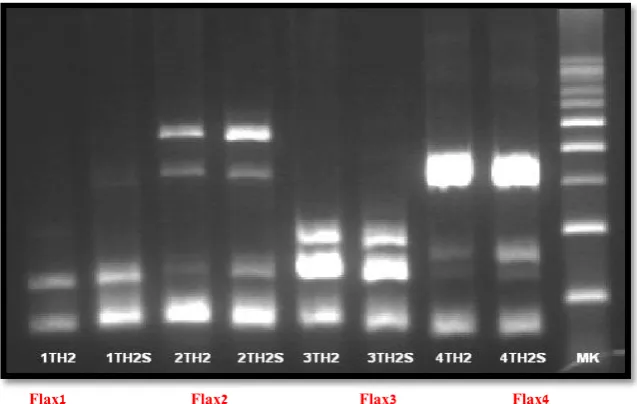
CD16 for NK progenitor lineage. As shown in Fig. 2, the amplification of the RT-PCR products
of GAPDH and a panel of transcription factors that regulate self-renewal and pluripotency in hESCs, Oct4, Sox2, OCN, Nestin, Nanog and DPM [17].
a b c d
Picture 1. Primary hPB-SCs 200x 12 days culture (a MSCs, b NSCs, c ESCs, d HSCs no stain)
a b
Fig. 1. a Immunophenotype of PB-SCs stem cells isolated by fibrin microbeads (FMB). FACS analysis of the immunophenotype profile for hematopoietic marker CD45, CD34 and CD14; for
mesenchymal antigens CD90, CD73, CD44 and CD105; for embryonic lineage CD133 and Nestin; for neural lineage SSEA3 and TR-1; b CD56-16 for NK progenitor lineage. Cells isolated
Fig. 2. Reverse transcription polymerase chain reactions (RT-PCRs) were performed on both adherent and suspended mononucleated cells. GAPDH and Sox2, Oct4, Nanog, Nestin, Dmp and osteocalcin (Ocn) were expressed in both types of mononucleated cells. A 800-bp DNA size marker was used to identify the approximate size of the RT-PCR products and GAPDH was used as internal control. All the RT-PCR products were completely whipped out of DNA remained and confirmed with mRNA sequencing to be 100% identical to the known sequences obtained from BLAST analysis. Flax1 indicates the expression of GAPDH 94 Kb, OCT4 144 Kb; Flax2 indicates the expression of GAPDH 94 Kb, Ocn 150 Kb, Nestin 496 Kb; Flax3 indicates the expression of GAPDH 94 Kb, Sox2 151 Kb, DMP 200 Kb; Flax4 indicates the expression of
GAPDH 94 Kb, Nanog 346 Kb
Table 1. HbA1c dynamics through treatment phases
Patient HbA1c level HbA1c dropped by HbA1c percentage drop
# Gender Age InD Med SC InD-Med Med-SC InD- SC InD-Med Med-SC InD- SC
1 Female 64 9.3 6.0 5.9 3.3 0.1 3.4 35% 2% 37% 2 Female 65 9.0 8.3 7.1 0.7 1.2 1.9 8% 14% 21% 3 Male 73 8.0 7.5 6.5 0.5 1.0 1.5 6% 13% 19% 4 Female 54 9.8 9.6 5.2 0.2 4.4 4.6 2% 46% 47% 5 Male 66 11.0 10.2 5.4 0.8 4.8 5.6 7% 47% 51% 6 Female 84 7.4 6.0 5.9 1.4 0.1 1.5 19% 2% 20% 7 Male 50 10.0 8.3 6.3 1.7 2.0 3.7 17% 24% 37% 8 Male 62 7.2 6.7 6.7 0.5 0.0 0.5 7% 0% 7% 9 Male 48 8.9 8.4 6.1 0.5 2.3 2.8 6% 27% 31% 10 Male 56 8.8 8.1 6.0 0.7 2.1 2.8 8% 26% 32% 11 Male 60 10.0 8.9 7.1 1.1 1.8 2.9 11% 20% 29% 12 Male 62 8.7 6.3 6.2 2.4 0.1 2.5 28% 2% 29% 13 Male 68 8.5 7.1 6.4 1.4 0.7 2.1 16% 10% 25% 14 Male 73 8.0 7.3 6.3 0.7 1.0 1.7 9% 14% 21% 15 Male 64 9.5 7.0 6.6 2.5 0.4 2.9 26% 6% 31% Mean: 63 8.9 7.7 6.2 1.2 1.5 2.7 14% 17% 29% St.Dev.: 9.3 1.0 1.3 0.5 0.9 1.5 1.3 10% 15% 11% Max: 84 11.0 10.2 7.1 3.3 4.8 5.6 35% 47% 51% Min: 48 7.2 6.0 5.2 0.2 0.0 0.5 2% 0% 7%
InD – initially diagnosed, Med – after conventional medications (oral and insulin injections), SC – after treatment with stem cells (tested one month after the treatment)
Table 1 also shows the HbA1c absolute and percentage drop in each phase of treatment.
Abbreviation “InD” stands for “Initially diagnosed”, “Med” stands for “After conventional
8 oral medication and insulin injections,” and “SC” for “After treatment with stem cells.” All our patients were treated with the conventional medication before they received the PB-SC treatment Thus, the SC phase always followed the Med phase.
Table 2 shows some other important parameters as the patients’ weight, cholesterol,
triglyceride, and blood pressure before and after the PB-SCs. Patients’ weight and age in Table 1
and Table 2 are shown at a time of the
first appointment for the treatment with stem cells.
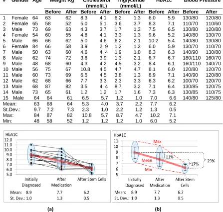
Fig. 3 shows the HbA1c reduction after the treatment with conventional medication including oral medication and insulin injections and after PB-SC treatment. The HbA1c dynamics for each of the fifteen patients along with the mean value for the group is shown in Fig. 3(a) while the dynamics of the mean level, standard deviation, max and min level of HbA1c for the group is shown in Fig. 3(b).
Table 2. HbA1c, weight, cholesterol, triglyceride, and blood pressure before and after PB-SC
# Gender Age Weight Kg Cholesterol (mmol/L)
Triglyceride (mmol/L)
HbA1c Blood Pressure
Before After Before After Before After Before After Before After
1 Female 64 63 62 8.3 4.1 6.2 1.3 6.0 5.9 130/80 120/80 2 Female 65 58 52 5.0 5.1 3.6 3.7 8.3 7.1 110/70 110/60 3 Male 73 69 63 4.3 3.7 1.7 1.3 7.5 6.5 130/80 120/80 4 Female 54 60 55 4.8 4.1 3.3 1.3 9.6 5.2 140/80 130/70 5 Male 66 66 62 5.0 4.6 6.2 2.1 10.2 5.4 140/80 130/80 6 Female 84 66 58 3.9 2. 9 1.2 1.2 6.0 5.9 130/70 110/70 7 Male 50 63 60 4.6 4. 4 1.9 1.0 8.3 6.3 140/90 130/80 8 Male 62 74 72 3.6 3.9 1.3 2.1 6.7 6.7 180/110 160/70 9 Male 48 68 60 4.3 4.2 4.5 3.2 8.4 6.1 160/110 140/70 10 Male 56 75 67 10.8 4.5 4.7 4.7 8.1 6.0 120/80 120/70 11 Male 60 73 69 6.5 4.5 3.8 1.3 8.9 7.1 140/90 120/80 12 Male 62 68 66 7.7 3.3 2.3 3.3 6.3 6.2 100/70 120/70 13 Male 68 87 82 3.5 4. 4 8.7 3.2 7.1 6.4 130/85 120/75 14 Male 73 65 61 1.2 1.2 1.7 1.6 7.3 6.3 130/85 110/75 15 Male 64 64 61 6.5 5.7 1.2 1.0 7.0 6.6 140/80 125/80 Mean: 63 68 64 5.3 4.0 3.7 2.2 7.7 6.2
St.Dev.: 9.7 7.2 7.3 2.3 1.0 2.2 1.2 1.3 0.5 Max: 84 87 82 10.8 5.7 8.7 4.7 10.2 7.1 Min: 48 58 52 1.2 1.2 1.2 1.0 6.0 5.2
Fig. 3. HbA1c dynamics (a) for each of the fifteen patients and (b) the dynamics of the mean level of HbA1c for the group
The patients quit all diabetes-related oral medications and insulin injections as soon as they had received the PB-SCs treatment. The dynamics of the results of the treatment with PB-SCs was checked in three follow-up HbA1c tests. The first follow-up was conducted in 1 month after the PB-SC treatment for the immediate “after the treatment” HbA1c results. The after the PB-SC treatment results are shown in the “SC” column in Table 1 and in the “after” column in Table 2. The second follow-up was done in 4-6 months and the third follow up was conducted in 12-18 months after the treatment. The dynamics of the results in all follow-up tests together with the initially diagnosed HbA1c level and the HbA1c after the conventional medication are shown in Table 3.
The follow-up tests have provided strong evidence that the treatment with PB-SC generates lasting results to the diabetes patients as shown in Table 3. Most patients have shown stable low HbA1c level through the follow-up tests. One patient (#2) showed significant increase of HbA1c and returned to full medication, both oral and insulin. However this patient failed to follow a LGI diet. Two more patients (#4 and #8) returned to full oral medication because their HbA1c level bounced back to 7.5 in the third follow-up test. One patient
(#9) was back on a low dose oral medication because ….
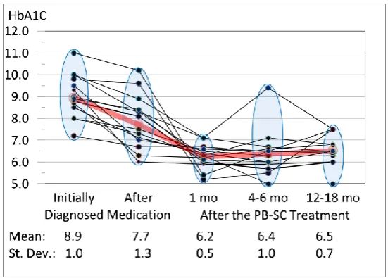
The mean values and mean percentage drop of HbA1c in different phases of the treatment are shown in Table 4 and illustrated in Fig. 4. Fig. 4 shows also the dynamics of the HbA1c level in all phases from initially diagnosed, after treatment with conventional medication and the PB-SCs with three follow-up tests for up to 18 months. As is evident from Table 4 and Fig. 2 the efficiency of the PB-SC treatment is much higher than the efficiency of treatment by conventional medication including insulin.
To analyze the statistical significance of the results, the following hypotheses were formulated and tested.
Hypothesis 1
The null hypothesis 1 (H10): The mean values of HbA1c before and after the treatment with the PB-SCs on the population represented by the given group of patients are the same.
H10 : µSC = µMed (1)
where H10 : µSC is the mean value of HbA1c after
the PB-SCs and µMed is the mean value of HbA1c
before the PB-SCs (the medication phase)
Table 3. The HbA1c dynamics through treatment phases and after treatment follow-ups
Patient HbA1c level
After treatment with PB-SC in follow-up tests # Gender Age InD Med 1 mo. 4-6 mo. 12-18 mo. Final Medication Status
1 Female 64 9.3 6.0 5.9 5.9 6.0 No Med.
2 Female 65 9.0 8.3 7.1 9.4 7.5 Back on full medication incl. inject 3 Male 73 8.0 7.5 6.5 6.3 6.3 No Med.
4 Female 54 9.8 9.6 5.2 5.5 7.5 Back on full oral medication 5 Male 66 11.0 10.2 5.4 6.5 6.4 No Med.
6 Female 84 7.4 6.0 5.9 5.9 6.0 No Med. 7 Male 50 10.0 8.3 6.3 5.0 5.8 No Med.
8 Male 62 7.2 6.7 6.7 6.5 7.5 Back on full oral medication 9 Male 48 8.9 8.4 6.1 6.0 6.5 Back on single low dose oral med. 10 Male 56 8.8 8.1 6.0 5.7 6.0 No Med.
11 Male 60 10.0 8.9 7.1 6.7 6.5 No Med. 12 Male 62 8.7 6.3 6.2 6.6 6.8 No Med. 13 Male 68 8.5 7.1 6.4 6.0 No show No Med. 14 Male 73 8.0 7.3 6.3 7.1 6.8 No Med. 15 Male 64 9.5 7.0 6.6 6.5 6.5 No Med.
Mean: 63 8.9 7.7 6.2 6.4 6.5 St.Dev.: 9.3 1.0 1.3 0.5 1.0 0.7 Max: 84 11.0 10.2 7.1 9.4 7.5 Min: 48 7.2 6.0 5.2 5.0 5.0
The alternative hypothesis 1 (H1Alt): The mean value of HbA1c after the treatment with PB-SCs is lower than the mean value of HbA1c before the treatment with PB-SCs on the population represented by the given group of patients.
H1Alt : µSC < µMed (2)
The null hypothesis was tested with the paired one-tailed Student’s t-test at α = 0.01. The test results are shown in Table 5.
Table 4. Mean value and standard deviation of HbA1c level for 15 patients at the time of initial diagnosis, after treatment with the conventional medication including insulin injections, and after the treatment with stem
cells in three follow-ups
Treatment phase Mean
value
Standard deviation
Initially diagnosed 8.9 1.0 After medication 7.7 1.3 After stem cells
(1 month)
6.2 0.5
After stem cells (4-6 month)
6.4 1.0
After stem cells (12-18 month)
6.5 0.7
Table 5. Paired two sample t-test for the null hypothesis 1
Paired T-test with α = 0.01
Variable 1 (HbA1c Before)
Variable 2 (HbA1c After)
Mean 7.7 6.2 Variance 1.69 0.30 Sample size 15
Hypothesized Mean difference
0
T 3.80
P (T ≤ t) one-tailed 0.001 The null hypothesis 1 is rejected
According to the t-test t = 3.81 and p = 0.00097, and the t-test result is significant at α = 0.01. Hence the null hypothesis was rejected and the alternative hypothesis was accepted. Thus, there is a significant difference between the measured values of HbA1c before and after the PB-SC treatment.
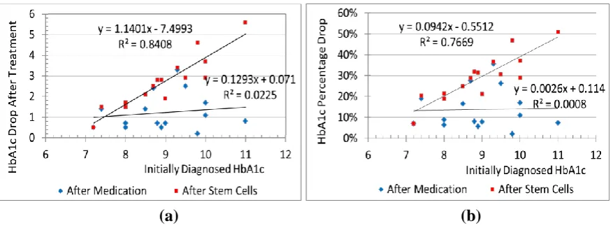
HbA1c level after treatment with the conventional medication as a function of the initially diagnosed level of HbA1c and Fig. 6 shows the absolute (a) and percentage (b) drop of the HbA1c level versus the initially diagnosed level of HbA1c. Similarly to Fig. 5 and Fig. 6, the dependencies in Fig. 7 and Fig. 8 provide strong evidence of high efficiency of the PB-SC treatment.
Fig. 4. HbA1c dynamics from initial diagnosis through medication and PB-SC treatment phases with three follow-up tests in 1 month, 4-6 months, and 12-18 months after the treatment
Fig. 5. HbA1c level after the treatment by conventional medication (Blue markers) and PB-SC (Red markets)
Fig. 6. HbA1c absolute (a) and percentage (b) drop after treatment vs the HbA1c level before the treatment
Fig. 7. HbA1c level after the treatment by conventional medication (Blue markers) and PB-SC (Red markets) versus the initially diagnosed HbA1c level
(a)
(b)
Fig. 8. HbA1c absolute (a) and percentage (b) drop after treatment vs the initially diagnosed HbA1c level
The analysis above indicates that treatment with the PB-SCs is more efficient than treatment with the traditional medication including oral medication and insulin injections. This conclusion is supported by a very high positive correlation between percentage drop of HbA1c level after the PB-SCs vs the level in the medication phase, r = 0.94 and high positive correlation between percentage drop of HbA1c level after the stem
cells vs the initially diagnosed HbA1c level, r = 0.88.
As shown in 9(a), the treatment efficiency in terms of HbA1c percentage drop after treatment with conventional medication relative to the initially diagnosed level against the patient’s age as shown in 9(b).
The analysis of relationships in 9(a) and (b) provides clear evidence that conventional medication is more efficient on elder patients while PB-SC is more efficient on younger patients. The HbA1c percentage drop after treatment with the conventional medication and the patient age shows low-medium positive correlation with the correlation coefficient, r = 0.27. On the other hand, the HbA1c percentage drop after treatment with the PB-SC and the patient’s age show a medium negative
correlation with the correlation coefficient, r = -0.47.
To summarize all our fifteen patients before the treatment with PB-SCs, were previously treated by oral medication and four of them were under insulin injection. In result of the treatment with the conventional medication but before the PB-SCs, their level of HbA1c had reduced from the
originally diagnosed mean level of 8.9% for the group (range from 7.2% to 11.0%) to the mean level 7.7% (range from 6.0% to 10.2%). The PB-SC treatment has reduced the mean HbA1c level in the group to 6.2% (range from 5.2% to 7.1%) as shown in Table 1. This HbA1c test was conducted one month after the PB-SC treatment.
4. DISCUSSION
4.1 The Relation between Peripheral
Blood Stem Cells and the Incretin
Domain, What’s the News?
The clinical use of PB-SCs and LGI diet seems to play an important role in the management of DM2. This venue together with the new outcomes obtained over the last decade that revealed the essential physiological role of incretin/GLP-1 in the evolving process of DM2, will better clarify new solid therapeutic strategies against diabetes progression. By this, we propose a possible explanation that may elucidate the way on how PB-SCs and LGI may exert a beneficial impact on DM2 through a direct or indirect induction on the incretin/GLP1-insulin axis.
Notably, there is high similarity between GLP-1 analogues and PB-SCs. Sustained pharmacological levels of GLP-1 are achieved by subcutaneous administration of GLP-1 analogues, while transient and better balanced physiological levels of glucose/insulin are attained following treatment with autologous SCs/LGI diet. Both therapeutic classes display a sustained and durable hypoglycemic action in patients with DM2. However, the GLP-1 incretin
(a)
(b)
Fig. 9. Correlation of HbA1c percentage drop after treatment with (a) conventional medications and (b) with stem cells against patient’s age
effect is known to be impaired in DM2 patients which consume high glycemic diet, and GLP-1 analogues and autologous SCs (from any sources and any type) revealed to lose their effectiveness over time in those individuals. The pathological mechanisms behind these observations is a sort of GLP1/insulin axis resistance due to a decrease in GLP-1 secretion from intestinal L-cells with a consequence decrease in GLP-1 plasma concentrations, combined or not with a reduced action of GLP-1 in the β-cell [49,50].
The key finding in this study is that, when diabetic individuals underwent autologous PB-SCs and basic life-style changes, reductions in compensatory hyper/hypoglycemia were only steadily reached when individuals consumed a low-GI diet for the whole post therapy period which lasted for > 1 year. We would like to stress the fact that, the daily glucose and HbA1c level did not change during the 2/3 week pre-period preparation characterized by low-GI diet, growth factors and bone marrow activators before stem cell intervention. Conversely, the effective initial change was seen only after the commencement of the infusion of autologous PB-SCs. These outcomes were shown to be independent of the body weight and fat mass and have to be related to changes in the gastrointestinal incretin/GLP-1. On the other hand, long term postprandial glucose response and HbA1c decreased only in those subjects who consumed a low-GI diet whereas a high-GI diet appears to progressively inactivate the activity of PB-SCs. When there is an inappropriate response to glucose, and compensatory hyper-insulinemia is altered this event is explained only by intake of high-GI food, which indicates an evident general dysfunction of β-α cell glucose and L-K incretin/GLP-1/GPI axis.
These data may clarify the specific influence of both low-GI diet/PB-SCs on the diminution of insulin secretion determined by reduced β-α cell glucose and L-K incretin/GLP-1 exposure. These outcomes are clinically significant with regards to both pharmacologic and stem cell management of individuals with DM-2. In the context of our current findings, we therefore propose that only a strict combination of diet and PB-SCs could control and reverse peripheral and hepatic/duodenal insulin resistance.
The treatment of 15 patients included in our study diagnosed with long term DM2 resulted in a significant drop of the HbA1c level for all patients together with better outcome in weight loss, cholesterol, triglycerides and blood pressure within a period of 4, 6 months and 1-1.5 year follow up (Table 2). The main endpoint included the discontinuation of oral anti-diabetic drugs and/or insulin injection. The patients that received therapy started to switch into LGI diet 2 weeks prior the cell treatment and started to reduce the amount of medication from the 3rd to 4th injection of autologous PB-SCs to complete stop by the end of the 10th injection, eventually patient’s blood sugar level and HbA1c tended to stabilize within a month after the 10th infusion treatment.
The efficiency of the PB-SC treatment can be analyzed by the comparison of the levels and the reduction of HbA1c after PB-SC treatment versus the levels and reduction in the phase of treatment by the conventional medication including oral medication and insulin injections. The PB-SCs work better with the higher level of HbA1c before the treatment than the traditional medication and are more efficient than conventional anti-diabetic medication (Figs. 3, 4).
(a)
(b)
Therefore, this procedure could exhibit favorable effects on the pathogenesis of metabolic
disorders in patients with DM2 by increasing insulin sensitivity as an
extrapancreatic activity.
5. CONCLUSION
Sequential aspects of glycemic variation, involving long term rise and fall of glucose, are associated with severe adverse effects in patients with diabetes. Currently, procedures to
quantify the risk of glycemic variation, both high and low, and the inconsistency that
includes its temporal aspects are now highly precise.
Thus, glucotoxicity is a complex disorder that affects the entire system and, a persistent stress might be the reason of a more muddy metabolic complication that need more than exercise and an LGI diet to be re-equilibrated. This assumption is sustained by the fact that chronic glucotoxicity accompanies the hyperglycemia/
hypoglycemia “rollercoaster effect” which has been shown to disrupt the GIP-incretin/
glucagone-insulin receptor expression [37-41] generating dysfunctions in pre and
postprandial incretin- glucagone- insulin responses intensifying the GIP glycation [42,43-50].
To conclude, we are well aware that the present study has several limitations. Small number of patients and, more investigation and further data are needed to confirm our hypothesis. Another limitation of the study was that assessment of a univocal lifestyle intervention that could not have been performed in a quantitative manner. Because the present results were achieved by investigating a small parameters it will be compulsory to investigate and confirm on long-term and chronic effects a larger and more specific blood factors before and after PB-SCs/LGI diet intervention and the effects on incretin/ glucagone-insulin sensitivity and HbA1c scale. However, in accordance with our analysis it was demonstrated the independent significance of clinically based PB-SCs/LGI diet therapy on the increased insulin sensitivity. Thus, we believe that this procedure could in part exhibit beneficial effects on insulin sensitivity in uncontrolled type DM2 patients. The authors would like to state that the current study is the continuation of a precedent published study on clinical application of PB-SCs on diabetic patients [51].
CONSENT
All authors declare that ‘written informed consent was obtained from the patient (or other approved parties) for publication of this paper and accompanying images.
COMPETING INTERESTS
Authors have declared that no competing interests exist.
REFERENCES
1. Körbling M, Freireich EJ. Twenty-five years peripheral blood stem cell transplantation. Blood. 2011;117:6411-6416.
2. Bond VP, Cronkite EP, Fliedner TM, Schork P. Deoxyribonucleic acid synthesizing cells in peripheral blood of normal human beings. Science. 1958; 128:202 203.
3. Swift MN, Taketa ST, Bond VP. Efficacy of hematopoietic protective procedure in rats x-irradiated with intestine shielded. Radiation Research. 1956;4:186-192. 4. Delassus S, Cumano A. Circulation of
hematopoietic progenitors in the mouse embryo. Immunity. 1996;4(1):97-106. 5. Goodman JW, Hodgson GS. Evidence for
stem cells in the peripheral blood of mice. Blood. 1962;19:702-714.
6. Cavins JA, Scheer SC, Thomas ED, Ferrebee JW. The recovery of lethally irradiated dogs given infusions of autologous leukocytes preserved at -80[deg]. Cord Blood. 1964;23:38-42. 7. Storb R, Epstein RB, Ragde H, Bryant J,
Thomas ED. Marrow engraftment by allogeneic leukocytes in lethally irradiated dogs. Blood. 1967;30:805-11.
8. Freireich EJ, Judson G, Levin RH. Separation and collection of leukocytes. Cancer Research. 1965;25:1516-20. 9. Micklem HS, Anderson N, Ross E. Limited
potential of circulating haemopoietic stem cells. Nature. 1975; 256:41-3.
10. McCredie KB, Hersh EM, Freireich EJ. Cells capable of colony formation in the peripheral blood of man. Science. 1971; 171:293-4.
12. Lie AKW, To LB. Peripheral blood stem cells: Transplantation and beyond. The Oncologist. 1997;2:40-49.
13. Ab Kadir R, Hisham Zainal Ariffin S, Megat Abdul Wahab R, Senafi S. Molecular characterization of human peripheral blood stem cells. South African Journal of Science. 2012;108(5/6):1-7.
14. El-Badri N, Gonheim MA. Mesenchymal stem cells in diabetes mellitus: Progress and challenges. Hindawi. 2013;1-7.
15. Shapiro AMJ, Lakey JRT, Ryan EA, et al. Islet transplantation in seven patients with type 1 diabetes mellitus using a glucocorticoid-free immunosuppressive regimen. The New England Journal of Medicine. 2000;230–238.
16. Jarajapu YPR, Grant MB. The promise of cell based therapies for diabetic complications, challenges and solutions. Circulation Research. 2010;106:854-869. 17. Gargiulo C, Pham VH, Hai NT, Nguyen
NCD, Pham VP, Abe K, Flores V, Shiffman M. Isolation and characterization of multipotent and pluripotent stem cells from human peripheral blood. Stem Cell Discovery. 2015;5(3):1-17.
18. Seewoodhary J, Evans PJ. Diabetes and stem cells: Endogenous effects and reparative mechanisms. British Journal of Diabetes & Vascular Disease. 2013;1-7. 19. DiPersio JF. Diabetic stem-cell
‘mobilopathy’. New England Journal of Medicine. 2011;365:2536-2538.
20. Saito H, Yamamoto Y, Hiroshi Y. Diabetes alters subsets of endothelial progenitor cells that reside in blood, bone marrow, and spleen. American Journal of Physiology and Cell Physiology. 2012; 302:C892-C901.
21. Volarevic V, Arsenijevic N, Lukic Nl, Stoj M. Concise review: Mesenchymal stem cells treatment of the complication of diabetes mellitus. Stem Cells. 2011;29:5-10.
22. Tang DQ, Cao LZ, Burkhardt BR, et al. In vivo and in vitro characterization of insulin-producing cells obtained from murine bone marrow. Diabetes. 2004; 53:1721–1732.
23. Chen LB, Jiang XB, Yang L. Differentiation of rat marrow mesenchymal stem cells into pancreatic islet beta-cells. World Journal of Gastroenterology. 2004;10:3016–3020. 24. Rezania A, Riede MJ, Wideman RD,
Karanu F. Production of functional glucagon-secreting α-cells from human
embryonic stem cells. Diabetes. 2011; 60(1):239-247.
25. Giovannucci E, Harlan DM, Archer MC, Bergenstal RM, Gapstur SM, et al. Diabetes and cancer: A consensus report. Diabetes Care. 2010;33:1674–1685. 26. Jarajapu YPR, Grant MB. The promise of
cell-based therapies for diabetic complications challenges and solutions. Circulation Research. 2010;106:854-869. 27. Van der Flier LG, Clevers H. Stem cells,
self-renewal, and differentiation in the intestinal epithelium. Annual Review of Physiology. 2009;71:241–260.
28. Volarevic V, Al-Qahtani A, Arsenijevic N, et al. Interleukin-1 receptor antagonist (IL-1Ra) and IL-1Ra producing mesenchymal stem cells as modulators of diabetogenesis. Autoimmunity. 2010; 43:255–263.
29. Xie QP, Huang H, Xu B, et al. Human bone marrow mesenchymal stem cells differentiate into insulin-producing cells upon microenvironmental manipulation in vitro. Differentiation. 2009;77:483–491.
30. Sun Y, Chen L, Hou XG, et al. Differentiation of bone marrow-derived mesenchymal stem cells from diabetic patients into insulin-producing cells in vitro. Chinese Medical Journal. 2007; 120:771– 776.
31. Holst JJ, Deacon C, Toft-Nielsen MB, Bjerre-Knudsen L. On the treatment of diabetes mellitus with glucagon-like peptide-1. Annual of New York Academy of Science. 1998;865:336–343.
32. Holst JJ, Gromada J. Role of incretin hormones in the regulation of insulin secretion in diabetic and non-diabetic humans. American Journal of Physiology and Endocrinology and Metabolism. 2004;287(2):E199–E206.
33. Stanger BZ, Datar R, Murtaugh LC, Melton DA. Direct regulation of intestinal fate by Notch. Proceedings of National Academy of Science U S A. 2005; 102(35):12443– 12448.
34. McDougall C, McKay GA, Fisher M. Drugs for diabetes. Part 6 GLP-1 receptor agonists. British Journal of Cardiology. 2011;18(4):167-169.
35. Jarajapu YPR, Grant MB. The promise of cell-based therapies for diabetic
complications challenges and solutions. Circulation Research. 2010;
36. Solomon TPJ, Haus JM, Kelly KR, Cook MD, et al. A low–glycemic index diet combined with exercise reduces insulin resistance, postprandial hyperinsulinemia, and glucose-dependent insulinotropic polypeptide responses in obese, prediabetic humans. The American Journal of Clinical Nutrition. 2010;92(6): 1359– 1368.
37. Del Prato S, Leonetti F, Simonson DC, Sheehan P, et al. Effect of sustained physiologic hyperinsulinaemia and hyperglycaemia on insulin secretion and insulin sensitivity in man. Diabetologia.
1994;37:1025–35.
38. Hribal ML, Perego L, Lovari S, et al. Hyperglycemia impairs insulin secretion by affecting insulin receptor expression, splicing, and signaling in RIN beta cell line and human islets of Langerhans. FASEB Journal. 2003;17:1340–2.
39. Poitout V, Robertson RP. Glucolipotoxiciy: Fuel excess and beta-cell dysfunction. Endocrinology Review. 2008;29:351–66. 40. Zhou J, Livak MF, Bernier M, et al.
Ubiquitination is involved in glucose-mediated downregulation of GIP receptors in islets. American Journal of Physiology, Endocrinology and Metabolism. 2007;293:E538–47.
41. Vollmer K, Gardiwal H, Menge BA, et al. Hyperglycemia acutely lowers the postprandial excursions of glucagon-like Peptide-1 and gastric inhibitory polypeptide in humans. Journal of Clininical Endocrinology and Metabolism. 2009;94:1379–85.
42. Mooney MH, Abdel-Wahab YH, Morgan LM, O'Harte FP et al. Detection of glycated gastric inhibitory polypeptide within the intestines of diabetic obese (ob/ob) mice. Endocrine. 2001;16:167–71.
43. Brand-Miller J, Hayne S, Petocz P, Colagiuri S. Low glycemic index diet in the management of diabetes a meta-analysis of randomized controlled trials. Diabetes Care. 2003;26(8):2261-2267.
44. American Diabetes Association: Evidence-based nutrition principles and recommendations for the treatment and prevention of diabetes and related complications. Diabetes Care. 2012; 25:202–212.
45. The Diabetes and Nutrition Study: Group of the European Association for the Study of Diabetes (EASD): Recommendations for the nutritional management of patients with diabetes mellitus. European Journal of Clinical Nutrition. 2000;54:353–355. 46. Giovannucci E, Harlan DM, Archer MC,
Bergenstal RM, Gapstur SM, et al. Diabetes and cancer: A consensus report. Diabetes Care. 2010;33:1674–1685. 47. Sanz C, Vázquez P, Blázquez C, Barrio
PA, et al. Signaling and biological effects of glucagon-like peptide 1 on the differentiation of mesenchymal stem cells from human bone marrow. American Journal of Physiology - Endocrinology and Metabolism. 2010;298(3).
48. Jinnouchi H, Sugiyama S,Yoshida A, Hieshima K, et al. Liraglutide, a glucagon-like peptide-1 analog, increased insulin sensitivity assessed by hyperinsulinemic-euglycemic clamp examination in patients with uncontrolled type 2 diabetes mellitus. Hindawi-Journal of Diabetes Research. 2015;1-8.
49. Roussel M, Mathieu J, Dalle S. Molecular mechanisms redirecting the GLP-1 receptor signalling profile in pancreatic β-cells during type 2 diabetes. Hormone Molecular Biology and Clinical Investigation. 2016; 26(2).
50. Nauck MA, Vardarli I, Deacon CF, Holst JJ, Meier JJ. Secretion of glucagon-like peptide-1 (GLP-1) in type 2 diabetes: What is up, what is down? Diabetologia. 2011;54(1):1-8.
51. Gargiulo C, Pham HV, Huynh DT, Nguyen CDK et al. Human peripheral blood stem cells can be a solution to diabetes mellitus type 2 a preliminary study on 14 patients. Journal of Stem Cell Research and Therapy. 2016;6:8.
_______________________________________________________________________________
© 2017 Gargiulo et al.; This is an Open Access article distributed under the terms of the Creative Commons Attribution License
(http://creativecommons.org/licenses/by/4.0), which permits unrestricted use, distribution, and reproduction in any medium,
provided the original work is properly cited.
Peer-review history: