_____________________________________________________________________________________________________ *Corresponding author: E-mail: [email protected];
(Past name: British Journal of Medicine and Medical Research, Past ISSN: 2231-0614, NLM ID: 101570965)
The Role of Indirect Immunofluorescence Methods
in the Detection of Autoimmune Antibodies in
Thyroid Disease
Nourdin Kadi
1*and Lina Kayali
11
Elba University, Syria.
Authors’ contributions
This work was carried out in collaboration between both authors. Author NK designed the study, performed the statistical analysis wrote the protocol and wrote the first draft of the manuscript. Author LK managed the analyses of the study and managed the literature searches. Both authors read and approved the final manuscript.
Article Information
DOI: 10.9734/JAMMR/2017/36949 Editor(s): (1) E. Umit Bagriacik, Department of Immunology, Gazi University, Turkey. Reviewers: (1) Emina Kasumagic-Halilovic, University Clinical Center Sarajevo, Bosnia and Herzegovina. (2)Liudmila Ivanova, Kuban State Medical University, Russia. Complete Peer review History:http://www.sciencedomain.org/review-history/21489
Received 25th September 2017 Accepted 18th October 2017 Published 20th October 2017
ABSTRACT
Aims: In this study, we evaluated the prevalence of antithyroglobulin antibodies (anti-Tg Ab) and antipyroxidase antibodies (anti-TPO Ab) in thyroid disease and investigated the role of indirect immunofluorescence methods in its detection.
Study Design: The study was carried out on 87 patients with thyroid disorders.
Place and Duration of Study: The patients were recruited from the Department of Internal Medicine and the Department of Surgery at Aleppo University Hospital. A group of 25 matched volunteers were included as a control group for the investigation. Data analyzed in this study was collected over the period from December 2005 to February 2006.
Methodology: Immunofluorescence methods were used to study the prevalence of antithyroglobulin antibodies (anti Tg Ab) and anti pyroxidase antibodies (anti TPO Ab) in patients with different thyroid diseases.
Results: We found that anti-Tg Ab was positive in 33% of patients with goiters, 40% of patients with hyperthyroidism, 73% of patients with Hashimoto's thyroiditis, and 25% of patients with
nodules. Anti-Tg Ab was positive in only 4% of the control group. Similarly, anti-TPO Ab was positive in 37% of patients with goiters, 73% of patients with hyperthyroidism, 88% of patients with Hashimoto's thyroiditis, and 50% of patients with nodules. Anti-TPO Ab was found positive in 8% of the control group.
Conclusion: Taken together, these results suggest anti-Tg Ab and anti-TPO Ab measurements are an important step in the evaluation and management of thyroid disorders. Anti-TPO Ab had the highest sensitivity in predicting thyroidal disorders. Immunofluorescence techniques, furthermore, were found to be a reliable method for the evaluation of these disorders.
Keywords: Thyroglobulin antibodies; autoimmune thyroid disorders; immunofluorescence; thyroid microsomal antibodies.
1. INTRODUCTION
Autoimmune thyroid disease (AITD) causes cellular damage and alters thyroid gland function by humoral and cell-mediated mechanisms. Cellular damage occurs when sensitized T-lymphocytes or autoantibodies bind to thyroid cell membranes, causing cell lysis and inflammatory reactions. Alterations in thyroid gland function result from the action of stimulating or blocking autoantibodies on cell membrane receptors [1,2]. Three principal thyroid autoantigens are involved
in AITD, namely thyroperoxidase (TPO),
thyroglobulin (Tg), and TSH receptor.
Traditional methods to measure thyroid
autoantibodies include immunofluorescence
techniques, using human or primate thyroid tissue, and passive (tanned) erythrocyte hemagglutination assays; newer techniques
include (ELISAs) and radioimmunoassys.
Thyroglobulin antibodies (Tg Abs) are circulating immunoglobulins directed against different epitopes of the thyroglobulin molecule [3].
Thyroid microsomal antibodies (TPO Abs) are circulating immunoglobulins directed against a component of the smooth endoplasmic reticulum of thyroid cells and are the earliest detectable abnormality in the course of developing
hypothyroidism, secondary to Hashimoto’s
thyroiditis [2,3]. Progression to hypothyroidism is directly correlated to the antibody titer and initial TSH levels [4,5].
Thyroglobulin is polymerized and degraded during the process of thyroid hormone synthesis and release [6]. Its use is limited to cancer patients [5]. In most cases, the diagnosis of Hashimoto's disease is not difficult. However, patients with chronic renal disease may have symptoms that mimic those of hypothyroidism; in addition, free T4 tests are not always reliable. Therefore, Tg/TPO Abs testing helps to confirm
the exact diagnosis. De Quervain's thyroiditis (granulomatous thyroiditis or subacute thyroiditis) is a rare disease. Both Tg Ab and TPO Abs are usually low or weakly positive in this condition and disappear with spontaneous healing of the gland [7].
2. MATERIALS AND METHODS
The current study was conducted on a group of 87 patients, with different thyroid disorders, from the Department of Internal Medicine and the Department of Surgery at Aleppo University Hospital. The data was collected over the period from December 2005 to February 2006. A group of 25 matched volunteers were included in the investigation as a control. Overweight patients (BMI > 33), patients with pituitary disease, patients on amiodarone or lithium therapy, and patients over 75 years old were excluded from the study.
We used an indirect immunofluoresence method (Euroimmun using Thyroid gland monkey with titer plane technique). Preparation of PBS Tween was carried out by dissolving 1 pack of PBS salt in 1 L of distilled water. 2 mL of Tween-20 was then added. 25 µm of diluted sample (10 µm of sample with 100 µm of PBS Tween) was applied to each reaction field of the reagent tray. Trays were incubated for 30 minutes at room temperature. Biochip slides were subsequently washed with PBS Tween solution for 5 min. 20 µl of fluorescein labeled anti-human globulin was added to each reaction field. Slides were incubated for 30 min (in a dark room). After incubation, the biochip slides were rinsed with PBS Tween for 5 minutes. Slides were covered with the prepared cover glasses. Fluorescence
intensity was read by the fluorescence
microscope at 495 nm. The same procedure was repeated with incremental dilutions (1/30-1/40).
3. RESULTS
Demographic data of the studied patients are depicted in Table 1. We found that thyroid disease was concentrated among patients in their 30s. Patients included in this study were further subdivided into six main categories as shown in Table 2.
Among the female patients, thyroid goiter accounts for 31% of cases, hyperthyroidism 25%, hypothyroidism 26%, and nodule 4%. In male patients, thyroid goiter accounts for 30% of cases, hyperthyroidism 25%, hypothyroidism 15%, and nodule 5%. In our study, the goiter represented a third of thyroid disease cases; this can likely be attributed to iodine deficiency in the local population. It’s worth noting, moreover, there is a notable prevalence of hyperthyroidism
(Grave's diseases) and hypothyroidism
(Hashimoto disease), as compared to thyroid nodules, among the patients studied. The most common cause of thyroid nodules is infection or adenoma.
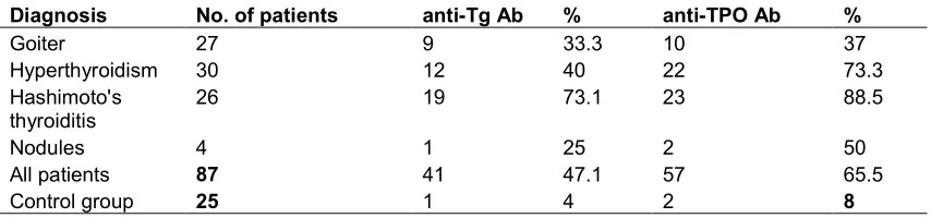
In Table 3, we show the prevalence of anti-Tg Ab and anti-TPO Ab by type of thyroid disorder. Anti-TG Abs were found to be most prevalent in hypothyroidism; this is because patients with hypothyroidism have an increase in TSH concentration and this is commonly associated with an increase in antithyroglobulin antibodies. Similarly, anti-TPO Abs were found to be the most prevalent in hypothyroidism; this can be attributed to the observation that anti-TPO Ab are typically the first antibodies to arise in this disease.
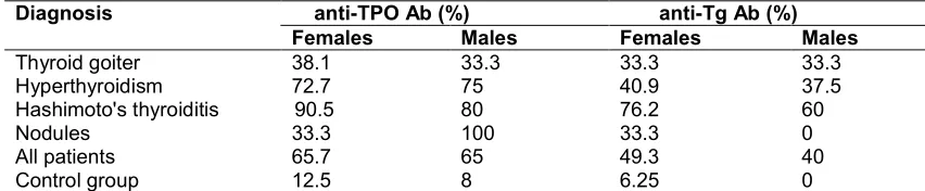
In Table 4, we show the distribution of anti-Tg Abs and anti-TPO Abs, by patient sex, in different types of thyroid diseases. The high prevalence of anti-TPO and anti-Tg Ab (65% and 47%, respectively) indicate that AITD is common in our area, especially hyperthyroidism (Grave's
diseases) and hypothyroidism (Hashimoto
disease). When we compare these results with those of the control group, we found that
the prevalence of antipyroxidase and
antithyroglobulin antibodies in the general
population is 8% and 4%, respectively.
Moreover, as might be expected, both are more common in females than males because AITD in general is more common among female patients.
Patients with positive anti-TPO Ab and anti-Tg Ab were subdivided depending on the antibodies titer, and the TSH concentration (Table 5). After
observation, it was found that the antibodies titer is related to the severity of the disease. When TSH is increased or decreased, beyond the normal range, the antibodies titer is increased which gives us a good evaluation of the case and this will help to control and treat the patient.
The correlation coefficient (r) between TSH and anti-TPO Ab was between 0.6 – 0.88, depending on the levels of TSH. The correlation coefficient between TSH and anti-Tg Ab was between 0.4 – 0.84, also depending on the levels of TSH. The diagnostic potential of antipyroxidase and antithyroglobulin antibodies in the detection of thyroid diseases is depicted in Table 6.
4. DISCUSSION
More than one mechanism is involved in the origin of thyroglobulin and thyroperoxidase antibodies, and this is consistent with antibody heterogeneity. Different groups have suggested that there could be an antigenic stimulation at the initial step of antibody production. It has been proposed that a viral infection such as subacute thyroiditis could induce occult antigenic changes within the thyroid. Other hypothesis referred to the abnormal antigenic stimulation in these patients, where thyroglobulin release in the bloodstream is accelerated, and iodinated forms, which are normally confined to the thyroid gland, are secreted. Another hypothesis suggests an immunoregulation disorder may play a role in the etiology of both Graves’ and Hashimoto's disease [8]. TSH is also thought to stimulate TPO Ab production in thyroid cells through a complex pathway involving cyclic AMP [9,10].
Table 1. Distribution of the patient and control groups according to sex and age (SD, standard deviation) Control group Patient group SD Mean age % No. SD Mean age % No. 12.8 41.9 36 9 13.9 36.4 23 20 Males 9.8 39.3 64 16 16.4 38.7 77 67 Females 100 25 100 87 Total
subacute thyroiditis. He demonstrated that 86% of patients with hypothyroidism have positive anti-TPO Ab and 71%have positive anti-Tg Ab, while 72% of patients with hyperthyroidism have positive TPO Ab and 44% have positive anti-Tg Ab. The similarity of results in these investigations can likely be attributed to the fact that all the studies, including ours, used indirect immunofluorescence techniques.
Table 2.Distribution of patients depending on clinical findings Percentage (%) Total number Diagnosis 25.28 22 Hyperthyroidism without goiter 24.13 21 Hypothyroidism without goiter 9.19 8 Hyperthyroidism with goiter 5.74 5 Hypothyroidism with goiter 31.03 27 Euthyroid with goiter 4.59 4 Euthyroid with nodules 100 87 All patients
Savoie JC, et al. [13]screened 629 patients with thyroid disease and 100 controls for TPO Ab and
anti-Tg Ab by hemagglutination. Thyroid
antibodies were present in 4% of control patients. The overall prevalence of thyroid antibodies in autoimmune thyroiditis were: TPO Ab 93%, anti-Tg Ab 78%. In Graves’ disease, anti-TPO Ab was 63%, while anti-Tg Ab was 33%. He found a slightly higher prevalence rate of anti-TPO Ab and anti-Tg Ab in hypothyroid patients (93% and 78%, respectively), compared to our study, but similar results in hyperthyroidism patients (63% and 33%, respectively). Mariotti S, et al. [14] investigated anti-TPO Ab in 596 patients with different thyroid disorders using monoclonal antibody-assisted RIA. Anti-TPO Abs was positive in 74% of patients with Graves' disease and 95% of patients with Hashimoto's. In contrast, anti-TPO Abs was detected in only 8.4% of the control group. Both Savoie and Mariotti used radioimmunoassay methods in their study to improve sensitivity. The correlation between the levels of TSH and the titer of anti-TPO Ab is consistent with Kontianen S, et al. [15], who demonstrated that 47% of patients with a titer of anti-TPO Ab more than 1/40 have abnormal TSH levels. Walsh JP, et al. [16] analyzed serum samples from 2115 adults for
FT4, TSH, TPO Ab, and Tg Ab by
immunochemiluminescent assays. He
demonstrated that 63% of patients with high concentration of TSH (more than 4 micro u/ml) have high titer of anti-TPO Ab and 60% of them have high titer of anti-Tg Ab.
Table 3.The prevalence of anti-Tg Ab and anti-TPO Ab according to pathology
% anti-TPO Ab
% anti-Tg Ab No. of patients
Table 4.Sex distribution of anti-Tg Ab and anti-TPO Ab in the study and control groups
anti-Tg Ab (%) anti-TPO Ab (%)
Diagnosis
Males Females
Males Females
33.3 33.3
33.3 38.1
Thyroid goiter
37.5 40.9
75 72.7
Hyperthyroidism
60 76.2
80 90.5
Hashimoto's thyroiditis
0 33.3
100 33.3
Nodules
40 49.3
65 65.7
All patients
0 6.25
8 12.5
Control group
Table 5.The prevalence of anti TPO and anti-Tg Ab depending on antibodies titer and their relation to TSH concentration
TSH concentration (micro unit /ml)
1/10 1/20 -1/30 1/40 anti-TPO
Ab (%)
anti- Tg Ab (%)
anti- TPO Ab (%)
anti-Tg Ab (%)
anti TPO Ab (%)
anti- Tg Ab (%)
Less than 0.27 27.2 16.6 27.3 16.6 45.5 50
Between 0.27- 4.2 58.4 60 25 60 16.6 10
More than 4.2 21.7 21 30.5 21 47.8 42.2
All patient 31.5 29.3 28 34.1 40.5 36.6
Control group 50 100 50 0 0 0
Table 6.The sensitivity, specificity, positive and negative predictive values of anti TPO Ab
and anti Tg Ab in the detection of thyroid diseases
Parameter Anti-TPO Ab (%)
Anti-TG Ab (%)
Sensitivity 80 55
Specificity 61 67
Positive predictive value
78 75
Negative predictive value
63 45
5. CONCLUSION
In conclusion, anti-TPO Ab and anti-Tg Ab are important measurements in the evaluation
and management of thyroid disorders.
Immunofluorescence techniques are reliable methods for the evaluation of these disorders, bearing in mind the mainstay of diagnosis and management relays on clinical presentation and physical findings.
CONSENT
It is not applicable.
ETHICAL APPROVAL
All authors hereby declare that all experiments have been examined and approved by the
appropriate ethics committee and have therefore been performed in accordance with the ethical standards laid down in the 1964 Declaration of Helsinki.
COMPETING INTERESTS
Authors have declared that no competing interests exist.
REFERENCES
1. Benveniste P, Row VV, Volpé R. Studies on the immunoregulation of thyroid autoantibody production in vitro. Clin Exp Immunol. 1985;61:274-282.
2. Kimura S, Kotani T, McBride OW. Human thyroid peroxidase: Complete cDNA and protein sequence, chromosome mapping, and identification of two alternately spliced mRNAs. Proc Natl Acad Sci. 1987;84:55-59.
3. Chiovato L, Fiore E, Vitti P. Outcome of thyroid function in Graves' patients treated with radioiodine: Role of thyroid-stimulating and thyrotropin-blocking antibodies and of radioiodine-induced thyroid damage. J Clin Endocrinol Metab. 1998;83:40-50.
4. Weetman AP. New aspects of thyroid
immunity. Horm Res. 1997;48:51-55. 5. Nordyke R, Gilbert F, Miyamoto L, Fleury
Hashimoto's thyroiditis. Arch Intern Med. 1993;153:862-5.
6. McLachlan S, Rapoport B. Genetic and epitopic analysis of thyroid peroxidase (TPO) autoantibodies: Markers of the human thyroid autoimmune response. Clin Exp Immunol. 1995;101:200-206.
7. Gilmour J, Brownlee Y, Foster P, Geekie
C, Kelly P, Robertson S, et al.
The quantitative measurement of
autoantibodies to thyroglobulin and thyroid peroxidase by automated microparticle based immunoassays in Hashimoto's disease, Graves' disease and a follow-up study on postpartum thyroid disease. Clin Lab. 2000;46:57-61.
8. DeGroot LJ, Larsen PR, Hennemann G. The Thyroid and Its Diseases. 6th ed. New York: Churchill Livingstone; 1996.
9. Laurence MD, Carole AS. Laboratory
support for the diagnosis and monitoring of thyroid disease. Clin Endocrinal. 2002;56: 67-9.
10. Doullay F, Ruf J, Codaccioni JL, Carayon P. Prevalence of autoantibodies to thyroperoxidase in various thyroid and
autoimmune diseases. Autoimmunity.
1991;9:237-44.
11. Gardas A, Lasterzebska-Bohaterewicz E, Wojciechowska W. Place of thyroglobulin
antibodies assay in laboratory diagnostic of autoimmune thyroid diseases. Endokrynol Pol. 2005;56:30-41.
12. Dai WX, Lian XL, Bai Y, Sun ML, Guo ZS. Clinical validity of anti-thyroperoxidase antibody and anti-thyroglobulin antibody. Clin Eendocrinal. 2004;26:677-81.
13. Savoie JC, Guillauseau C, Eyquem A, Viencens M, Laurent MF, Leger AF. Value of the detection of antithyroid antibodies in thyroid pathology. Pathol Biol. 1985;33: 653-8.
14. Mariotti S, Caturegli P, Piccolo P, Barbesino G, Pinchera A. Antithyroid peroxidase autoantibodies in thyroid diseases. Clin Endocrinal. 1990;71:661-9. 15. Kontianen S, Melamies L, Miettinen A,
Weber T. Thyroid autoantibodies in serum samples with abnormal TSH levels. APMIS: Acta Pathologica, Microbiologica, et Immunologica Scandinavica. 1994;102: 716-20.
16. Walsh JP, Oleary PC, Feddema PH,
Micheangeli VP, Leedman J, Chew GT, et al. Investigations of thyroid
hormones and antibodies based on a community health survey: the Busselton thyroid study. Clin Endocrinal. 2006;64: 97-104.
_________________________________________________________________________________
© 2017 Kadi and Kayali; This is an Open Access article distributed under the terms of the Creative Commons Attribution License (http://creativecommons.org/licenses/by/4.0), which permits unrestricted use, distribution, and reproduction in any medium, provided the original work is properly cited.
Peer-review history: