Diabetic Retinopathy Detection Using Tensor
Flow Based on Machine Learning
M.Rajeswari1, R.J.Nithya sri2, P.Santhiya3, P.Saranya4
Asst. Professor, Dept. of IT, PIT, Chennai, India1
UG Scholar, Dept. of IT, PIT, Chennai, India2,3,4
ABSTRACT: Diabetic retinopathy is a main source of visual impairment among working-age grown-ups. It affect the blood vessels of the light-sensitive tissue at the back of the eye (retina). Early screening and treatment of DR prevents vision deterioration, however the recommendation of yearly screening is often not being met. Hemorrhages, hard Exudates, and Micro-aneurysms (HEM) that appear in the retina are the early signs of DR. Early diagnosis of HEM is crucial to prevent blindness. Textures features such as Local Binary pattern (LBP), Local Ternary Pattern (LTP) and Local Energy-based Shape Histogram (LESH) have been widely used in the past as a technique for DR detection. This paper focuses on a machine learning solution that identifies vascular diseases of the retina affected with diabetes mellitus using TensorFlow. So we use A histogram binning scheme along with K-Means algorithm for features representation is proposed. KNN and SVM for classification. The experimental results show that K-Means, KNN and SVM is the best performing algorithm with an obtained accuracy of 97.608 % for extraction of features and in detection of diabetic retinopathy which makes the diagnosis and screening of retinal images
KEYWORDS: SVM (support vector machine),K-means algorithm, diabetic retinopathy, Machine learning, K-nearest neighbors algorithm.
I. INTRODUCTION
The eye is one of the most important and sensitive part of the human body. The inner part of the eye, facing the lens, includes retina, optic disc, macula, and fovea. It can be seen during an eye exam by looking through the pupil. The retina is sensitive to light. Ophthalmologists examine retinal images manually. These images, known and retinal fundus images are captured by specialized cameras and are used in medical diagnosis.
ISSN(Online): 2319-8753 ISSN (Print): 2347-6710
International Journal of Innovative Research in Science,
Engineering and Technology
(A High Impact Factor, Monthly, Peer Reviewed Journal)
Visit: www.ijirset.com
Vol. 8, Issue 3, March 2019
and KNN for image classification. In the result of diabetic retinopathy automatic detection using Support Vector Machines (SVM) algorithm and it has an accuracy of 97.608 %.
II. RELATED WORK
Some work has been proposed to detect DR using texture features to preserve the HEM structure. In [2], the authors use Local Binary Patterns (LBP) for the detection of hemorrhages and exudates (HMA). The tests were performed on a database of 89 images. They obtained an average accuracy of 86.15% and an AUC of 0.87. In [3], the authors used fluorescein angiography (FA) fundus images to detect micro-aneurysms (MA). They used Radon transform (RT) and multi-overlapping windows. Tests were conducted on three databases of respectively 120, 50, and 22 images. The authors report the obtained best results for the two first databases with respectively sensitivity and specificity of 94% and 75% for the first database and 100% and 70% for the second one. MA can be easily detected using this imaging modality, but the procedure needs the administration of some injections to the patient, making this approach less interesting as it can cause non-desirable health effects. The best way to examine the retinal fundus is still to use standard retinal fundus imaging technologies. In [4], discrete wavelet transforms are used. The wavelet decomposition was performed up to the second level. Eight energy features were extracted. Two features from the coefficients of two levels and six energy values in three orientations (horizontal, vertical, and diagonal). Classification was performed using Support Vector Machines (SVM) with different kernels. The authors collected 240 retinal fundus images (120 normal and the remaining with different degrees of DR). They reported a result for accuracy, sensitivity, and specificity of 99% using SVM with polynomial kernel of order 3. The results of these research works are interesting, however the number of images used for experimental tests is small. In recent years, deep learning techniques were proposed for DR detection. Gulshan et al. [5] trained an Inception-v3 Convolutional Neural Networks (CNN) using a dataset of 128,175 retinal images, they report an AUC performance of 0.991. Ting et al. [6] trained a deep convolutional network in 71,896 retinal images and obtained an AUC of 0.936. They experimented using 10 additional datasets, and obtained an AUC ranging from 0.889 to 0.983. Deep learning techniques are data hungry and need large image numbers for training. In this work we use an available public dataset that has a limited number of images. Past work dealing with DR using texture features, has mainly focuses on Local Ternary Patterns (LTP) and Local Energy-based Shape Histogram (LESH) with obtained accuracy 0.904 using SVM.
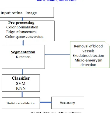
III. PROPOSED SYSTEM
Fig. 1 Block Diagram of Proposed System
ISSN(Online): 2319-8753 ISSN (Print): 2347-6710
International Journal of Innovative Research in Science,
Engineering and Technology
(A High Impact Factor, Monthly, Peer Reviewed Journal)
Visit: www.ijirset.com
Vol. 8, Issue 3, March 2019
into k number of bunch. Let p(x, y) be an info pixels to be group and ck be the bunch focuses. The calculation for k-means13 bunching is following as:
1. Introduce number of group k and focus.
2. For every pixel of a picture, figure the Euclidean separation d, between the middle and every pixel of a picture utilizing the connection given underneath.
d = ||p(x, y) – ck||
3. Relegate every one of the pixels to the closest focus dependent on separation d.
4. After the sum total of what pixels have been relegated, recalculate new position of the middle.
5. Rehash the procedure until it full fills the resistance or blunder esteem.
6. Reshape the group pixels into picture.
Yield of the K-Means demonstrate the compacted picture. The compacted picture looks near the first one which implies we're ready to hold most of the qualities of the first picture.
Fig. 2 Shows Difference between Orginal Picture and Compacted picture
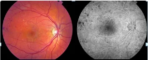
IV. EXPERIMENTAL RESULTS
Fig. 3 Shows the ouput of Diabetic Retinopathy
V. CONCLUSION
The degree for this undertaking later on is to expand the exactness via preparing the model on machines with better handling force. We intend to give fast and available analysis to individuals from varying backgrounds, to keep this illness from further disintegrating to a point where it winds up untreatable. Using retinal fundus images can help automate the diagnosis. Micro hemorrhages and aneurysms, known as HEM, are the early signs of diabetic retinopathy (DR) and are difficult to identify because of their similarities with normal parts of a healthy human. Other problems such as non-uniform lighting, low contrast, etc. can lead to a bad diagnosis. Texture based techniques for DR detection were proposed in the past. Most of these techniques use LBP, LTP and LESH and wavelets for feature extraction from retinal images. In this work, we propose the use K-Means algorithm for feature extraction. These features extracted from the retinal fundus images are used to learn signs of HEM and differentiate between DR and non-DR. SVM and KNN is used to classify these features. Experimental tests with the CSV dataset show that k-mean, SVM and KNN perform well in detecting DR with an accuracy 97.608 % compared to the previous work.
REFERENCES
[1] S. Mohammadian, A. Karsaz and Y. M. Roshan, "A comparative analysis of classification algorithms in diabetic retinopathy screening," 2017 7th International Conference on Computer and Knowledge Engineering (ICCKE), Mashhad, pp. 84-89, 2017.
[2] Dr. Sunil Bhutada1, Dr. Ch. Mukundha2, G. Shreya3, Ch. Lahari4 1,2 Professor, “Convolutional Neural Networks for Automatic Classification of Diabetic Retinopathy” International Research Journal of Engineering and Technology (IRJET) e-ISSN: 2395-0056 Volume: 05 Issue: 04 | Apr-2018
[3] M. N. Ashraf, Z. Habib and M. Hussain, "Texture Feature Analysis of Digital Fundus Images for Early Detection of Diabetic Retinopathy,“2014 11th International Conference on Computer Graphics, Imaging and Visualization, Singapore, pp. 57-62, 2014.
[4] M. Tavakoli, R. Shahri, H. Pourreza, A. Mehdizadeh, T. Banaee and M. BahreiniToosi, "A complementary method for automated detection ofmicroaneurysms in fluorescein angiography fundus images to assess diabetic retinopathy," Pattern Recognition, vol. 46, no. 10, pp.
2740-2753, 2013.
[5] K. Noronha, U. R. Acharya, K. P. Nayak, S. Kamath, and S. V. Bhandary, "Decision support system for diabetic retinopathy using discrete wavelet transform," ProcInstMechEng H, vol. 227, no. 3, pp. 251–261, 2013.