HIGHLIGHTED ARTICLE
GENETICS | INVESTIGATION
Comprehensive Genetic Analysis of Paralogous
Terminal Septin Subunits Shs1 and Cdc11 in
Saccharomyces cerevisiae
Gregory C. Finnigan,* Julie Takagi,†Christina Cho,‡and Jeremy Thorner1
*Division of Biochemistry, Biophysics and Structural Biology, Department of Molecular and Cell Biology, University of California, Berkeley, California 94720-3202,†Department of Microbiology and Immunology, University of California School of Medicine, San Francisco, California 94158-2200, and‡Harvard School of Dental Medicine, Boston, Massachusetts 02115
ABSTRACT Septins are a family of GTP-binding proteins considered to be cytoskeletal elements because they self-assemble into
filaments and other higher-order structuresin vivo. In budding yeast, septins establish a diffusion barrier at the bud neck between a mother and daughter cell, promote membrane curvature there, and serve as a scaffold to recruit other proteins to the site of cytokinesis. However, the mechanism by which any septin engages a partner protein has been unclear. The two most related and recently evolved subunits appear to beCdc11andShs1, and the basic building blocks for assembling septin structures are hetero-octameric rods (Cdc11– Cdc12–Cdc3–Cdc10–Cdc10–Cdc3–Cdc12–Cdc11andShs1–Cdc12–Cdc3–Cdc10–Cdc10–Cdc3–Cdc12–Shs1). Loss ofCdc11is not nor-mally tolerated, whereas cells lackingShs1do not appear grossly abnormal. We established several different sensitized genetic backgrounds whereinShs1is indispensable, which allowed us to carry out thefirst comprehensive and detailed genetic analysis ofShs1in vivo. Our analysis revealed several novel insights, including: (i) the sole portion ofShs1essential for its function is a predicted coiled-coil-forming segment in its C-terminal extension (CTE); (ii) the CTE ofCdc11shares this function; (iii) this role for the CTEs ofCdc11andShs1is quite distinct from that of the CTEs ofCdc3andCdc12; and (iv) heterotypicCdc11andShs1junctions likely occurin vivo.
Related article inGENETICS:Finnigan, G. C.et al., 2015 The Carboxy-Terminal Tails of Septins Cdc11 and Shs1 Recruit Myosin-II Binding Factor Bni5 to the Bud Neck inSaccharomyces cerevisiae. Genetics200:843–862.
KEYWORDSyeast; cytoskeleton; complexes;filaments; mutants
S
EPTINS are GTP-binding proteins conserved acrossEukarya (except higher plants) (Panet al.2007; Nishihama et al. 2011). This protein family wasfirst identified because the corresponding loci were among the temperature-sensitive (ts) cell division cycle (cdc) mutations isolated inSaccharomyces cerevisiae(Hartwell 1978). At the restrictive temperature,cdc3, cdc10,cdc11, andcdc12 mutants continued to bud, replicate, and segregate their chromosomes, yet failed to execute cell division, resulting in the formation of chains of multinucleate cells (Hartwell 1971). Shifting a ts allele of any of these four homologous gene products to restrictive temperaturepre-vented formation of what appeared to be rings of highly ordered membrane-associated filaments at the bud neck (Byers and Goetsch 1976). Antibody decoration (Haarer and Pringle 1987; Ford and Pringle 1991; Kimet al.1991) and, later, use of fusions to greenfluorescent protein (GFP) (Cidet al.1998) demonstrated that these four proteins colo-calized with and were likely constituents of thesefilaments. Because loss of the function of these proteins prevents cyto-kinesis and cell septation, and they seemed to be integral components of the filamentous structures erected at the bud neck, they were dubbed septins (Sanders and Field 1994; Pringle 2008). Indeed, subsequent purification of these pro-teins from yeast (Frazieret al.1998), and their production as recombinant proteins in bacteria (Verseleet al.2004; Versele and Thorner 2004; Farkasovskyet al.2005), demonstrated that Cdc3, Cdc10, Cdc11, and Cdc12 were necessary and sufficient for the formation of long-pairedfilamentsin vitro (Bertinet al.2008) that closely resemble those observed at Copyright © 2015 by the Genetics Society of America
doi: 10.1534/genetics.115.176495
Manuscript received March 17, 2015; accepted for publication May 8, 2015; published Early Online May 12, 2015.
Supporting information is available online athttp://www.genetics.org/lookup/suppl/ doi:10.1534/genetics.115.176495/-/DC1.
1Corresponding author: Department of Molecular and Cell Biology, Barker Hall, Rm.
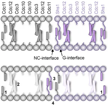
the bud neck in vivo(Bertin et al.2012; Onget al.2014). Detailed ultrastructural analysis led to the discovery that the septin complex that is the fundamental building block of the yeastfilaments is a linear, hetero-octameric rod, with the orderCdc11–Cdc12–Cdc3–Cdc10–Cdc10–Cdc3–Cdc12– Cdc11 (Bertinet al. 2008), which polymerizes end-on-end via Cdc11–Cdc11 interaction. Subsequently, the nonpolar, linear hetero-octamer with twofold rotational symmetry was found to be characteristic of mammalian septin complexes too (Mostowy and Cossart 2012; Fung et al. 2014). Each septin subunit comprises a Ras-related GTP-binding domain (Sirajuddin et al. 2007) preceded by an N-terminal extension of variable length and followed by a C-terminal extension (CTE) of variable length (Verseleet al.2004; Weirichet al.2008).
However, when the sequence of the entire S. cerevisiae genome was determined (Meweset al.1997), it was revealed that three other genes highly related toCDC3,CDC10,CDC11, andCDC12were present. Two of them,SPR3andSPR28, encode proteins expressed exclusively in MATa/MATa cells and only under conditions that induce meiosis and spor-ulation (Neiman 2011; Brar et al. 2012). The third,SHS1/
SEP7(Carrollet al.1998; Minoet al.1998), encodes a protein expressed in mitotically growing haploids and diploids, but, unlike loss ofCdc3,Cdc10,Cdc11, orCdc12, even the com-plete absence of Shs1 seemed to have only a very modest effect on cell growth and morphology (Iwaseet al.2007; Garcia et al. 2011). Biochemical and ultrastructural analysis demon-strated that Shs1 occupies the same terminal position in the linear hetero-octamer as Cdc11, and that suchShs1–Cdc12– Cdc3–Cdc10–Cdc10–Cdc3–Cdc12–Shs1rods do not polymerize into long-paired filaments in vitro, but rather assemble into much more elaborate structures (rings, spirals, and gauzes, depending on the conditions) (Garciaet al.2011). Nonethe-less, these findings left open the question of what the two alternative terminal subunits,Cdc11andShs1, each contrib-ute to the supramolecular architecture of septin structures and to the physiological function of those structures in vivo. Titra-tion ofShs1-capped hetero-octamers with increasing amounts of Cdc11-capped hetero-octamers reduces the thickness and increases the diameter of the rings formed in vitro (Garcia et al. 2011). Consistent with a role for Shs1-capped rods in modulating the plasticity of septin-based structures, the Shs1 ortholog inCandida albicanscontributes to septin ring dynamics (Gonzalez-Novoet al.2008) and theShs1ortholog inAshbya gossypiihas been implicated in scaling the size of the septin ringin vivo(Meserollet al.2012). Moreover, in keeping with a primarily regulatory role, Shs1 undergoes extensive post-translational modification during passage through the cell cy-cle, including SUMOylation (Johnson and Blobel 1999) and phosphorylation (Mortensen et al. 2002; Dobbelaere et al. 2003; Smolkaet al.2006; Egelhoferet al.2008).
In contrast to cells lackingShs1, cells lackingCdc11either are, depending on strain background, inviable (Verseleet al. 2004) or exhibit extremely slow growth and a highly elon-gated morphology (Frazieret al.1998). Viablecdc11Dcells propagate somewhat better on galactose medium than on
glucose medium for reasons that are not understood (McMurray et al.2011). Remarkably, however, in a strain background where cells lackingCdc11are dead,cdc11Dshs1Ddouble mutants are viable and, to survive, require that the resulting heterohexamers polymerize via a nonnativeCdc12–Cdc12interaction (McMurray et al.2011). Conversely, in a strain background wherecdc11D cells are able to propagate, overexpression ofSHS1kills them, but not otherwise isogenic CDC11+ cells (Iwase et al.2007).
These results indicate that Shs1-capped hetero-octamers alone are not sufficient for viability and, when no competingCdc11is present,Shs1caps theCdc12subunit and prevents polymeriza-tion of Cdc12–Cdc3–Cdc10–Cdc10–Cdc3–Cdc12complexes, in agreement with the observation thatShs1-capped rods are unable to assemble intofilamentsin vitro(Garciaet al.2011). Taken together, these results indicate that the ability to form filaments is needed for the execution of some essential func-tion(s) mediated by the septins (McMurrayet al.2011).
Thus, althoughCdc11and Shs1occupy the same terminal position, share greater similarity to each other than do any other pair ofS. cerevisiaeseptin subunits as judged by pairwise sequence comparisons, and seem to have evolved relatively re-cently from a common ancestral gene (Panet al.2007), they appear to have diverged in certain aspects of their function. As one approach to discern both the unique and shared functions ofCdc11andShs1, especiallyin vivo, genetic analysis is a po-tentially useful tool. However, until recently, and regardless of strain background, loss of Shs1has not evoked a readily dis-cernible phenotype that would aid in carrying out such an analysis. As described here, we have now established several different conditions under which absence of Shs1 is lethal, which allowed us to carry out extensive mutational dissection of this gene product. In this way, we were able to pinpoint previously uncharacterized elements in this septin that are re-quired for its function and to compare their roles to those of the corresponding elements inCdc3,Cdc10,Cdc11, andCdc12.
Materials and Methods
Yeast strains and plasmids
PCR using synthetic oligonucleotides containing tails with sequence homology to each of the fragments so as to assemble them in the desired order and to ensure their insertion down-stream of the plasmid-borne promoter. The resulting linear PCR product and the gapped plasmid were then introduced by DNA-mediated transformation using a modified lithium acetate procedure (Eckert-Bouletet al.2012) into appropriate yeast recipients, THS4218 (SF838–1DaHIS4 his3D::HygR) or
THS4213 (SF838–1DaHIS4), and selected on an appropriate drop-out or drug-containing medium. The resulting plasmids were rescued from yeast, propagated inEscherichea coliTOP10 (Lucigen Corp., Middleton, WI), purified (Sambrook and Russell 2001), and confirmed via automated DNA sequence analysis conducted by the University of California (UC) Berkeley DNA Sequencing Facility. For use in strain construction, the entire cassette (promoter through the drug marker) in such a plasmid was amplified by PCR and used for integrative transformation (Rothstein 1991). This strategy was used to integrate each of the septin alleles generated in this study into the corresponding endogenous chromosomal locus under control of its native pro-moter. Successful yeast strain construction was confirmed using isolated genomic DNA and multiple diagnostic PCR reactions to assess each affected loci after every successive round of integra-tion. Plasmids used in this study are listed in Table 2 and were constructed usingin vivoligation and homologous recombina-tion in yeast, as described above.
Culture conditions
Yeast were cultured in YP medium (1% yeast extract, 2% peptone) containing either 2% dextrose/glucose (YPD) or 2% galactose (YPGal), cultured in synthetic drop-out medium supplemented with the appropriate amino acids and contain-ing either glucose or galactose, or cultured in synthetic drop-out medium supplemented with the appropriate amino acids containing raffinose (2%) and sucrose (0.2%) (Shermanet al. 1986). Growth assays were performed by spotting onto agar plates5ml offivefold serial dilutions of an overnight culture of the strain(s) to be tested (first spot being roughly equiva-lent to 1A600 nmunit of cells. For growth tests on any medium containing 5-fluoro-orotic acid (5-FOA; Oakwood Products Inc., West Columbia, SC) (final concentration of 0.5 mg/ml of medium, heated to 75°for 30 min andfilter sterilized rather than autoclaved), yeast strains were always grown overnight in medium selective for the presence of theURA3 -marked plasmid (as well as any other plasmids present). The only exception to the preceding pregrowth regimen was for strains lackingCDC10, in which the yeast were grown over-night at 20°–25°in YPGal medium prior to spotting to allow for more efficient loss of theURA3-marked covering plasmid (McMurrayet al.2011). For strains that did not contain any URA3-marked covering plasmid, cultures were grown in YPD medium at the indicated temperature; if a strain harbored any additional plasmid(s), it was grown in minimal medium supplemented with appropriate nutrients to maintain selec-tion. For strains that required testing at high temperature (37°), strains were counterselected for all covering plasmids
(at permissive/low temperature) and growth assays were per-formed on appropriate medium without the need for 5-FOA selection. Growth results were scored after incubating the plates for 2–5 days, depending on the strains being tested.
Fluorescence and DIC microscopy
For imaging, exponentially growing cells were washed with water, resuspended in water, and immediately examined using an Olympus BH-2 upright fluorescence microscope (Olympus, Tokyo, Japan) equipped with a 1003objective, illuminated with a SOLA light engine (Lumencore, Beaverton, OR) and images recorded with a CoolSNAP MYO CCD cam-era (Photometrics, Tuscon, AZ). Images were analyzed using Micro-Manager software (Edelstein et al.2010) and ImageJ (National Institutes of Health). All images grouped together in any givenfigure were always scaled identically and always adjusted identically for brightness using Photoshop (Adobe). The cell perimeter was determined using either a DIC image of the samefield or an overexposedfluorescence image.
Results
Shs1 is essential for viability in four different genetic backgrounds
The products of four septin genesCDC3,CDC10,CDC11, and CDC12are essential for cell growth, normal morphology, and completion of cytokinesis in S. cerevisiae(Hartwell 1971; Hartwellet al.1974). By contrast, absence of thefifth, mitot-ically expressed septin geneSHS1, a close paralog ofCDC11 (Panet al.2007), causes rather minor and subtle phenotypes (Iwaseet al.2007; Egelhoferet al.2008; Garciaet al.2011; Butteryet al.2012). We explored whether compromising the function of another septin might provide a sensitized genetic background wherein presence ofShs1becomes essential for via-bility. We found,first, that cells of the S288C lineage (Mortimer and Johnston 1986) lacking Shs1 and endogenously ex-pressing a derivative ofCdc11tagged at its C terminus with a fluorescent protein tag, mCherry (mC) (Shaner et al. 2004), reproducibly grew somewhat less robustly than oth-erwise identical cells endogenously expressing eitherCdc3– mC orCdc10–mC (Figure 1A). To examine the efficiency of incorporation of such tagged subunits into the septin struc-tures at the bud neck, cells endogenously expressing either Cdc10–mC orCdc11–mC (and coexpressing a second copy of WT CDC10 or WT CDC11, respectively, from a URA3 -marked CENplasmid) were viewed by fluorescence micros-copy, and no dramatic differences were observed in cells lackingShs1, as compared to theSHS1+control cells (Figure
Table 1 Yeast strains used in this study
Strain Genotype Reference
BY4741 MATaleu2Dura3Dmet15Dhis3D Brachmannet al.(1998)
GFY-38 BY4741;shs1D::KanR This study
GFY-42 BY4741;cdc10D::CDC10::mCherry::SpHIS5 This study
GFY-6 BY4741;cdc10D::CDC10::mCherry::SpHIS5 shs1D::KanR This study
GFY-768 BY4741;cdc11D::CDC11::mCherry::SpHIS5 This study
GFY-769 BY4741;cdc11D::CDC11::mCherry::SpHIS5 shs1D::HygR This study
GFY-29 BY4741;cdc3D::CDC3::mCherry::SpHIS5 This study
GFY-30 BY4741;cdc3D::CDC3::mCherry::SpHIS5 shs1D::KanR This study
GFY-87 BY4741;cdc10D::KanRshs1D::SHS1::eGFP::NatR+ pJT2022 This study
GFY-137 BY4741;cdc10D::KanRshs1D::HygR+ pJT2022 This study
GFY-293 BY4741;cdc11D::cdc11(D357-415)::mCherry::SpHIS5 shs1D::SHS1::eGFP::NatR+ pJT1520 This study
GFY-166 BY4741;cdc11D::cdc11(D357-415)::mCherry::SpHIS5 shs1D::HygR+ pJT1520 This study
GFY-302a BY4741;cdc12D::cdc12(K391ND392-407)::HygRshs1D::SHS1::eGFP::NatR+ pJT1622 This study
GFY-139a BY4741;cdc12D::cdc12(K391ND392-407)::HygRshs1D::KanR+ pJT1622 This study
YFR-387b BY4742;hof1D::KanR+ pJT4836 This study
GFY-934 BY4742;hof1D::KanRshs1D::HygR+ pJT4836 This study
GFY-95c BY4741;cdc10D::KanRshs1D::shs1(T6A S447A)::eGFP::NatR+ pJT2022 This study
GFY-96c BY4741;cdc10D::KanRshs1D::shs1(T6A T386A S416A S441A S447A)::eGFP::NatR+ pJT2022 This study
GFY-97c BY4741;cdc10D::KanRshs1D::shs1(T386A S416A S441A S447A S460A T462A S519A S520A
S521A S522A S525A S545A)::eGFP::NatR+ pJT2022
This study
GFY-191c BY4741;cdc10D::KanRshs1D::shs1(S350A T351A T386A S416A S441A S447A T454A S460A
T462A S519A S520A S521A S522A S525A S529A S530A T539A T541A S545A)::eGFP::NatR+
pJT2022
This study
GFY-189d BY4741;cdc10D::KanRshs1D::shs1(S259A)::eGFP::NatR+ pJT2022 This study
GFY-88e BY4741;cdc10D::KanRshs1D::shs1(T6D)::eGFP::NatR+ pJT2022 This study
GFY-192e BY4741;cdc10D::KanRshs1D::shs1(T6D T386D S416D S441D S447D)::eGFP::NatR+ pJT2022 This study
GFY-90e BY4741;cdc10D::KanRshs1D::shs1(T386D S416D S441D S447D S460D T462D S519D S520D
S521D S522D S525D S545D)::eGFP::NatR+ pJT2022
This study
GFY-91e BY4741;cdc10D::KanRshs1D::shs1(S259D)::eGFP::NatR+ pJT2022 This study
GFY-310e BY4741;cdc10D::KanRshs1D::shs1(S221D)::eGFP::NatR+ pJT2022 This study
GFY-188 BY4741;cdc10D::KanRshs1D::shs1(S221A)::eGFP::NatR+ pJT2022 This study
GFY-160 BY4741;cdc11D::CDC11::mCherry::SpHIS5Rshs1D::SHS1::eGFP::NatR+ pJT1520 This study
GFY-147 BY4741;cdc11D::KanRshs1D::SHS1::eGFP::NatR+ pJT1520 This study
GFY-164 BY4741;cdc11D::CDC11::mCherry::SpHIS5Rshs1D::HygR+ pJT1520 This study
GFY-163f BY4741;cdc11D::KanRshs1D::HygR+ pJT1520 This study
GFY-437g BY4741;cdc11D::NatRshs1D::HygRcdc12D::cdc12(W267A)::KanR+ pJT1520/pJT1622 This study
GFY-369 BY4741;cdc10D::KanRshs1D::shs1(G30D)::eGFP::NatR+ pJT2022 This study
GFY-249 BY4741;cdc10D::KanRshs1D::shs1(R13A R14A K15A K16A K19A R20A)::eGFP::NatR+ pJT2022 This study
GFY-93 BY4741;cdc10D::KanRshs1D::shs1(D2-18)::eGFP::NatR+ pJT2022 This study
GFY-58 BY4741;cdc11D::CDC11::mCherry::SpHIS5 + pJT1520 This study
GFY-121 BY4741;cdc11D::cdc11(G29D)::mCherry::KanR+ pJT1520 This study
GFY-308 BY4741;cdc11D::cdc11(G29D)::mCherry::KanRshs1D::HygR+ pJT1520 This study
GFY-375 BY4741;cdc11D::cdc11(G29D)::mCherry::KanRshs1D::shs1(G30D)::eGFP::NatR+ pJT1520 This study
GFY-397 BY4741;cdc11D::KanRshs1D::shs1(G30D)::eGFP::NatR+ pJT1520 This study
GFY-843h BY4741;cdc11D::KanRshs1D::shs1(G30D)::eGFP::NatRcdc12D::cdc12(W267A)::SpHIS5 + pJT1520/
pJT1622
This study
GFY-246i BY4741;cdc11D::cdc11(R12A K13A R14A K15A K18A R19A)::mCherry::SpHIS5 + pJT1520 This study
GFY-247 BY4741;cdc11D::cdc11(R12A K13A R14A K15A K18A R19A)::mCherry::SpHIS5 shs1D::HygR+
pJT1520
This study
GFY-307 BY4741;cdc11D::cdc11(R12A K13A R14A K15A K18A R19A)::mCherry::SpHIS5 shs1D::shs1(R13A R14A K15A K16A K19A R20A)::eGFP::NatR+ pJT1520
This study
GFY-253 BY4741;cdc11D::KanRshs1D::shs1(R13A R14A K15A K16A K19A R20A)::eGFP::NatR+ pJT1520 This study
GFY-123 BY4741;cdc11D::cdc11(D2-18)::mCherry::SpHIS5R+ pJT1520 This study
GFY-165 BY4741;cdc11D::cdc11(D2-18)::mCherry::SpHIS5Rshs1D::HygR+ pJT1520 This study
GFY-161 BY4741;cdc11D::cdc11(D2-18)::mCherry::SpHIS5Rshs1D::shs1(2-18D)::eGFP::NatR+ pJT1520 This study
GFY-148 BY4741;cdc11D::KanRshs1D::shs1(D2-18)::eGFP::NatR+ pJT1520 This study
GFY-94 BY4741;cdc10D::KanRshs1D::shs1(D349-551)::eGFP::NatR+ pJT2022 This study
GFY-98 BY4741;cdc10D::KanRshs1D::shs1(D465-496)::eGFP::NatR+ pJT2022 This study
GFY-178j BY4741;cdc10D::KanRshs1D::shs1(Y465A R468A I472A R475A L479A L482A S486A E489A
R493A E496A)::eGFP::NatR+ pJT2022
This study
GFY-122 BY4741;cdc11D::cdc11(D357-415)::mCherry::SpHIS5 + pJT1520 This study
Table 1,continued
Strain Genotype Reference
GFY-162 BY4741;cdc11D::cdc11(D357-415)::mCherry::SpHIS5 shs1D::shs1(D349-551)::eGFP::NatR+
pJT1520
This study
GFY-149 BY4741;cdc11D::KanRshs1D::shs1(D349-551)::eGFP::NatR+ pJT1520 This study
GFY-378j BY4741;cdc11D::cdc11(E377A V380A L384A K387A L391A R394A L398A I401A L405A E408A
K412A E415A)::mCherry::SpHIS5 + pJT1520
This study
GFY-383j BY4741;cdc11D::cdc11(E377A V380A L384A K387A L391A R394A L398A I401A L405A E408A
K412A E415A)::mCherry::SpHIS5 shs1D::HygR+ pJT1520
This study
GFY-475j BY4741;cdc11D::cdc11(E377A V380A L384A K387A L391A R394A L398A I401A L405A E408A
K412A E415A)::mCherry::SpHIS5 shs1D::shs1(Y465A R468A I472A R475A L479A L482A S486A E489A R493A E496A)::eGFP::NatR+ pJT1520
This study
GFY-233 BY4741;cdc10D::KanRshs1D::shs1(D342-352)::eGFP::NatR+ pJT2022 This study
GFY-234 BY4741;cdc10D::KanRshs1D::shs1(D353-379)::eGFP::NatR+ pJT2022 This study
GFY-235 BY4741;cdc10D::KanRshs1D::shs1(D380-404)::eGFP::NatR+ pJT2022 This study
GFY-236 BY4741;cdc10D::KanRshs1D::shs1(D405-429)::eGFP::NatR+ pJT2022 This study
GFY-237 BY4741;cdc10D::KanRshs1D::shs1(D430-450)::eGFP::NatR+ pJT2022 This study
GFY-519 BY4741;cdc10D::KanRshs1D::shs1(D342-386)::eGFP::NatR+ pJT2022 This study
GFY-503 BY4741;cdc10D::KanRshs1D::shs1(D387-436)::eGFP::NatR+ pJT2022 This study
GFY-514 BY4741;cdc10D::KanRshs1D::shs1(D342-436)::eGFP::NatR+ pJT2022 This study
GFY-381 BY4741;cdc11D::cdc11(D301-313)::mCherry::SpHIS5 shs1D::HygR+ pJT1520 This study
GFY-398 BY4741;cdc11D::cdc11(D314-329)::mCherry::SpHIS5 shs1D::HygR+ pJT1520 This study
GFY-399 BY4741;cdc11D::cdc11(D330-343)::mCherry::SpHIS5 shs1D::HygR+ pJT1520 This study
GFY-382 BY4741;cdc11D::cdc11(D344-357)::mCherry::SpHIS5 shs1D::HygR+ pJT1520 This study
GFY-513 BY4741;cdc11D::cdc11(D301-329)::mCherry::SpHIS5 shs1D::HygR+ pJT1520 This study
GFY-524 BY4741;cdc11D::cdc11(D330-357)::mCherry::SpHIS5 shs1D::HygR+ pJT1520 This study
GFY-512 BY4741;cdc11D::cdc11(D301-357)::mCherry::SpHIS5 shs1D::HygR+ pJT1520 This study
GFY-676 BY4741;cdc11D::cdc11(D301-357)::mCherry::SpHIS5 shs1D::shs1(342-436D)::eGFP::NatR+
pJT1520
This study
GFY-126 BY4741;cdc3D::CDC3::mCherry::SpHIS5 + pJT2059 This study
YMVB33 BY4741;cdc3D::KanR+ pJT2059 McMurrayet al.(2011)
GFY-501 BY4741;cdc3D::cdc3(D440-520)::mCherry::SpHIS5 + pJT2059 This study GFY-764j BY4741;cdc3D::cdc3(E467A F471A H474A K478A L481A L485A L488A K492A L495A I499A
V506A)::mCherry::SpHIS5 + pJT2059
This study
GFY-494 BY4741;cdc3D::cdc3(D412-423)::mCherry::SpHIS5 + pJT2059 This study GFY-493 BY4741;cdc3D::cdc3(D424-435)::mCherry::SpHIS5 + pJT2059 This study GFY-507 BY4741;cdc3D::cdc3(D436-450)::mCherry::SpHIS5 + pJT2059 This study GFY-511 BY4741;cdc3D::cdc3(D412-450)::mCherry::SpHIS5 + pJT2059 This study
GFY-251 BY4741;cdc12D::CDC12::mCherry::KanR+ pJT1622 This study
YMVB61 BY4741;cdc12D::KanR+ pJT1622 McMurrayet al.(2011)
GFY-515 BY4741;cdc12D::cdc12(D339-407)::mCherry::KanR+ pJT1622 This study
GFY-535j BY4741;cdc12D::cdc12(E368A I371A R375A L478A L482A,I485A V489A L492A V496A L499A
K403A L406A)::mCherry::KanR+ pJT1622
This study
GFY-516 BY4741;cdc12D::cdc12(D315-326)::mCherry::KanR+ pJT1622 This study
GFY-498 BY4741; cdc12D::cdc12(D327-338)::mCherry::KanR+ pJT1622 This study
GFY-510 BY4741; cdc12D::cdc12(D339-353)::mCherry::KanR+ pJT1622 This study
GFY-525 BY4741; cdc12D::cdc12(D315-353)::mCherry::KanR+ pJT1622 This study
GFY-179 BY4741;cdc10D::KanRshs1D::shs1(Y465A)::eGFP::NatR+ pJT2022 This study
GFY-180 BY4741;cdc10D::KanRshs1D::shs1(R468A)::eGFP::NatR+ pJT2022 This study
GFY-181 BY4741;cdc10D::KanRshs1D::shs1(I472A)::eGFP::NatR+ pJT2022 This study
GFY-362 BY4741;cdc10D::KanRshs1D::shs1(R475A)::eGFP::NatR+ pJT2022 This study
GFY-368 BY4741;cdc10D::KanRshs1D::shs1(L479A)::eGFP::NatR+ pJT2022 This study
GFY-366 BY4741;cdc10D::KanRshs1D::shs1(L482A)::eGFP::NatR+ pJT2022 This study
GFY-367 BY4741;cdc10D::KanRshs1D::shs1(S486A)::eGFP::NatR+ pJT2022 This study
GFY-363 BY4741;cdc10D::KanRshs1D::shs1(E489A)::eGFP::NatR+ pJT2022 This study
GFY-391 BY4741;cdc10D::KanRshs1D::shs1(R493A)::eGFP::NatR+ pJT2022 This study
GFY-370 BY4741;cdc10D::KanRshs1D::shs1(E496A)::eGFP::NatR+ pJT2022 This study
GFY-544k BY4741;cdc11D::cdc11(1-308)::shs1(349-551)::mCherry::KanRshs1D::HygR+ pJT1520 This study
GFY-540k BY4741;cdc11D::cdc11(D357-415)::mCherry::SpHIS5 shs1D::shs1(1-348)::cdc11(309-415)::eGFP::
NatR+ pJT1520
This study
functionally deficient when Shs1 is present (Supporting Information,Figure S1A), our results revealed thatCdc11is somewhat impaired when mC is fused to its CTE because over-all septin function became sensitive to the absence ofShs1.
To explore this possibility further, we perturbed Cdc11 even more by fusing mC to a Cdc11derivative in which its CTE had been deleted. Even though the CTE ofCdc11is not required for cell viability whenShs1is present (Verseleet al. 2004), or forfilament formationin vitro(Bertinet al.2008), we found, in agreement with our expectation, that cells ex-pressing Cdc11(DCTE)–mC as the sole source of this septin were unable to propagate at all whenShs1was absent, but grew essentially normally whenShs1was present (Figure 1C, top). In a similar fashion, we systematically explored other known circumstances that partially cripple the function of septins and were able to find three other conditions under which expression ofSHS1becomes essential for cell viability:
i. On galactose medium and at 25°, cells lackingCdc10are able to survive (McMurrayet al.2011), but, we found that, under the same conditions, such cells were not viable if
they also lackedShs1(Figure 1C, top middle), confirming an observation made by Iwaseet al.(2007). EitherShs1or Shs1–eGFP was able to restore viability (Figure S1B). ii. Likewise, a frameshift mutation inCDC12that generates
a temperature-sensitive substitution-truncation allele [cdc12-6; Cdc12(K391N E392Stop)] (Johnsonet al. 2015) grows well at 25°(Dobbelaere and Barral 2004; Gladfelter et al. 2005; Nagaraj et al. 2008; Weems et al. 2014), whereas we found that such cells were inviable at this otherwise permissive temperature if they also lacked Shs1(Figure 1C, bottom middle).
iii. Finally, in agreement with an observation made by Meitinger et al.(2013), we found that cells lackingHof1, a bud neck-localized, F-BAR- and SH3 domain-containing pro-tein that regulates actomyosin ring dynamics (Nishihama et al.2009; Meitingeret al.2011), are inviable ifShs1is absent (Figure 1C, bottom).
Collectively, these results indicated that, far from being dispensable,Shs1does contribute significantly to optimal septin functionin vivoand provided the means to delineate Table 1,continued
Strain Genotype Reference
GFY-542k BY4741;cdc11D::cdc11(1-308)::shs1(349-551)::mCherry::KanRshs1D::shs1(D349-551)::eGFP::NatR+
pJT1520
This study
GFY-692k BY4741;cdc11D::cdc11(1-308)::shs1(349-551)::mCherry::KanRshs1D::shs1(1-348)::
cdc11(309-415)::eGFP::NatR+ pJT1520
This study
GFY-548k BY4741;cdc10D::KanRshs1D::shs1(1-348)::cdc11(309-415)::eGFP::NatR+ pJT2022 This study
aIt was reported that thecdc12-6allele is a frameshift mutation arising from insertion of a single A into a tract of seven adenines (nucleotides 1167–1173) in theCDC12ORF (Longtineet al.2000); however, the original sequencing data (B. Haarer, M. Longtine, and J. Pringle, personal communication) show thatcdc12-6is a frameshift arising from deletion of a single A from this same adenine tract, resulting in a substitution mutation (K391N) immediately followed by a stop codon (TAG), thereby causing truncation of the remainder of the protein (D392–407). As documented elsewhere, loss of the C-terminal residues (rather than the K391N substitution) is responsible for the ts phenotype conferred bycdc12-6(Johnsonet al.2015). Ourcdc12-6construct arises from removal of a single A (nucleotide 1173) from theCDC12ORF, producing the frameshift that results in substitution mutation K391N immediately followed by the nonsense codon that truncates the remainder of the protein and has theADH1transcriptional terminator sequence and aHygRdrug resistant marker inserted immediately after nucleotide 1224.
bJTY5682 (hof1D::KanR/HOF1heterozygous diploid) was transformed with pJT4836 (pRS316–HOF1), sporulated, and the resulting tetrads dissected to isolate strain YFR-387. cEachshs1phosphonull mutant was amplified, respectively, from genomic DNA of yeast strains DK966, DK912, DK1033, and DK985 (Egelhoferet al.2008). dTo introduce a mutation into an ORF,e.g., Shs1(S221A) encoded by GFY-188, a modified Quikchange PCR protocol (Zhenget al.2004) was carried out using as the
template the gene of interest inserted into a TOPO-II vector (Life Technologies, Inc.). To introduce multiple mutations,e.g., Shs1(R13A R14A K15A K16A K19A R20A) encoded by GFY-249, successive rounds of Quikchange were used.
eEachshs1phosphomimetic allele was PCR amplified from previously constructed expression vectors harboring these mutations (Garciaet al.2011).
fTo create GFY-163, thecdc11D::SkHIS3locus from JT3253 was cloned into a TOPO-II vector with 300 bp of itsflanking DNA both 59and 39; the knock-out cassette is in the reverse orientation to that of theCDC11ORF. Following subcloning into pRS315, theSkHIS3cassette was excised and the resulting gapped plasmid cotransformed with a PCR product containing the KanRcassetteflanked by sequences homologous to thoseflanking theCDC11ORF.In vivoligation in yeast generatedcdc11D::KanRin which the inserted
cassette has the same orientation as theCDC11ORF. Thecdc11D::KanRcassette and itsflanking DNA were amplified by PCR, treated withDpnI enzyme, and transformed into WT yeast (BY4741) that also contained aURA3-marked plasmid (pJT1520) expressing WT untaggedCDC11. Proper substitution of the endogenousCDC11locus with the deletion cassette in KanRclones was confirmed in two ways. First, clonal isolates were tested for inviability at 30°upon streaking twice on 5-FOA medium (to select against the
covering plasmid). Second, genomic DNA was prepared from the apparentcdc11D::KanRisolates and analyzed by PCR using primers internal to theKanRgene and primers upstream of the 59-flanking region of thecdc11D::KanRconstruct. Finally, a confirmedcdc11D::KanRisolate was transformed with DNA containing theshs1D::HygRknock-out cassette and 500 bp of itsflanking regions both 59and 39, which was generated by PCR amplification of DNA from strain JT5324, yielding strain GFY-163.
gTo construct strain GFY-437, aMATacdc11D::NatRshs1D::HygRdouble mutant (harboring aURA3-markedCENplasmid expressing WTCDC11) was mated against aMATacdc12D::cdc12(W267A)::KanRstrain (harboring aURA3-markedCENplasmid expressing WTCDC12), the latter generated from sporulation and dissection of JT3619 and a subsequent drug marker swap). After mating, NatRHygRKanRdiploids were selected on medium containing Geneticin (Gibco/Life Technologies, Grand Island,
NY), ClonNat (Werner BioAgents, Jena, Germany), Hygromycin (EMD Millipore Biosciences, Billerica, MA), sporulated, the resulting tetrads dissected, and the desired cdc11D::NatRshs1D::HygRcdc12D::cdc12(W267A)::KanRhaploid spores selected using the same three antibiotics. Thefinal spore clone chosen must contain at least the coveringCDC11plasmid (but may also contain theCDC12plasmid).
hTo create strain GFY-843, the marker in GFY-843 was swapped (cdc11D::NatRtocdc11D::KanR) before integratingshs1(G30D)::eGFP::NatRat theSHS1locus. iFor GFY-246, thecdc11gene was amplified from YCp111/cdc11AS (McMurrayet al.2011).
jThe following strategy was used to construct strains expressing derivatives of Cdc3, Cdc11, Cdc12, and Shs1 in which 10–12 residues corresponding to the predicted coiled-coil-forming segment in the CTE were substituted with Ala (“CC-A”mutants). A 455-bp fragment of each septin gene including the Ala substitutions was synthesized (GenScript, Piscataway, NJ). The fragment containing the mutations was PCR amplified with primers that had upstream homology to the remaining gene sequence and downstream homology to link the fragment to either eGFP or mCherry, and thenin vivoligation with the remainder of each septin gene containing overlap to the synthetic mutant fragment was performed to reassemble each of the septin genes.
specific domains and/or residues withinShs1that contribute to its physiological role, which we could corroborate in several independent genetic backgrounds. (SeeTable S1.)
For simplicity and clarity, we are documenting our results by showing primarily data obtained using theShs1-dependent growth ofcdc10Dcells on Gal medium at 25°. However, as we also show in multiple instances, the same phenotypes were manifest in one or more of the other sensitizedShs1 -dependent genetic backgrounds just described, thereby verifying all of our primary conclusions in more than one cellular context. In all cases, our mutants were integrated and expressed from the corresponding endogenous chromo-somal locus.
Phosphorylation of its CTE is not required for Shs1 function
Thefirst question we chose to address in this regard pertained to the numerous sites of cell-cycle-dependent phosphorylation inShs1that have been previously reported (Mortensenet al. 2002; Dobbelaereet al. 2003; Egelhoferet al. 2008), but whose functional significance, if any, has not yet been dis-cerned. Others have generated nonphosphorylatable (Ser-or Thr-to-Ala) mutations and/(Ser-or phosphomimetic (Ser- (Ser-or Thr-to-Asp) alleles at positions determined to be phosphory-lated eitherin vitroorin vivo(Dobbelaereet al.2003; Egelhofer et al.2008; Garciaet al.2011). Hence, one set ofShs1 muta-tions we tested were nonphosphorylatable alleles (Egelhofer et al. 2008): P1(A), a single mutation (T6A) near theShs1 N terminus; P2(A), T6A plus four sites in the CTE (T386A, S416A, S441A and S447A); P3(A), P2 plus eight more sites in the CTE (S460A, T462A, S519A, S520A, S521A, S522A, S525A and S545A); P4(A) 19 sites exclusively in the CTE (S350A, T351A, T386A, S416A, S441A, S447A, T454A, S460A, T462A, S519A, S520A, S521A, S522A, S525A, S529A, S530A,
T539A, T541A and S545A); and, a single mutation (S259A) in the globular GTP-binding domain located at neither its G or NC interfaces, based on homology modeling (Bertin et al.2010). Adjacent monomers in linear septin complexes interact by two alternating contact modes, a‘‘G interface’’(involving res-idues in and around the GTP-binding pockets), and an ‘‘NC interface’’ (involving residues in and around the N- and C-terminal segments of the GTP-binding domains).Cdc12 interacts withCdc3by an NC interface (Bertinet al.2008) and must therefore interact withShs1via a G interface. An-other set of Shs1 mutations we tested were corresponding phosphomimetic alleles: P1(D), P2(D), P3(D), and, S259D (Garcia et al.2011). A third pair of alleles we tested were S221A and S221D at a site phosphorylated by Rad53in re-sponse to DNA damage (Enserinket al. 2006; Smolkaet al. 2006; Smolkaet al.2007; Albuquerqueet al.2008).
Using three different genetic backgrounds in which growth is dependent on the presence of functional Shs1, we found that, like WTShs1, all of the nonphosphorylatable alleles and all of the phosphomimetic alleles at putative CDK andGin4 sites were able to support growth (Figure 1, D, E, and F). The sole exception was the P3(D), which appeared to be severely crippled for function, but only in thecdc10Dbackground (Figure 1D). This result suggests that, in the absence of Cdc10uniquely, the cumulative effect on the electrostatic and presumably protein-binding properties of the CTE of Shs1(due to introduction of 13 nonnative negatively charged residues) is deleterious to overall septin function. For this reason, and because it would contain 19 new negatively charged residues, we did not construct and test a P4(D) al-lele. It is important to note, however, that the lack of com-plementation by P3(D) was not due to lack of stable protein because it (and all of the other alleles examined here) was expressed in cdc10D cells at a level equivalent to WTShs1
Table 2 Plasmids used in this study
Plasmid Description Reference
pSB1/JT1520 pRS316URA3 CDC11 Verseleet al.(2004)
pMVB57/JT2022 YCplac33URA3 CDC10 McMurrayet al.(2011)
pMVB39/JT1622 YCplac33URA3 CDC12 Verseleet al.(2004)
pMVB100/JT2059 YCplac33URA3 CDC3 Verseleet al.(2004)
pJT4836 pRS316prHOF1::HOF1 This study
pGF-IVL1 pRS315prCDC11::CDC11::mCherry::SpHIS5 This study
pRS315 CEN,LEU2 Sikorski and Hieter (1989)
pGF–preIVL6 pRS315prCDC10::CDC10::mCherry::SpHIS5 This study
pGF–IVL370 pRS315prCDC10::CDC10::shs1(349-551)::eGFP::NatR This study
pGF–IVL371 pRS315prCDC10::CDC10::cdc11(309-415)::GFP::NatR This study
pGF–preIVL59 pRS315prSHS1::SHS1::eGFP::NatR This study
pGF–preIVL60 pRS315shs1D::shs1(T6D)::eGFP::NatR This study
pGF–preIVL21 pRS313prSHS1::shs1(T6D T386D S416D S441D S447D)::eGFP::NatR This study
pGF–preIVL22 pRS313prSHS1::shs1(T386D S416D S441D S447D S460D T462D S519D S520D S521D S522D S525D S545D)::eGFP::NatR
This study
pGF–preIVL23 pRS313prSHS1::shs1(S259D)::eGFP::NatR This study
pGF–preIVL62 pRS315prSHS1::shs1(T6A S447A)::eGFP::NatR This study
pGF–preIVL63 pRS315prSHS1::shs1(T6A T386A S416A S441A S447A)::eGFP::NatR This study
pGF–preIVL64 pRS315prSHS1::shs1(T386A S416A S441A S447A S460A T462A S519A S520A S521A S522A S525A S545A)::eGFP::NatR
This study
and localized at the bud neck (data not shown). Thus, phos-phorylation at none of the CDK (Cdc28andPho85) orGin4 sites previously mappedin vivoandin vitrois required for Shs1function in the cell, contrary to prior claims for some regulatory role for these modifications (Mortensen et al. 2002; Dobbelaereet al.2003; Egelhoferet al.2008). Sim-ilarly, even the P4(A) mutant was able to support growth inhof1Dcells (Figure S1E), contrary to a suggestion that le-thality ofhof1Dshs1Dcells is somehow connected to a role for this septin subunit as a substrate forGin4(Meitingeret al. 2013).
In marked contrast, one allele that mimics a permanently phosphorylated state at just a single residue, S221D, another site located on the surface of the GTP-binding domain away
from either the G or NC interface, was unable to support growth in any of the four Shs1-dependent strain back-grounds, whereasShs1(S221A) did (Figure 1D andFigure S1C). Again, this lack of complementation was not due to lack of expression or proper folding becauseShs1(S221D) was produced at a level equivalent to WTShs1and incorpo-rated at the bud neck (Figure S1D). Moreover,Shs1(S221D) appeared to be competent to associate withCdc12as assessed by the independent criterion of blocking the growth of a cdc11Dshs1Ddouble mutant as effectively as WTShs1or Shs1(S221A) (Figure S1C, bottom). Taken together, these findings are consistent with the possibility that cell cycle delay and morphological elongation elicited by DNA damage may arise, at least in part, from transient Rad53-mediated Figure 1Sensitized genetic backgrounds permit analysis of Shs1 function. (A) Overnight cultures of the indicated genotypes (strains BY4741, GFY-38, GFY-42, GFY-6, GFY-768, GFY-769, GFY-29, and GFY-30) were spotted infive-fold serial dilu-tions onto rich medium plates and grown at 30°for 2 days. (B) Exponentially growing cultures of the indicated genotype, in which the mCherry-tagged septin was expressed from its endogenous locus and the cells also expressed a plasmid-borne copy of the corresponding WT septin, were examined first byfluorescence microscopy (left ) and then, after selection for loss of the covering plasmid on 5-FOA medium, by DIC microscopy (right). Dotted white line, cell periphery; arrowheads, cells with abnormal morphology. (C) Otherwise isogenic strain pairs of the indicated genotype containing or lacking SHS1, respectively, and harboring a
URA3-marked CEN plasmid expressing the indi-cated WT gene—GFY-293 and GFY-166 (top), GFY-87 and GFY-137 (second row), GFY-302 and GFY-139 (third row), and YFR-387 and GFY-934 (bottom)—were grown overnight in SD–Ura at 30°, spotted as in A onto the appropriate medium with glucose (D, dextrose) as the carbon source in the absence (left) and presence (right) of 5-FOA to select against the coveringURA3-marked plasmid, and incubated at 30°for 3 days, except thecdc10D
phosphorylation of S221, thereby altering someShs1-dependent aspect of septin architecture at the bud neck with an ensuing temporary disruption to septin function (Enserinket al.2006; Smolkaet al.2006).
Cdc11 and Shs1 occupy the terminal position within a septin hetero-octamer in vivo
Among the budding yeast septins, Cdc11and Shs1are the most closely related subunits, most likely arising from a rel-atively recent gene duplication within the fungal clade (Pan et al. 2007). Biochemical analysis of purified recombinant S. cerevisiaeseptin complexes demonstrated thatShs1is able to occupy only the terminal position in septin hetero-octamers (Garciaet al.2011), the same position occupied byCdc11 (Bertinet al.2008). Thus,in vitro, bothCdc11andShs1 are capable of binding toCdc12via a G interface interaction, thereby capping each end of the linear rod. Given their po-tential to serve overlapping and/or competing roles, we sought to further examineCdc11andShs1functionin vivo. First, however, we wanted to ensure that the integrated alleles and new strains uniquely generated for this study re-capitulated our prior conclusions about the interplay between Cdc11 and Shs1 obtained by using other strains and con-structs (McMurrayet al.2011). Here, otherwise isogenic cells of different genetic constitution where any allele of interest is expressed from its endogenous chromosomal locus were cov-ered by a URA3-marked plasmid expressing WT CDC11, which was selected against on 5-FOA medium to reveal any growth phenotype. As we have shown before (McMurray et al. 2011), cells lackingCDC11are inviable (Figure 2A, compare top line to second lane), whereas loss of SHS1 showed very little growth defect under standard conditions (30°), even in cells where the sole source ofCdc11isCdc11– mC (Figure 2A, compare top line to third line), although the cells become somewhat more elongated (Figure 2B, compare left top to right top). Strikingly, as we have also shown before (McMurrayet al.2011), removal ofSHS1restores viability to cells lackingCdc11(Figure 2, compare top line to fourth line down), even though their morphology is grossly abnormal (Figure 2B). We attributed the viability ofcdc11Dshs1Dcells to the capacity of Cdc12–Cdc3–Cdc10–Cdc10–Cdc3–Cdc12 heterohexamers to formfilaments, albeit of nonnative structure, via homotypicCdc12–Cdc12G interface contact (McMurray et al.2011); correspondingly, as observed previously (McMurray et al.2011), mutation of a residue inCdc12critical for such interaction (W267A) killscdc11Dshs1Dcells (Figure 2A, com-parefifth line to fourth line), whereas it has no effect on CDC11+cells (Figure 2A, compare left to right side offifth line).
By the same reasoning, the explanation for the inviability of cdc11DSHS1 cells is that the presence ofShs1 caps the ends ofCdc12–Cdc3–Cdc10–Cdc10–Cdc3–Cdc12 heterohex-amers via a G interface withCdc12and blocks the formation of anyfilaments becauseShs1–Cdc12–Cdc3–Cdc10–Cdc10– Cdc3–Cdc12–Shs1hetero-octamers are incapable offilament formation, at leastin vitro(Garciaet al.2011). If this expla-nation is correct, thencdc11Dcells expressing anyShs1
mu-tant unable to form a G interface withCdc12should also be viable, a premise not previously directly tested. Unlike elim-ination of the hydrophobic contact, Cdc12(W267A), which only cripples homotypicCdc12–Cdc12interaction, others have shown that a gain-of-charge mutation in the nucleo-tide-binding P-loop (Figure S2A), Cdc11(G29D), although not sufficient to dissociateCdc11fromCdc12per se(Nagaraj et al. 2008; Weemset al.2014), is sufficient to make the G interface betweenCdc11andCdc12temperature sensitive for function [presumably because it interferes with propagation of GTP binding-induced conformational changes necessary for some aspect of proper septin architecture (Sirajuddin et al. 2009)]. The corresponding position in Shs1 is G30. Clearly, survival of CDC11–mC shs1D cells required that Cdc11 be able to form a proper G interface with Cdc12 be-cause, in the context of endogenously expressed Cdc11–mC, the G29D mutation was sufficient to cause inviability even at 30°(Figure 3A, compare second line to third line). Consistent with Cdc11(G29D) remaining bound toCdc12(and thereby capping the ends ofCdc12–Cdc3–Cdc10–Cdc10–Cdc3–Cdc12 heterohexamers in a nonfunctional manner), such cells remain inviable even in the absence of Shs1 or in the presence of Shs1(G30D) (Figure 3A, compare second line to fourth and fifth lines). However, most tellingly and as predicted, in the absence of any competing Cdc11, crippling the G interface in Shs1with the G30D mutation is sufficient to rescue the viabil-ity ofcdc11D cells (Figure 3A, compare sixth line to seventh line), just like a complete null (shs1D) (Figure 2). Thisfinding suggests that this P-loop alteration is sufficient to dissociate Shs1 from the ends of Cdc12–Cdc3–Cdc10–Cdc10–Cdc3– Cdc12 heterohexamers, allowing for their polymerization by homotypicCdc12–Cdc12interaction. Consistent with that view, presence of Cdc12(W267A) madecdc11D shs1(G30D) cells inviable (Figure 3A, compare seventh line to eighth line). These data provide strong evidence that in the cell, just as observedin vitro(Garciaet al.2011),Cdc11andShs1 represent alternative terminal subunits in septin hetero-octamers. Moreover, as assessed in the context of one of the genetic backgrounds whereShs1function is required for vi-ability (cdc10Dcells at 25°on Gal medium) (Figure 1C), Shs1(G30D) was unable to support detectable growth, con-firming that it is a loss-of-function allele (Figure 3B).
Assessment of the roles of thea0 helix and its basic residues in septin function
(Zhanget al.1999; Bertinet al.2010). We perturbed this segment ofShs1in several ways and expressed the resulting mutant alleles from the chromosomalSHS1locus in ourShs1-dependent genetic backgrounds to assess their impact onShs1function.
First, when the six Lys and Arg residues in the predicted a0 ofShs1were replaced with Ala, the residue that possesses the greatest a-helix-forming propensity (Myerset al. 1997), thus maintaining structure but eliminating charge, this mu-tant (designated Shs1–NT-A) was clearly nonfunctional be-cause it was no longer able to support the growth ofcdc10D cells at 25°on Gal medium (Figure 3B). We had previously observed that conversion of all four basic residues to Ala in a0 ofCdc10itself did not compromise its function under the same conditions (McMurray et al. 2011); however, upon revisiting this issue, we found that Cdc10–NT-A could not support growth at 30°or higher temperature (Figure S2B) and, even though it was well expressed and competent to be incorporated at the bud neck, it caused markedly aberrant cell morphology if present as the sole source ofCdc10when the cells were shifted to 30°(Figure S2C). It was somewhat surprising, therefore, that an N-terminal deletion (D2-18) in Shs1, including most of itsa0, was able to support growth of thecdc10Dcells at 25°on Gal medium (Figure 3B). However, when the two remaining basic residues not removed by this deletion were mutated to Ala, the resultingShs1(D2-18 K19A R20A) mutant, like the Shs1–NT-A mutant, was unable to support growth of the cdc10D cells at 25°on Gal medium (Figure S3A). These results dramatically highlight the impor-tance of positive charge in this region for retention ofShs1 (andCdc10) function.
We then tested whether the same was true of the tract of basic residues in thea0 inCdc11. As expected, when the six Lys and Arg residues in the predicteda0 ofCdc11–mC were replaced with Ala, this mutant (designatedCdc11–NT-A-mC) was clearly nonfunctional (Figure 3C, compare third line to first line). This behavior was not due to competition from Shs1 because cells expressing Cdc11–NT-A-mC as the sole source of Cdc11were inviable even when Shs1was absent (Figure 3C, compare fourth line to third line) or lacked its own basic tract ina0 (Figure 3C, comparefifth line to third line). In agreement with other evidence that Shs1–NT-A is otherwise expressed, folded, and able to associate with Cdc12, it was capable, like WTShs1, of blocking the growth
ofCdc11-deficient cells (Figure 3C, compare seventh line to sixth line). We were somewhat surprised, therefore, when we observed that a Cdc11(D2-18 R19A)–mC, a mutant that should mimicCdc11–NT-A–mC with regard to its loss of pos-itive charge, was able to support growth (Figure S3B, com-pare third line to first line). In this case, however, we suspected that the positive residues inShs1might serve a par-tially redundant function for recruitment of septin complexes to the cytosolic side of the PM (Bertin et al. 2010; Bridges et al. 2014). Consistent with this view, cells expressing Cdc11(D2-18 R19A)–mC were inviable whenShs1was ab-sent (Figure S3B, compare fourth line to third line) or carried the equivalent alterations in its N-terminal region (Figure S3B, comparefifth line to third line), even thoughShs1(D 2-18 K19A R20A) was produced, folded, and able to associate withCdc12, because like WTShs1orShs1–NT-A, its expres-sion blocked growth of Cdc11-deficient cells (Figure S3B, compare seventh line to sixth line).
One explanation for the apparent“rescue”ofCdc11(D 2-18 R19A)-mC by the presence of Shs1 (and its own set of basic residues) might be that at some frequency in normal septin structures, Shs1 and Cdc11 associate via an NC in-terface and that an intact N terminus in one subunit is suf-ficient to maintain that contact. This proposal is completely analogous to another, previously described situation where only one septin subunit need have an intactDa0 to make a productive NC interface. Specifically, when expressed as the sole source ofCdc10, neitherCdc10(Da0) norCdc10(I22E), another mutant that disrupts a primary contact at the NC interface, was able to support growth; however, when coex-pressed with WTCdc10,Cdc10(Da0) was not toxic and was efficiently incorporated into the septin structures at the bud neck (McMurrayet al.2011). In agreement with this possi-bility, cells expressingCdc11(D2-18) are viable in the pres-ence of Shs1, but not when Shs1 was absent or, most tellingly, when combined withShs1(D2-18) (Figure 3D, com-pare fourth andfifth lines to third line), despite the fact that Shs1(D2-18) was produced, folded, and able to associate withCdc12because its expression blocked growth ofCdc11 -deficient cells (Figure S3, compare seventh line to sixth line). Also, it should be recalled that expression ofShs1(D2-18) in cdc10Dcells (which have WTCDC11) allowed for growth at 25°on Gal medium (Figure 3B). Thus, as long as either of the Figure 2 Either Shs1 or Cdc11 can occupy the terminal position in septin hetero-octamers. (A) Left, strains of the indicated genotype (160, 147, 164, GFY-163, and GFY-437) harboring aURA3-markedCEN plas-mid expressing WTCDC11were propagated, spotted and tested for ability to grow on medium lacking and contain-ing 5-FOA to select for loss of the covercontain-ing CDC11 -expressing plasmid, as in Figure 1C. Right, scheme of septin constitution; Cdc11-capped octamers in gray and Shs1-capped octamers in purple. For clarity, the internal subunits (Cdc12, Cdc3, and Cdc10) are colored according to the identity of the terminal subunit. Dashed circles, subunit that is absent; red asterisk,
cdc12-W267Aallele. (B) The topfive strains from A were propagated twice on medium containing 5-FOA at 20°to select for cells lacking the covering plasmid, then samples of exponentially-growing cultures (20°) were examined by DIC microscopy. After this selection, thecdc11Dshs1Dcdc12-W267A
two alternative terminal subunits retained an intacta0, cells were viable. Collectively, these results provide genetic evidence that heterotypic NC junctions betweenCdc11and Shs1 may occurin vivo.
To obtain additional insight about their properties, cells coexpressing paired alleles of bothCdc11(tagged with mC) andShs1(tagged with eGFP) from the corresponding chro-mosomal loci were propagated in the presence of aCEN plas-mid expressing untaggedCDC11(required for their viability) and examined byfluorescence microscopy.Cdc11(Da0) and Shs1(Da0) presumably have a defective NC interface and will be unable to interact in that mode. However, they each have an intact G interface and retain, collectively, 3 of their 12 Lys and Arg residues. Hence, we expected that they would com-pete well with the untagged Cdc11 for association with Cdc12 and become incorporated into the septin structures at the bud neck, as observed (Figure 3E). Conversely, for Cdc11(G29D) andShs1(G30D), which have a crippled G in-terface, elevated cytoplasmicfluorescence was observed, sug-gesting that they competed less well with untaggedCdc11for incorporation at the bud neck, as anticipated (Figure 3E). Strikingly, retention ofa0, but substitution of all 12 Lys and Arg residues inCdc11andShs1combined, caused the most severe reduction in the efficiency of their incorporation at the bud neck (Figure 3E). This latter finding suggests that re-cruitment of Cdc11- and Shs1-containing complexes to the cytosolic side of the PM is a prelude to their assembly into filaments and higher-order structuresin vivo, consistent with recent imaging studiesin vivo(Bridgeset al.2014) and with in vitro studies wherein association with lipid monolayers
promoted septinfilament formation (Bertinet al.2010). Thus, the importance of positive charge ina0, at least forCdc11and Shs1(and, likely,Cdc10;Figure S2) (Figures 3 andFigure S3), seems primarily to be in concentrating septin complexes at the PM, thereby increasing the rate of their incorporation into polymers.
The CTE of Cdc11 and Shs1 share a role critical for optimal septin function
One feature conserved throughout the phylogeny of the septin protein family in opisthokont eukaryotes, from fungi to humans (Panet al.2007; Nishihamaet al.2011; Hall and Russell 2012; Fung et al. 2014), is the presence of a long CTE appended to the globular Ras-related GTP-binding do-main (Sirajuddinet al. 2007; Weirich et al.2008). Asfirst recognized in sequence comparisons of an Aspergillus nidu-lans septin (AspB) to other septin orthologs (Momany and Hamer 1997), the CTE always contains a segment of signif-icant length wherein the spacing of overtly hydrophobic res-idues (and occasional polar resres-idues whose arms are of sufficient length to have significant hydrophobicity) matches the 4–3 repeat characteristic of sequences able to form ei-ther homotypic or heterotypic coiled coils (Harbury et al. 1995; Parry et al. 2008). Of the five mitotically expressed S. cerevisiae septins, Cdc10 is the only subunit that lacks such a CTE. Genetic analysis andin vitrobiochemical studies have demonstrated that the CTEs of Cdc3 and Cdc12 are essential for cell viability (Versele et al.2004), needed for high-affinityCdc3–Cdc12pairing (presumably via an NC in-terface) when these subunits are coexpressed as recombinant Figure 3 Characterization of Shs1 and Cdc11 alleles affecting their G and NC interfaces. (A) Strains of the indicated genotype, including the
cdc11(G29D)and shs1(G30D)alleles, which alter a residue in the P-loop of the GTP-binding domain and harboring a plasmid expressing WT CDC11
(GFY-58, GFY-164, GFY-121, GFY-308, GFY-375, GFY-147, GFY-397, and GFY-843), were propa-gated, spotted, and tested for ability to grow on medium either lacking or containing 5-FOA to se-lect for loss of the coveringCDC11-expressing plas-mid, as in Figure 1C. (B) Strains of the indicated genotype expressing the indicated shs1 alleles and harboring a plasmid expressing WT CDC10
proteins in bacteria (Verseleet al.2004), and requiredin vitro for pairedfilament formation in solution whenCdc3,Cdc10, Cdc11, andCdc12are coexpressed (Bertinet al.2010). More-over, although not necessary for assembly of hetero-octamers per se, in the absence of the CTEs of Cdc3 andCdc12, the hetero-octamers formed are markedly less stable than WT hetero-octamers (Bertin et al. 2010). On the basis of such evidence, and ultrastructure analysis by electron microscopy (Bertin et al. 2008), it appears that the CTEs of Cdc3 and Cdc12 form a parallel heterotypic coiled coil projecting or-thogonal to the hetero-octameric rod, which associates in register with the same coiled coil in a laterally adjacent hetero-octamer, forming an antiparallel four-helix bundle whose formation promotes highly cooperative polymeriza-tion of hetero-octamers into paired filaments. Thus, at least in the long paired filaments formed from purified recombi-nantCdc11–Cdc12–Cdc3–Cdc10–Cdc10–Cdc3–Cdc12–Cdc11 hetero-octamers in solution, the coiled coil formed between the CTEs ofCdc3andCdc12serves the same role as the rungs in a ladder.
By contrast, the CTE ofCdc11is not required for stable hetero-octamer formation (Verseleet al. 2004; Bertinet al. 2008) or for the formation of long-pairedfilamentsin vitro (Bertinet al.2008, 2010), and cells expressingCdc11 lack-ing its CTE are viable, although they do grow with markedly elongated buds and display a defect in cytokinesis (Versele et al.2004). Our observations that fusion of mC to the CTE of Cdc11-sensitized growth of the cells to the absence of Shs1(Figure 1A), and that removal of the CTE ofCdc11in the same context made cell viability completely dependent on the presence ofShs1(Figure 1B), suggested that the CTEs of Cdc11 andShs1might have a common function. The latter sensitized strain, as well as the three other genetic back-grounds we devised that also depend on Shs1expression for growth, provided the means for detailed genetic anal-ysis of the CTE ofShs1.
Indeed, just like a null mutation (shs1D), a derivative of Shs1lacking its CTE (D349–551) (Figure S4A), designated shs1(DCTE), and a derivative in which only its predicted coiled-coil-forming segment (D465–496) (Figure S4A) was deleted, designatedshs1(DCC), were unable to support the growth ofcdc10D cells at 25°on Gal medium (Figure 4A). Likewise, and just like an shs1D mutation,shs1(DCTE) was unable to support the growth of cells expressing Cdc11 (DCTE)–mC as the sole source ofCdc11(Figure 4B). Even an otherwise normalShs1in which just 10 side chains in the center of the predicted coiled-coil-forming segment (Figure 4C) were mutated to Ala, designatedshs1–CC-A, was unable to support the growth of thecdc10Dtester cells (Figure 4A). The inability ofshs1(DCTE)to function was not due to lack of expression because it was able to prevent the growth of cells lackingcdc11D(Figure 4B) and because, when tagged with eGFP and expressed from its chromosomal locus in the presence of aCENplasmid expressing untaggedCDC11 (re-quired for viability) and examined by fluorescence micros-copy,shs1(DCTE)(andshs1–CC-A) were efficiently incorporated
at the bud neck, as expected for a properly folded septin subunit (Figure 4D). Interestingly, when 12 side chains in the C-terminal two-thirds of the predicted coiled-coil-forming segment in Cdc11(Figure 4C) were mutated to Ala inCdc11–mC, it was able to support the growth of cells lackingShs1or expressing Shs1–CC-A (Figure 4E), unlikeCdc11(DCTE)–mC (Figure 1C and Figure 4B). Thus, the function of the CTE ofCdc11was preserved in Cdc11–CC-A, which implicates the upstream one-third of the predicted coiled-coil-forming segment as the most critical for its physiological role, consistent with the fact that this portion of the coiled coil is somewhat more highly conserved than the portion we mutated (Figure 4C).
Predicted coiled-coil region is the critical functional element in Shs1 and Cdc11 CTE
Compared to its predicted coiled-coil-forming segment (CC element), the remainder of the CTE in bothShs1andCdc11is very poorly conserved (Figure S4, A and B). Yet, the CC ele-ment inShs1is always situated about50–150 residues from its C terminus, whereas that inCdc11is situated right at its C terminus. To determine whether the position of the CC ele-ment relative to the body of the globular GTP-binding domain, and the difference in its location between Shs1and Cdc11, was important for its physiological function, we generated deletions that removed various segments of the intervening “spacer”sequence, or the entire spacer, between the end ofa6 in the G domain and the CC element in Shs1 and Cdc11 (Figure S4, A and B ), and tested their ability to complement. Unlike absence of Shs1, or removal of its entire CTE (in-cluding its CC element), versions of Shs1retaining the CC element, but lacking any segment, or the entirety, of the in-tervening spacer sequence were all able to support the growth ofcdc10D cells on Gal medium at 25°(Figure 5A). Likewise, in contrast toCdc11(DCTE)–mC, which is unable to support the growth ofshs1Dcells, versions ofCdc11retaining the CC element, but lacking any segment, or the entirety, of the intervening spacer sequence were all able to support the growth ofshs1D cells (Figure 5B). Moreover, unlike the in-viability of any cell that expressed versions of bothCdc11and Shs1 that lack their CTEs (Figures 4B andFigure S5), cells expressing versions ofCdc11andShs1that each retain only their CC elements grew robustly (Figure 5C).