| INVESTIGATION
Persistence of RNAi-Mediated Knockdown in
Drosophila
Complicates Mosaic Analysis Yet Enables
Highly Sensitive Lineage Tracing
Justin A. Bosch, Taryn M. Sumabat, and Iswar K. Hariharan1 Department of Molecular and Cell Biology, University of California, Berkeley, California 94720
ABSTRACTRNA interference (RNAi) has emerged as a powerful way of reducing gene function inDrosophila melanogastertissues. By expressing synthetic short hairpin RNAs (shRNAs) using the Gal4/UAS system, knockdown is efficiently achieved in specific tissues or in clones of marked cells. Here we show that knockdown by shRNAs is so potent and persistent that even transient exposure of cells to shRNAs can reduce gene function in their descendants. When using the FLP-out Gal4 method, in some instances we observed unmarked“shadow RNAi”clones adjacent to Gal4-expressing clones, which may have resulted from brief Gal4 expression following recombination but prior to cell division. Similarly, Gal4 driver lines with dynamic expression patterns can generate shadow RNAi cells after their activity has ceased in those cells. Importantly, these effects can lead to erroneous conclusions regarding the cell autonomy of knockdown phenotypes. We have investigated the basis of this phenomenon and suggested experimental designs for eliminating ambiguities in interpretation. We have also exploited the persistence of shRNA-mediated knockdown to design a sensitive lineage-tracing method, i-TRACE, which is capable of detecting even low levels of past reporter expression. Using i-TRACE, we demonstrate transient infidelities in the expression of some cell-identity markers near compartment boundaries in the wing imaginal disc.
KEYWORDSRNAi; shRNA; Gal4/UAS; lineage tracing; compartment boundary
R
NA interference (RNAi) is an endogenous gene-silencing mechanism in eukaryotic cells that has been harnessed as a powerful reverse genetics tool (Hannon 2002). RNAi is initiated by short interfering RNAs (siRNAs) or microRNAs (miRNAs) that target messenger RNAs for degradation or translational inhibition in a sequence-specific manner (Wilson and Doudna 2013). Importantly, RNAi can be artificially in-duced by gene-specific hairpin RNAs that are processed into siRNAs (Fireet al.1998; Paddisonet al.2002). These RNAi reagents, along with completely sequenced genomes, have enabled experimenters to perform loss-of-function studies in diverse organisms (Mohret al.2014).An important consideration for knockdown experiments is whether RNAi-mediated knockdown is sustained or transient.
InCaenorhabditis elegans(Sijenet al.2001) and plants (Vaistij et al.2002), siRNAs undergo amplification by RNA-dependent RNA polymerases (RdRPs), leading to a long-lasting RNAi response. In contrast,Drosophilaand vertebrates do not have RdRP homologs (Zonget al.2009) and RNAi is normally tran-sient (Chiet al.2003; Roignantet al.2003). The development of transgenic strategies to express RNA hairpins has overcome this problem, and RNAi can be induced, sustained, and/or repressed using different promoter sequences (Perrimon et al.2010; Livshits and Lowe 2013). This ability to control RNAi in a temporal mannerin vivohas proven essential for generating reversible phenotypes (Livshits and Lowe 2013) and for dissecting the biological functions of pleiotropic genes (Perrimonet al.2010).
InDrosophila, accurate control of where and when RNAi occurs is critical for evaluating the effects of knockdown in specific cell populationsin vivo(Perrimonet al.2010). Spa-tiotemporal control of RNAi-mediated knockdown is most of-ten accomplished using the Gal4/UASsystem (Fischeret al. 1988; Brand and Perrimon 1993), where cell/tissue-specific Gal4 transgenes drive co-expression of hairpin RNAs and cel-lular markers (e.g.,UAS-GFP) underUAScontrol. These hairpin Copyright © 2016 by the Genetics Society of America
doi: 10.1534/genetics.116.187062
Manuscript received January 14, 2016; accepted for publication March 9, 2016; published Early Online March 15, 2016.
Available freely online through the author-supported open access option.
Supplemental material is available online atwww.genetics.org/lookup/suppl/doi:10. 1534/genetics.116.187062/-/DC1.
1Corresponding author: University of California, 361 LSA, Berkeley, CA 94720.
transgenes are available either as long double-stranded RNAs (dsRNAs) or as short hairpin RNAs (shRNAs) embedded within amiR-1microRNA backbone (Perrimonet al.2010), with the latter thought to be more effective at gene silencing (Niet al. 2011). Gal4 transgenes are also used as reporters of endoge-nous gene expression (Fischeret al.1988; Brand and Perrimon 1993), and, for many Gal4 lines, expression may dynamically change on a timescale of hours or days during development (Yehet al.1995; Evanset al.2009), homeostasis (Micchelli and Perrimon 2006; Buchonet al.2009), or environmental changes (Halfon et al.1997; Agaisse et al. 2003). Several studies in mammalian cell culture andin vivomodels have shown that protein levels do not recover immediately after turning off RNAi, usually requiring.2 days (Guptaet al.2004; Dickins et al.2005; Bartlett and Davis 2006; Zhanget al.2007; Baccarini et al.2011). Despite the known potential for RNAi persistence to occur, no studies to date have documented or addressed how this can affect Gal4-regulated knockdown experiments that require precise temporal and spatial resolutionin vivo.
Here, we demonstrate inDrosophilatissues that even transient production of shRNAs leads to persistent gene knockdown after Gal4 expression has ceased. We show that this phenomenon can, in the context of common experimen-tal designs, lead to false interpretations about the identity of cells undergoing knockdown, and we provide experimental workarounds to address this issue. Furthermore, we exploit RNAi persistence to develop a novel lineage-tracing tool called i-TRACE that we demonstrate can be used to identify instances where even brief changes in gene expression have occurred during the generation of specific cell lineages.
Materials and Methods
Drosophila genetics
Crosses were maintained on standardfly food at 25°unless otherwise noted.
Most transgenic stocks were obtained or derived from the Bloomington Stock Center and are listed here with corresponding stock numbers (BL#): ptc-Gal4(BL2017), en-Gal4(BL30564),dpp-Gal4(BL1553),nub-Gal4(BL25754), ap-Gal4(BL3041),UAS-GFP(BL6874),UAS-RFPnls(BL30556), UAS-mCD8.ChRFP (BL27391), UAS-GFP-shRNA#1 Chr. II (BL41557), UAS-GFP-shRNA#1 Chr. III (BL41556), UAS-GFP-dsRNA(BL9330),UAS-RFP-shRNA(BL35785),UAS-crb-shRNA (BL40869),UAS-crb-dsRNA(BL27697),hsp70-GFP(BL51354), ubi-GFPnls (BL5189), ubi-RFPnls (BL34500), UAS-Nslmb-vhhGFP4 (BL38421), tub-Gal80ts (BL7108), G-TRACE (BL28281), hsFLP (BL8862), Act5c-FRT-CD2-FRT-Gal4 (BL4780), and Act5c-FRT-y+-FRT-Gal4 (BL3953). Addi-tional stocks with BL#s are listed in Table S1 and Table S2.
The remaining stocks used originated from the publications noted:ci-Gal4(Crokeret al.2006),hh-Gal4(Tanimotoet al. 2000), esg-Gal4(Micchelli and Perrimon 2006),FRT40A MARCM (Lee and Luo 1999), andFRT40A(Xu and Rubin 1993).
For experiments involving FLP-out Gal4 induction of shRNAs in clones (Figure 1; Supplemental Material, Figure S1), different combinations of transgenes produce shadow RNAi clones (genotypes written as Chr. X; Chr. II; Chr. III): GFPRNAi (Figure 1B;Figure S1, B and C);hsFLP/+;ubi-GFP/+; Act5c-FRT-CD2-FRT-Gal4, UAS-RFP/UAS-GFP-shRNA; RFP RNAi (Figure S1, A, D, and F);hsFLP/+;Act5c-FRT-y+-FRT-Gal4, UAS-GFP/ubi-RFP;UAS-RFP-shRNA/+;crbRNAi (Figure 1, C and D); andhsFLP/+; +/+;Act5c-FRT-CD2-FRT-Gal4,UAS-GFP/ UAS-crb-shRNA.
For experiments involving knockdown of different genes using theptc-Gal4RNAi persistence tester (Figure S3,Table S2), the following crossing scheme was used:yv; UAS-gene-shRNA(Chr. III) Xw;ptc-Gal4, UAS-GFP, ubi-RFP; UAS-RFP-shRNA.
For i-TRACE analysis ofenhancer-Gal4lines, the following crossing schemes were used: enhancer-Gal4 Xw;UAS-RFP, ubi-GFP; UAS-GFP-shRNA; andenhancer-Gal4Xw;UAS-GFP, ubi-RFP; UAS-RFP-shRNA. iTRACE tester stocks will be made available through the Bloomington Stock Center.
Dissections, antibody staining, and microscopy
Unless otherwise noted, all tissues were dissected with forceps in glass well dishes with 13PBS. Tissues werefixed in 4% paraformaldehyde in 13PBS for 20 min. After washing in 13 PBS, tissues were stained with DAPI (1 ng/ml) in 13PBS for 1 hr, washed with 13PBS, and mounted onto slides with Vectashield mounting media (Vector Labs) or SlowFade Gold mounting medium (Life Technologies). Mounted samples were imaged on a Zeiss 700 or 780 confocal microscope. Confocal slices were processed with ImageJ software (NIH). For wing imaginal discs, wandering third instar larvae were bisected and inverted to expose the imaginal discs to fixative. For immunostaining of wing discs,fixed carcasses with attached wing discs were permeabilized with PBS+0.1% X100 for 20 min, blocked with PBS+0.1% Triton-X100+5% normal goat serum for 1 hr, and incubated with primary antibodies diluted in blocking solution overnight at 4°. Samples were washed three times in PBS+0.1% Triton-X100 for 15 min each. Subsequent steps involving staining using secondary antibodies were the same as primary anti-bodies. Antibodies used were the following: mouse anti-Arm (1:100, N2 7A1; Developmental Studies Hybridoma Bank) and rat anti-Crb (1:500) (Richardet al.2006).
For adult midguts, females 1 week post eclosion were starved for 4 hr to purge any gut contents that are autofluorescent. This was performed by placing adults into empty vials containingfilter paper soaked with 4% sucrose. Adult midguts were dissected from decapitated animals by gently pulling out the gut and placing it intofixative.
For experiments requiring heat-shock induction of the hsp70-GFP transgene, crosses were incubated at 37° for 30 min, returned to 25°, and dissected 2 hr later. Non-heat-shocked controls were kept at 25°until dissection.
For heat-shift experiments involving tub-Gal80ts, eggs from crosses were initially incubated at 18° (permissive temperature, Gal4 off). Vials were incubated at 29° (non-permissive temperature, Gal4 on) for 16 hr until dissected as wandering third instar larvae. Controls were kept at the same temperature throughout development (18°or 29°).
Data availability
The authors state that all data necessary for confirming the conclusions presented in the article are represented fully within the article.
Results
Transient expression of shRNAs causes persistent knockdown in unmarked“shadow RNAi”cells
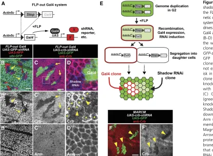
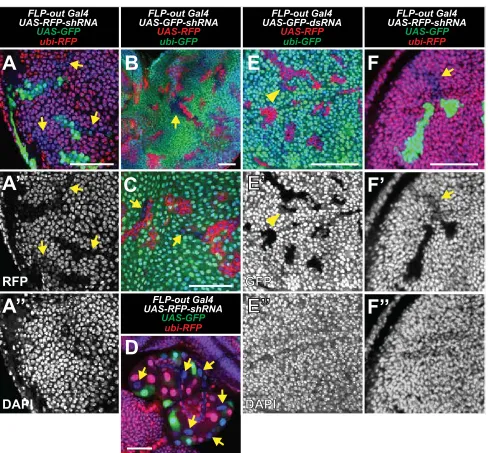
The FLP-out Gal4 system (Pignoni and Zipursky 1997) can be used to induce RNAi in a clonal lineage of cells that stably express Gal4. Clones are generated using a heat-shock-inducible FLP transgene, which catalyzes the removal of a transcriptional stop upstream of the Gal4-coding sequence
(Figure 1A). While using this system, we unexpectedly found that clonal expression of shRNAs causes knockdown in cells that do not express Gal4. For example, in larvae that ubiqui-tously express GFP (ubi-GFP), we generated Gal4 clones that express shRNA targeting GFP (UAS-GFP-shRNA) and red fluo-rescent protein (UAS-RFP) and dissected wing discs 48 hr after clone induction (ACI). As expected, RFP-expressing clones knock down GFP (Figure 1B). However, we also ob-served patches of cells that knock down GFP but do not ex-press RFP. We refer to this unexpected cell type as“shadow RNAi”cells since these cells exhibit knockdown of their target gene but do not express Gal4 as assessed by the absence of RFP expression.
Importantly, wefind that shadow RNAi cells are produced when shRNAs target two other genes,ubi-RFP(Figure S1A) and the endogenous genecrumbs(crb) (Figure 1, C and D). Furthermore, crbshadow RNAi cells exhibited a knowncrb
mutant phenotype characterized by altered localization of Crb where they contact wild-type cells (Figure 1D) (Pellikka et al.2002; Chenet al.2010; Hafeziet al.2012). In addition, shadow RNAi cells were readily observed in other larval tissues (Figure S1, B–D) and using independently derived transgenes (seeMaterials and Methods). These results sug-gest that production of shadow RNAi cells may be an inher-ent phenomenon when using the FLP-out Gal4 system, as
opposed to sporadic effects such as chromosomal instability or epigenetic silencing of transgenes.
We note that tests of three other endogenous genes (fat,
gigas, and dachshund) did not obviously generate shadow RNAi cells (Table S1; not shown). In addition, when we re-peated FLP-out Gal4 experiments using dsRNAs targeting GFP (UAS-GFP-dsRNA), we found that shadow RNAi cells were not clearly visible and may have exhibited only weak knockdown (Figure S1E). Therefore, shadow RNAi cells may manifest only when targeting particular genes or when using certain RNAi reagents (seeDiscussion).
Several observations of shadow RNAi cells hint at a mech-anism by which they are generated. Shadow RNAi cells nearly always appear as cohesive groups in contact with Gal4 clones (Figure 1, B and C,Figure S1), which is a well-documented behavior of sister clones in the imaginal disc (Xu and Rubin 1993). Furthermore, in cases where shadow RNAi cells ex-hibit partial knockdown of the target gene (Figure 1B), each cell within a cohesive group shows the same level of knock-down, suggesting a synchronized reversal of RNAi over time. Indeed, we find that knockdown in shadow RNAi cells is barely visible at 72 hr ACI (Figure S1F), suggesting that knockdown is not sustained as in Gal4-expressing clones. These observations suggest that shadow RNAi cells produced using the FLP-out Gal4 system are a sister lineage to Gal4 clones and that knockdown persists for up to 3 days after being transiently induced.
To explain our observations with the FLP-out Gal4 system, we propose that shRNAs are transiently expressed in an ancestral mother cell that gave rise to Gal4-expressing clones and sister shadow RNAi clones. This event could occur during G2 when cells have duplicated their genome if one of two Act-FRT-stop-FRT-Gal4transgenes undergoes recombination and
briefly expresses Gal4 before cell division (Figure 1E). In contrast, recombination during G1, or recombination of both Act-FRT-stop-FRT-Gal4 transgenes, would not be expected to generate shadow RNAi clones. To test this model, we performed clonal RNAi experiments using the MARCM (Mosaic Analysis with a Repressible Cell Marker) system, which restricts Gal4 activity until after two daughter cells are produced and the levels of the Gal80 repressor in the cytoplasm decay (Lee and Luo 1999). Consistent with this hypothesis, when using MARCM to express shRNAs that tar-getcrb, wefind that Crb protein is knocked down only in the Gal4 clone (Figure 1F). In addition, this result rules out the possibility that shRNAs or Gal4 are transferred from the Gal4 clone into shadow RNAi clones.
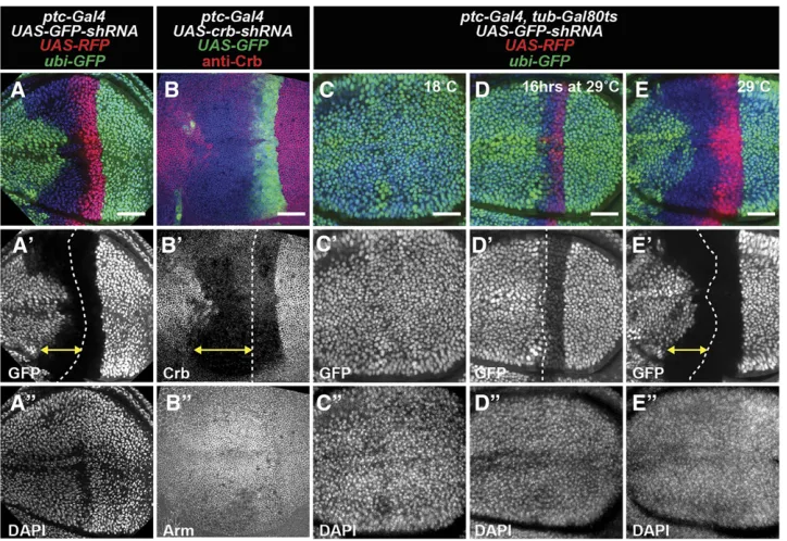
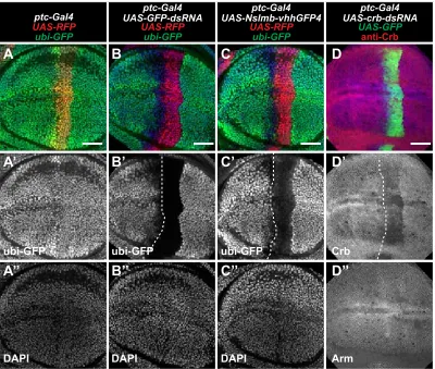
Since our model predicts that transient expression of shRNAs causes persistence of RNAi-mediated knockdown, we wanted to verify this using an independent method. patched-Gal4 (ptc-Gal4) is a commonly used enhancer trap line that expresses Gal4 in theptcexpression pattern (Hinz et al. 1994). In early wing disc development, ptc-Gal4 is expressed in all cells of the anterior compartment and later becomes restricted to a thin stripe of anterior cells that bor-der the posterior compartment (Phillipset al.1990; Evans et al. 2009). When we used ptc-Gal4 to express shRNAs targetingGFP(Figure 2A) orcrb(Figure 2B), we observed knockdown of the target gene within cells of the stripe cur-rently expressing Gal4, as well as cells far anterior to the stripe that no longer express Gal4 (assessed by afluorescent protein expressed under UAS control). In contrast, dsRNAs targeting GFP transcript or a nanobody fusion that de-grades GFP protein (Caussinuset al.2012) cause knockdown of GFPfluorescence mainly within theptc-expressing stripe, although some cells immediately anterior to the stripe have
Figure 2 Gene knockdown in shadow RNAi cells caused by dynamic expression of ptc-Gal4. (A–E) Wing imaginal discs with RNAi under control of ptc-Gal4. (A) ptc-Gal4 expression of RFP (red) and GFP-shRNA cause knockdown of GFP (green). Cell nuclei labeled with DAPI (blue). (B) ptc-Gal4 expression of GFP (green) and crb-shRNA cause knockdown of Crb protein (red). Arm staining (blue) shows cell membrane. (C–E) Temperature control of ptc-Gal4 expression with tub-Gal80ts. ptc-Gal4
reduced GFP levels (Figure S2, B and C). Similarly, dsRNAs that targetcrbcause knockdown only within theptc -express-ing stripe (Figure S2D). To directly test if past expression of ptc-Gal4in more anterior regions of the wing disc is required to generate shadow RNAi cells, we used a temperature-sensitive Gal80 transgene (McGuire et al.2003) to restrict expression of Gal4 to a 16-hr window immediately preceding dissection (Figure 2D). Under these conditions, shadow RNAi cells are not observed, suggesting that the shadow RNAi cells were generated by prior expression of the shRNA in those cells.
Investigation of mechanisms contributing to the persistence of RNAi-mediated knockdown
Our observation that it takes3 days to reverse the effects of GFP knockdown is consistent with reports in mammalian cell culture and in vivomouse models (Guptaet al. 2004; Dickins et al. 2005; Bartlett and Davis 2006; Zhang et al. 2007; Baccariniet al.2011), although our experiments were performed at a comparably lower temperature (25°). In these mammalian systems, it is generally thought that rever-sal from RNAi occurs by siRNA degradation and/or dilution with cell divisions (Dickinset al.2005; Baccariniet al.2011). Yet, considering this explanation, we were surprised by the high degree of persistent GFP knockdown following a short pulse of shRNA expression (Figure 1, B–D). Therefore, we considered the possibility that RNAi was being actively main-tained in some manner.
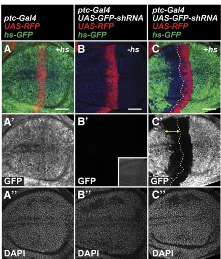
Active maintenance of RNAi has been demonstrated in different species, such as RNAi amplification in C. elegans (Sijenet al.2001; Alderet al.2003) or RNAi-induced tran-scriptional silencing (RITS) (Verdelet al.2004) inS. pombe. In addition, Piwi-interacting RNAs (piRNAs) target transcripts via an amplifying“ping-pong”cycle (Brenneckeet al.2007). Initiation of each of these mechanisms requires the presence of target transcripts. Therefore, we tested whether RNAi persis-tence inDrosophilatissues occurs when the target gene is not expressed until immediately before dissection. This was ac-complished using a heat-shock-inducible GFP transgene (hs-GFP) that is highly expressed when animals are incubated at 37°(Figure 3). Usingptc-Gal4to expressGFP-shRNAin a hs-GFPbackground, and inducing GFP expression 2 hr before dissection, wefind that GFP knockdown occurs in theptcstripe (RFP+) as well as in cells far anterior (RFP2) (Figure 3C). We do not detect GFPfluorescence without heat shock and ob-serve tissue autofluorescence only at higher exposure settings (Figure 3B9). These results suggest that previous expression of transcripts is not required for RNAi persistence in shadow RNAi cells.
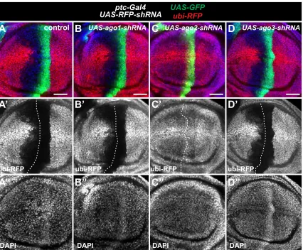
We also systematically tested the requirement of genes that might promote RNAi persistence based on mecha-nisms that operate in other systems. This was accomplished by knocking down each gene while monitoring transient knockdown of a ubiquitously expressed RFP (ubi-RFP) using the ptc-Gal4 expression system. Our goal was to identify genes that are selectively required for RNAi persistence in
cells anterior to theptcstripe. We testedDrosophila ortho-logs of genes involved in RITS, chromatin-remodeling genes, and machinery involved in miRNA, siRNA, and piRNA processing. With one exception, none of the genes when knocked down abolished persistent RNAi of the ubi-RFP reporter gene (Figure S3; Table S2). The exception wasAgo2RNAi, which nearly abolishes RFP knockdown in all cells expressing ptc-Gal4(Figure S3C). This result is consistent with the known role of Ago2 to bind siRNAs and coordinate RNAi-induced silencing complex (RISC) degrada-tion of target transcripts (Ni et al.2011). In summary, our results favor a model where the persistence of RNAi is simply the result of a slow rate of degradation of shRNAs and/or their siRNA derivatives.
i-TRACE: a novel lineage analysis tool based on RNAi
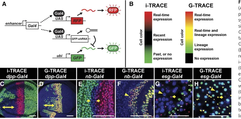
Since even transient expression of an shRNA could generate persistent knockdown (Figure 1, B and C), we explored its use as a lineage-tracing tool. To facilitate RNAi-based lineage tracing with Gal4 lines, we constructed afly strain containing three transgenes: (1) a reporter of Gal4 activity (e.g.,UAS-RFP), (2) a ubiquitously expressed target gene (e.g.,ubi-GFP), and (3) a Gal4-controlled shRNA (e.g.,UAS-GFP-shRNA) (Figure 4A). Therefore, when this triple-transgenic line is crossed
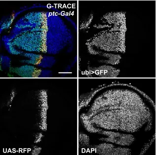
with a Gal4 line, F1progeny will contain cells and tissues that report real-time Gal4 expression (RFP+, GFP2) and recent Gal4 expression (RFP2, GFP2) (Figure 4B). Since exoge-nousfluorescent transgenes are used, the tissues being ana-lyzed are wild type and antibody staining is not necessary. We refer to this system as i-TRACE (RNAi-Technique for R eal-timeAndClonalExpression), which shares a similar naming convention with G-TRACE, a recombination-based lineage-tracing technique (Evanset al.2009). We compared i-TRACE with G-TRACE using several well-characterized Gal4 lines.
dpp-Gal4expresses in the anterior wing disc at early de-velopmental stages and becomes restricted to a thin stripe of cells at the border between anterior and posterior com-partments (Masucciet al.1990; Evanset al.2009). Using i-TRACE, we observed large regions of the anterior wing disc that previously expresseddpp-Gal4(Figure 4C). Using G-TRACE (Figure 4D), wefind that the region of lineage-traced cells is patchier and restricted to a smaller domain. Results withptc-Gal4are comparable todpp-Gal4as they express in similar domains (Figure S4). nubbin-Gal4 (nub-Gal4) ex-presses in the wing disc pouch, and the outer edge of this domain is thought to shift throughout larval development (Zirin and Mann 2007). Using i-TRACE, we confirmed this phenomenon byfinding a thin ring of cells outside of the nub-Gal4 domain that previously expressed Gal4 (Figure 3E). In contrast, when using G-TRACE, this ring of past expression is not visible (Figure 3F). Thus, in at least these two cases, i-TRACE appears more sensitive than G-TRACE.
escargot-Gal4(esg-Gal4) expresses in two cell types of the adult midgut: intestinal stem cells and their immediate de-scendants called enteroblasts (EBs) (Micchelli and Perrimon 2006). EBs give rise to two differentiated cell types that no longer express esg-Gal4: enterocytes and enteroendocrine cells. Together, these four cell types compose the entire midgut
epithelium. Using i-TRACE withesg-Gal4, we observed that all cells of the midgut are GFP2(Figure 3G). These cells include enterocytes, which are discernible by their large nuclear size (Micchelli and Perrimon 2006). In contrast, mus-cle cells that surround the midgut epithelium express GFP, confirming that animals contain theubi-GFPtransgene. This result supports the model that differentiated cell types in the midgut epithelium are descendants of a lineage that expressed esg-Gal4. Using G-TRACE with esg-Gal4 demon-strates similar results to i-TRACE (Figure 3H).
In summary, our analysis of several Gal4 lines using the i-TRACE system suggests that it is a useful tool for simulta-neously visualizing past and present gene expression.
Reversible changes in compartment identity markers are revealed using i-TRACE
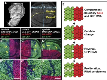
During animal development, boundaries between gene ex-pression domains are important to physically separate cells of different function (Dahmannet al.2011). In theDrosophila wing disc, four compartments are separated by two bound-aries, the anterior/posterior (A/P) boundary, and the dorsal/ ventral (D/V) boundary (Figure 5A). The A/P boundary is specified during embryogenesis and the D/V boundary at the end of thefirst larval instar. Lineage-tracing techniques have demonstrated that cells initially specified in one compart-ment do not normally switch identities (Garcia-Bellidoet al. 1973). We set out to test this model by analyzing the expres-sion patterns of several compartment-specific Gal4 lines with i-TRACE.
The A/P boundary is specified by the selector gene
engrailed(en) (Kornberget al.1985), which expresses in all cells of the posterior compartment and activates transcrip-tion of hedgehog (hh) (Tabata et al.1992). Using i-TRACE to analyze hh-Gal4, we observed present expression in the
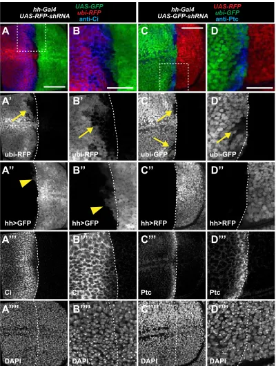
posterior compartment of the third instar wing disc (Figure 5B), consistent with previous studies (Tanimotoet al.2000). Surprisingly, in all discs imaged (.20), we also observed patches of shadow RNAi cells in the anterior compart-ment (Figure 5B), indicating that hh-Gal4 was previously expressed in these cells. These shadow RNAi patches were always adjacent to the A/P boundary and expressed anterior identity genes (Figure S5). Furthermore, we occasionally found that a subset of anterior shadow RNAi cells actively expressedhh-Gal4(Figure S5, A and B;Figure S6, A and B). To verify our results via a different method, we used G-TRACE to analyze pasthh-Gal4expression in the wing disc. Again, wefind patches of cells that previously expressed hh-Gal4 in the anterior compartment (Figure S6, C and D), although at a much lower frequency (1 disc of 10). This is consistent with the reduced sensitivity of G-TRACE in detecting past expression. These results suggest that at least some anterior cells in the wing disc expresshh-Gal4 at some point in development.
During late third instar wing development,enexpression expands into a small region of the anterior compartment that borders the posterior compartment (Blair 1992). We wondered whether this anteriorenexpression could be re-sponsible for activating hh-Gal4 in anterior cells as seen with i-TRACE. To test this possibility, we examined a devel-opmental time series to determine when anterior shadow RNAi cells form inhh-Gal4i-TRACE wing discs. Wefind that anterior hh-Gal4 shadow RNAi cells are first visible in the second instar and early third instar (Figure S7, A–D). We also find similar results withen-Gal4i-TRACE (Figure S7, G–J), where the appearance of anterior shadow RNAi cells pre-cedes the late third instar expression ofen-Gal4in anterior
cells (Figure S7, K and L). Furthermore, the anterioren ex-pression domain, which extends mostly along the dorsal/ ventral boundary, does not obviously overlap with the loca-tion and shape ofhh-Gal4shadow RNAi patches (Figure S8). These results suggest thaten-Gal4andhh-Gal4are expressed in anterior cells at a time point much earlier than previously described.
To determine if other markers of compartment identity transiently express outside of their canonical compartment, we analyzed the expression patterns of additional Gal4 lines with i-TRACE in the third instar wing disc. cubitus interruptus(ci), an essential component of thehh pathway, is repressed in the posterior compartment by en and thus is expressed only in the anterior compartment (Eaton and Kornberg 1990). Using i-TRACE to analyzeci-Gal4, wefind the expected current expression in the anterior compartment, but also evidence of past expression in cells of the posterior compartment (Figure 5C). In addition, a subset of posterior shadow RNAi cells actively express ci-Gal4 (Figure 5C9).
apterous(ap) is a selector gene expressed in the dorsal com-partment of the wing disc (Blairet al.1994). Using i-TRACE to analyzeap-Gal4, we observe cells in the ventral compart-ment that previously expressed Gal4 (Figure 5D). In sum-mary, our results with i-TRACE suggest that the expression of each of four different compartment-specific Gal4 lines (hh-Gal4, en-Gal4, ci-Gal4, and ap-Gal4) is not completely re-stricted to its specific compartment.
Several similarities in the characteristics of shadow RNAi patches produced from different compartment Gal4 lines suggest that they are clones that originate close to the com-partment boundary. First, these cells appear as cohesive groups with similar levels of knockdown, suggesting that they
belong to a shared clonal lineage that underwent several cell divisions after expression of Gal4 (Xu and Rubin 1993). Sec-ond, these patches are frequently elongated in the proximo/ distal direction, an indicator that there is significant prolifer-ation after the labeling event (Baena-Lopez et al. 2005). Third, these patches lie in proximity to the compartment boundary defined by the particular Gal4 line. These results suggest that cells located at wing-disc compartment bound-aries can transiently express at least some markers of the opposite compartment (Figure 5E).
Discussion
In this study, we show that transient expression of shRNAs in Drosophila tissues can cause persistent knockdown in cells that outlasts co-expressed marker transgenes. We term this effect“shadow RNAi,”since cells with persistent knockdown are not discernible without visualizing target gene expres-sion. Although this effect was obvious when targeting three different genes,GFP,RFP, andcrb, it is possible that other genes may behave differently. Indeed, we were unsuccessful in observing shadow RNAi cells for three other genes (fat,
gigas, anddachshund) in the wing disc using FLP-out Gal4 (Table S1; not shown). While these could represent technical failures, it is also possible that gene-specific factors influence the susceptibility to shadow RNAi, such as transcript/protein expression levels or stability. Similarly, different RNAi re-agents may or may not cause shadow RNAi. For both GFP andcrb, we found that an shRNA transgene was much more effective than a long dsRNA transgene in generating shadow RNAi (see Table S1). This difference may simply be explained by better knockdown efficiency using shRNAs com-pared to dsRNAs, as has been observed previously (Niet al. 2011). Alternatively, shRNAs, which are embedded in a miR-1microRNA backbone (Niet al.2011), might be more stable in cells than long dsRNAs or produce greater numbers of siRNAs. Importantly, it is possible that other hairpin trans-genes, derived from different sources or that target different regions of a transcript, may behave differently.
Since shadow RNAi cells can have mutant phenotypes, as we showed with crb (Figure 1D), it is important that re-searchers take this phenomenon into consideration, espe-cially when drawing conclusions about the cell autonomy of mutant phenotypes caused by RNAi-induced knockdown. For some experiments, simply identifying where shadow RNAi cells are located may allow a proper interpretation of results. To test if an shRNA generates shadow RNAi cells in vivo, it is critical to visualize target gene expression while conducting knockdown. Although we used antibodies to de-tect protein levels,in situ hybridization to detect transcript levels may also be effective. Complementary to testing an shRNA, a Gal4 line can be assayed with i-TRACE to determine if it causes persistent RNAi of afluorescent reporter transgene. We also suggest methods to prevent the generation of shadow RNAi cells. For example, including a temperature-sensitive Gal80 transgene can allow more refined temporal
control over when Gal4 is turned on (e.g., Figure 2, C–E), thus giving shadow RNAi cells less time to form. Alternatively, based on our experiments with GFP and crb knockdown, using long dsRNAs instead of shRNAs seems to prevent for-mation of shadow RNAi cells. If performing clonal RNAi ex-periments, we recommend using the MARCM system since this prevents the phenomenon of shadow RNAi clones. Fur-thermore, shadow RNAi cells are not predicted to occur when using FLP-out Gal4 in nonproliferative tissues since we sug-gest that transient expression of Gal4 before cell division is required for their generation (Figure 1E).
As an outcome of our work describing RNAi persistence in vivo, we developed the i-TRACE system as a novel method to monitor dynamic gene expression from Gal4 reporter lines. The i-TRACE systemfills an important gap in existing genetic methods. For example, real-time detection of Gal4 expression is accomplished with a reporter under UAS control (Fischer et al.1988; Brand and Perrimon 1993) but cannot be used to report past expression of Gal4. Conversely, recombination-based methods are used to stably mark cell lineages that pre-viously expressed Gal4 (Evanset al.2009), but can overlook short-term changes in gene expression that occur after stable recombination. The i-TRACE system can be used as a lineage-tracing tool for visualizing recent gene expression, since re-porter knockdown in marked cells reverses after72 hr. In addition, in at least some situations, the i-TRACE system ap-pears to be a more sensitive reporter of past Gal4 expression than G-TRACE.
Only rarely has a switch in compartment identity been observed near lineage-restricted boundaries, such as in the Drosophila embryo (Gettings et al. 2010) and in the wing discs during regeneration (Herrera and Morata 2014). Our data demonstrate that cells located at lineage-restricted boundaries of the wing disc can transiently express Gal4 re-porters of the opposite compartment identity (Figure 5E), raising the possibility that boundary cells may be less com-mitted to their respective compartmental identities than pre-viously thought, although they ultimately seem to maintain their originally fated compartmental identities. An important caveat is that Gal4 reporter transgenes might not accurately reflect transcription of the endogenous gene. Therefore, it remains unknown whether boundary cells express endog-enous identity genes of the opposite compartment and whether this results in transient cell-fate changes. Careful imaging of endogenous compartment identity gene ex-pression in developing wing discs may help resolve this issue. Furthermore, other possibilities such as direct trans-fer of Gal4 or shRNAs between cells at the boundary also merit consideration.
Acknowledgments
American Cancer Society Research Professor Award 120366-RP-11-078-01-DDC (to I.K.H.). J.A.B. was funded by the Cancer Research Coordinating Committee of the University of California and T.M.S. was funded by National Science Foundation Graduate Research Fellowship.
Literature Cited
Agaisse, H., U. M. Petersen, M. Boutros, B. Mathey-Prevot, and N. Perrimon, 2003 Signaling role of hemocytes in Drosophila JAK/STAT-dependent response to septic injury. Dev. Cell 5: 441–450.
Alder, M. N., S. Dames, J. Gaudet, and S. E. Mango, 2003 Gene silencing in Caenorhabditis elegans by transitive RNA interfer-ence. RNA 9: 25–32.
Baccarini, A., H. Chauhan, T. J. Gardner, A. D. Jayaprakash, R. Sachidanandamet al., 2011 Kinetic analysis reveals the fate of a microRNA following target regulation in mammalian cells. Curr. Biol. 21: 369–376.
Baena-Lopez, L. A., A. Baonza, and A. Garcia-Bellido, 2005 The orientation of cell divisions determines the shape of Drosophila organs. Curr. Biol. 15: 1640–1644.
Bartlett, D. W., and M. E. Davis, 2006 Insights into the kinetics of siRNA-mediated gene silencing from live-cell and live-animal bioluminescent imaging. Nucleic Acids Res. 34: 322–333. Blair, S. S., 1992 Engrailed expression in the anterior lineage
compartment of the developing wing blade of Drosophila. De-velopment 115: 21–33.
Blair, S. S., D. L. Brower, J. B. Thomas, and M. Zavortink, 1994 The role of apterous in the control of dorsoventral com-partmentalization and PS integrin gene expression in the devel-oping wing of Drosophila. Development 120: 1805–1815. Brand, A. H., and N. Perrimon, 1993 Targeted gene expression as
a means of altering cell fates and generating dominant pheno-types. Development 118: 401–415.
Brennecke, J., A. A. Aravin, A. Stark, M. Dus, M. Kellis et al., 2007 Discrete small RNA-generating loci as master regulators of transposon activity in Drosophila. Cell 128: 1089–1103. Buchon, N., N. A. Broderick, S. Chakrabarti, and B. Lemaitre,
2009 Invasive and indigenous microbiota impact intestinal stem cell activity through multiple pathways in Drosophila. Genes Dev. 23: 2333–2344.
Caussinus, E., O. Kanca, and M. Affolter, 2012 Fluorescent fusion protein knockout mediated by anti-GFP nanobody. Nat. Struct. Mol. Biol. 19: 117–121.
Chen, C. L., K. M. Gajewski, F. Hamaratoglu, W. Bossuyt, L. Sansores-Garcia, C. Tao, and G. Halder, 2010 The apical-basal cell polar-ity determinant Crumbs regulates Hippo signaling in Drosophila. Proc. Natl. Acad. Sci. USA 107: 15810–15815.
Chi, J. T., H. Y. Chang, N. N. Wang, D. S. Chang, N. Dunphyet al., 2003 Genomewide view of gene silencing by small interfering RNAs. Proc. Natl. Acad. Sci. USA 100: 6343–6346.
Croker, J. A., S. L. Ziegenhorn, and R. A. Holmgren, 2006 Regulation of the Drosophila transcription factor, Cubitus interruptus, by two conserved domains. Dev. Biol. 291: 368–381.
Dahmann, C., A. C. Oates, and M. Brand, 2011 Boundary forma-tion and maintenance in tissue development. Nat. Rev. Genet. 12: 43–55.
Dickins, R. A., M. T. Hemann, J. T. Zilfou, D. R. Simpson, I. Ibarra
et al., 2005 Probing tumor phenotypes using stable and regulated synthetic microRNA precursors. Nat. Genet. 37: 1289–1295. Eaton, S., and T. B. Kornberg, 1990 Repression of ci-D in posterior
compartments of Drosophila by engrailed. Genes Dev. 4: 1068– 1077.
Evans, C. J., J. M. Olson, K. T. Ngo, E. Kim, N. E. Lee et al., 2009 G-TRACE: rapid Gal4-based cell lineage analysis in Dro-sophila. Nat. Methods 6: 603–605.
Fire, A., S. Xu, M. K. Montgomery, S. A. Kostas, S. E. Driveret al., 1998 Potent and specific genetic interference by double-stranded RNA in Caenorhabditis elegans. Nature 391: 806–811. Fischer, J. A., E. Giniger, T. Maniatis, and M. Ptashne, 1988 GAL4
activates transcription in Drosophila. Nature 332: 853–856. Garcia-Bellido, A., P. Ripoll, and G. Morata, 1973 Developmental
compartmentalisation of the wing disk of Drosophila. Nat. New Biol. 245: 251–253.
Gettings, M., F. Serman, R. Rousset, P. Bagnerini, L. Almeidaet al., 2010 JNK signalling controls remodelling of the segment boundary through cell reprogramming during Drosophila mor-phogenesis. PLoS Biol. 8: e1000390.
Gupta, S., R. A. Schoer, J. E. Egan, G. J. Hannon, and V. Mittal, 2004 Inducible, reversible, and stable RNA interference in mammalian cells. Proc. Natl. Acad. Sci. USA 101: 1927–1932. Hafezi, Y., J. A. Bosch, and I. K. Hariharan, 2012 Differences in
levels of the transmembrane protein Crumbs can influence cell survival at clonal boundaries. Dev. Biol. 368: 358–369. Halfon, M. S., H. Kose, A. Chiba, and H. Keshishian, 1997 Targeted
gene expression without a tissue-specific promoter: creating mo-saic embryos using laser-induced single-cell heat shock. Proc. Natl. Acad. Sci. USA 94: 6255–6260.
Hannon, G. J., 2002 RNA interference. Nature 418: 244–251. Herrera, S. C., and G. Morata, 2014 Transgressions of
compart-ment boundaries and cell reprogramming during regeneration in Drosophila. eLife 3: e01831.
Hinz, U., B. Giebel, and J. A. Campos-Ortega, 1994 The basic-helix-loop-helix domain of Drosophila lethal of scute protein is sufficient for proneural function and activates neurogenic genes. Cell 76: 77–87.
Kornberg, T., I. Siden, P. O’Farrell, and M. Simon, 1985 The en-grailed locus of Drosophila: in situ localization of transcripts reveals compartment-specific expression. Cell 40: 45–53. Lee, T., and L. Luo, 1999 Mosaic analysis with a repressible cell
marker for studies of gene function in neuronal morphogenesis. Neuron 22: 451–461.
Livshits, G., and S. W. Lowe, 2013 Accelerating cancer modeling with RNAi and nongermline genetically engineered mouse mod-els, pp. 991–1005. Cold Spring Harb. Protoc. 2013: pii: pdb. top069856.
Masucci, J. D., R. J. Miltenberger, and F. M. Hoffmann, 1990 Pattern-specific expression of the Drosophila decapentaplegic gene in ima-ginal disks is regulated by 39cis-regulatory elements. Genes Dev. 4: 2011–2023.
McGuire, S. E., P. T. Le, A. J. Osborn, K. Matsumoto, and R. L. Davis, 2003 Spatiotemporal rescue of memory dysfunction in Drosophila. Science 302: 1765–1768.
Micchelli, C. A., and N. Perrimon, 2006 Evidence that stem cells reside in the adult Drosophila midgut epithelium. Nature 439: 475–479.
Mohr, S. E., J. A. Smith, C. E. Shamu, R. A. Neumuller, and N. Perrimon, 2014 RNAi screening comes of age: improved tech-niques and complementary approaches. Nat. Rev. Mol. Cell Biol. 15: 591–600.
Ni, J. Q., R. Zhou, B. Czech, L. P. Liu, L. Holderbaumet al., 2011 A genome-scale shRNA resource for transgenic RNAi in Drosoph-ila. Nat. Methods 8: 405–407.
Perrimon, N., J. Q. Ni, and L. Perkins, 2010 In vivo RNAi: today and tomorrow. Cold Spring Harb. Perspect. Biol. 2: a003640. Phillips, R. G., I. J. Roberts, P. W. Ingham, and J. R. Whittle,
1990 The Drosophila segment polarity gene patched is in-volved in a position-signalling mechanism in imaginal discs. De-velopment 110: 105–114.
Pignoni, F., and S. L. Zipursky, 1997 Induction of Drosophila eye development by decapentaplegic. Development 124: 271–278. Richard, M., F. Grawe, and E. Knust, 2006 DPATJ plays a role in
retinal morphogenesis and protects against light-dependent de-generation of photoreceptor cells in the Drosophila eye. Dev. Dyn. 235: 895–907.
Roignant, J. Y., C. Carre, B. Mugat, D. Szymczak, J. A. Lepesant
et al., 2003 Absence of transitive and systemic pathways al-lows cell-specific and isoform-specific RNAi in Drosophila. RNA 9: 299–308.
Sijen, T., J. Fleenor, F. Simmer, K. L. Thijssen, S. Parrish et al., 2001 On the role of RNA amplification in dsRNA-triggered gene silencing. Cell 107: 465–476.
Tabata, T., S. Eaton, and T. B. Kornberg, 1992 The Drosophila hedgehog gene is expressed specifically in posterior compart-ment cells and is a target of engrailed regulation. Genes Dev. 6: 2635–2645.
Tanimoto, H., S. Itoh, P. ten Dijke, and T. Tabata, 2000 Hedgehog creates a gradient of DPP activity in Drosophila wing imaginal discs. Mol. Cell 5: 59–71.
Vaistij, F. E., L. Jones, and D. C. Baulcombe, 2002 Spreading of RNA targeting and DNA methylation in RNA silencing requires
transcription of the target gene and a putative RNA-dependent RNA polymerase. Plant Cell 14: 857–867.
Verdel, A., S. Jia, S. Gerber, T. Sugiyama, S. Gygiet al., 2004 RNAi-mediated targeting of heterochromatin by the RITS complex. Science 303: 672–676.
Wilson, R. C., and J. A. Doudna, 2013 Molecular mechanisms of RNA interference. Annu. Rev. Biophys. 42: 217–239.
Xu, T., and G. M. Rubin, 1993 Analysis of genetic mosaics in de-veloping and adult Drosophila tissues. Development 117: 1223– 1237.
Yeh, E., K. Gustafson, and G. L. Boulianne, 1995 Greenfl uores-cent protein as a vital marker and reporter of gene expression in Drosophila. Proc. Natl. Acad. Sci. USA 92: 7036–7040. Zhang, J., C. Wang, N. Ke, J. Bliesath, J. Chionis et al., 2007 A
more efficient RNAi inducible system for tight regulation of gene expression in mammalian cells and xenograft animals. RNA 13: 1375–1383.
Zirin, J. D., and R. S. Mann, 2007 Nubbin and Teashirt mark barriers to clonal growth along the proximal-distal axis of the Drosophila wing. Dev. Biol. 304: 745–758.
Zong, J., X. Yao, J. Yin, D. Zhang, and H. Ma, 2009 Evolution of the RNA-dependent RNA polymerase (RdRP) genes: duplica-tions and possible losses before and after the divergence of major eukaryotic groups. Gene 447: 29–39.
GENETICS
Supporting Information www.genetics.org/lookup/suppl/doi:10.1534/genetics.116.187062/-/DC1
Persistence of RNAi-Mediated Knockdown in
Drosophila
Complicates Mosaic Analysis Yet Enables
Highly Sensitive Lineage Tracing
Justin A. Bosch, Taryn M. Sumabat, and Iswar K. Hariharan
Figure S1. Additional data relevant to Figure 1 (A)
Wing imaginal disc with FLP-out clones expressing GFP
(green) and RFP-shRNA, with knockdown of ubi-RFP (red). Arrows indicate shadow RNAi clones.
(B-D)
Larval tissues with shadow RNAi clones, indicated by arrows,
(B)
eye imaginal disc,
(C)
lymph gland,
(D)
prothoracic gland.
(E)
Wing imaginal disc with FLP-out clones expressing RFP (red) and GFP-dsRNA, with
knockdown of ubi-GFP (green). Arrowhead indicates possible shadow RNAi cells.
(F)
Clones induced 72hrs
before dissection. Arrow indicates shadow clone. Cell nuclei labeled with DAPI (blue). Scale bars are 50µm.
E
E’
E’’
A
GFP
DAPI
RFP
DAPI
A’
B
D
C
A’’
FLP-out Gal4
UAS-RFP UAS-GFP-dsRNA
ubi-GFP FLP-out Gal4
UAS-RFP UAS-GFP-shRNA
ubi-GFP
FLP-out Gal4
UAS-GFP UAS-RFP-shRNA
ubi-RFP
FLP-out Gal4
UAS-GFP UAS-RFP-shRNA
ubi-RFP FLP-out Gal4
UAS-GFP UAS-RFP-shRNA
ubi-RFP
F
F’
Figure S2. Additional data relevant to Figure 2 (A-D)
Wing imaginal discs expressing ptc-Gal4.
(A-C)
Expression of RFP (red) in an ubi-GFP background. Cell nuclei labeled with DAPI (blue).
(A)
Control disc that
does not express GFP-shRNA.
(B)
Expression of GFP-dsRNA.
(C)
Expression of Nslmb-vhhGFP4 (deGradFP).
vhhGFP4 is a nanobody that binds to GFP protein, and Nslmb is a truncated form of the E3 ubiquitin ligase
slmb that contains the F-box domain (Caussinus et al., 2012). Therefore, expression of Nslmb-vhhGFP4 causes
ubiquitination of GFP and degradation via the proteasome.
(D)
Expression of GFP (green) and crb-dsRNA, and
antibody staining for Crb (red). Cell membranes labeled with Arm staining (blue). Scale bars are 50µm.
A
DAPI DAPI DAPI
ubi-GFP ubi-GFP ubi-GFP
A’
A’’
B
B’
B’’
C
C’
C’’
Arm Crb
D
D’
D’’
ptc-Gal4
UAS-RFP ubi-GFP
ptc-Gal4
UAS-RFP
UAS-GFP-dsRNA
ubi-GFP
ptc-Gal4
UAS-RFP
UAS-Nslmb-vhhGFP4
ubi-GFP
ptc-Gal4
UAS-GFP UAS-crb-dsRNA
Figure S3. Additional data relevant to Figure 3 (A-D)
Wing imaginal disc with ptc-Gal4 expression of GFP
(green) and RFP-shRNA, in an ubi-RFP background. Cell nuclei labeled with DAPI (blue).
(A)
Control disc.
Expression of
(B)
ago1-shRNA,
(C)
ago2-shRNA, or
(D)
ago3-shRNA. Scale bars are 50µm.
ptc-Gal4
control
A
A’
A’’
B
B’
B’’
C
C’
C’’
D
D’
D’’
ubi-RFP
DAPI
ubi-RFP
DAPI
ubi-RFP
DAPI
ubi-RFP
DAPI
UAS-ago1-shRNA UAS-ago2-shRNA UAS-ago3-shRNA
UAS-GFP
Figure S4. Additional data relevant to Figure 4.
G-TRACE analysis of ptc-Gal4 in the wing imaginal disc.
Current expression indicated by RFP (red), recombined lineage expression indicated by GFP (green). Cell
nuclei labeled with DAPI (blue).
Scale bar is 50µm.
ptc-Gal4
G-TRACE
DAPI
UAS-RFP
Figure S5. Additional data relevant to Figure 5.
Anterior shadow RNAi cells produced from hh-Gal4 express
anterior cell identity markers in the wing imaginal disc.
(A-D)
i-TRACE analysis with hh-Gal4. Arrows
indicate shadow RNAi cells in anterior compartment.
(A-B)
hh-Gal4 drives expression of GFP and RFP-shRNA
in an ubi-RFP background. Anti-Ci staining (blue) in the anterior compartment). Arrowheads indicate current
expression of hh-Gal4 in anterior cells.
(B)
Enlargement of box in
A
.
(C-D)
hh-Gal4 drives expression of RFP
and GFP-shRNA in an ubi-GFP background. Anti-Ptc staining (blue) in anterior cells that border the posterior
compartment.
(D)
Enlargement of box in
C
. Scale bars are 50µm in
A
and
C
, and 25µm in
B
and
D
.
ubi-RFP
hh>GFP
Ci
DAPI
A
A’
A’’
A’’’
A’’’’
B
B’
B’’
B’’’
B’’’’
C
C’
C’’
C’’’
C’’’’
D
D’
D’’
D’’’
D’’’’
ubi-RFP
hh>GFP
Ci
DAPI
ubi-GFP
hh>RFP
Ptc
DAPI
ubi-GFP
hh>RFP
Ptc
DAPI
hh-Gal4 UAS-GFP
UAS-RFP-shRNA ubi-RFPanti-Ci hh-Gal4
Figure S6. Additional data relevant to Figure 5. (A-B)
hh-Gal4 analyzed with i-TRACE. A subset of cells
within anterior shadow RNAi patches exhibit low level current expression of hh-Gal4. hh-Gal4 drives
expression of GFP (green) and RFP-shRNA in a ubi-RFP background. Arrows indicate anterior shadow RNAi
cells. Arrowheads indicate anterior cells that currently express hh-Gal4.
(B)
Enlargement of box in
A
. The
white dotted line in
B’’
and
B’’’
outlines anterior shadow RNAi cells
(C-D)
hh-Gal4 analyzed with G-TRACE.
RFP marks currently expressing cells, GFP marks past expressing cells. Arrows indicate anterior cells that are
GFP+ but RFP-.
(D)
Enlargement of box in
C
. Cell nuclei labeled with DAPI (blue).
Scale bars are 50µm in
A
and
C
, and 25µm in
B
and
D
.
A
A’
A’’
A’’’
A’’’’
B
B’
B’’
B’’’
B’’’’
C
C’
C’’
C’’’
D
D’
D’’
D’’’
hh>GFP
ubi-RFP
DAPI
hh>GFP
hh>GFP
ubi>GFP
UAS-RFP
ubi-RFP
DAPI
hh>GFP
DAPI
ubi>GFP
UAS-RFP
DAPI
hh-Gal4 UAS-GFPUAS-RFP-shRNA ubi-RFPDAPI hh-Gal4
Figure S7. Additional data relevant to Figure 5.
Developmental time-series of wing imaginal discs from
stages L2 to L3. Arrows indicate shadow RNAi cells.
(A-F)
i-TRACE analysis of hh-Gal4.
(G-L)
i-TRACE
analysis of en-Gal4.
(A-B, G-H)
stage L2 wing discs.
(C-D, I-J)
early stage L3 wing discs.
(E-F, K-L)
late
stage L3 wing discs. Cell nuclei labeled with DAPI (blue). All scale bars are 50µm.
hh-Gal4
UAS-GFP
UAS-RFP-shRNA
ubi-RFP
en-Gal4
UAS-GFP
UAS-RFP-shRNA
ubi-RFP
A
B
C
D
E
F
G
H
I
Figure S8. Additional data relevant to Figure 5.
Anterior shadow RNAi cells produced from hh-Gal4 are
distinguishable from anterior expression of En in the late 3
rdinstar wing disc.
(A-B)
i-TRACE analysis with
hh-Gal4. Arrows indicate shadow RNAi cells in anterior compartment. Arrowheads indicate anterior En expression.
(B)
Enlargement of box in
A
. Cell nuclei labeled with DAPI (blue).
Scale bars are 50µm in
A
, and 25µm in
B
.
A
A’
A’’
A’’’
A’’’’
B
B’
B’’
B’’’
B’’’’
hh-Gal4
UAS-RFP
UAS-GFP-shRNA
ubi-GFP
anti-En
En
En
RFP
RFP
GFP
GFP
Target
Knockdown
type Genotype BL #
FLP-out Gal4
phenotype Figure ptc-Gal4 phenotype Figure
ubi-GFP dsRNA
w[1118];
P{w[+mC]=UAS-GFP.dsRNA.R}142 9330
rare and faint shadow
RNAi cells Fig. S1
faint shadow RNAi cells
anterior to ptc stripe Fig. S2
ubi-GFP shRNA
y[1] sc[*] v[1]; P{y[+t7.7]
v[+t1.8]=VALIUM20-EGFP.shRNA.1}attP2 41556
obvious shadow RNAi
clones Fig. 1
obvious shadow RNAi
cells anterior to ptc stripe Fig. 2
hs-GFP shRNA
y[1] sc[*] v[1]; P{y[+t7.7]
v[+t1.8]=VALIUM20-EGFP.shRNA.1}attP40 41555 - -
obvious shadow RNAi
cells anterior to ptc stripe Fig. 3
ubi-GFP deGradFP w[*]; P{w[+mC]=UAS-Nslmb-vhhGFP4}3 38421 - -
faint shadow knockdown
cells anterior to ptc stripe Fig. S2
ubi-RFP shRNA
y[1] sc[*] v[1]; P{y[+t7.7]
v[+t1.8]=VALIUM20-mCherry}attP2 35785
obvious shadow RNAi
clones Fig. S1
obvious shadow RNAi
cells anterior to ptc stripe Fig. S3
crb dsRNA
y[1] v[1]; P{y[+t7.7]
v[+t1.8]=TRiP.JF02777}attP2 27697 - -
no shadow RNAi cells
anterior to ptc stripe Fig. S2
crb shRNA
y[1] sc[*] v[1]; P{y[+t7.7]
v[+t1.8]=TRiP.HMS02036}attP2 40869
obvious shadow RNAi
clones Fig. 1
obvious shadow RNAi
cells anterior to ptc stripe Fig. 2
gigas shRNA
y[1] sc[*] v[1]; P{y[+t7.7]
v[+t1.8]=TRiP.HMS01217}attP2/TM3, Sb[1] 34737
no shadow clones observed, not in figures
data not
shown - -
ft shRNA
y[1] sc[*] v[1]; P{y[+t7.7]
v[+t1.8]=TRiP.HMS00932}attP2 34970
no shadow clones observed, not in figures
data not
shown - -
dac shRNA
y[1] sc[*] v[1]; P{y[+t7.7]
v[+t1.8]=TRiP.HMS01435}attP2 35022
no shadow clones observed, not in figures
data not
shown - -
Gene
Function
RNAi Phenotype Bloomington #
TRiP #
shRNA version
ago1
miRNA associated,
RISC component
none
33727
HMS00610
VALIUM20
ago2
siRNA associated,
RISC component
Abolishes RNAi
of RFP reporter
34799
HMS00108
VALIUM20
ago3
piRNA pathway
none
34815
HMS00125
VALIUM20
eIF-2gamma
S. pombe RITS
homologue
none
33401, 32914
HMS00279,
HMS00704