| INVESTIGATION
Repression of Germline Genes in
Caenorhabditis
elegans
Somatic Tissues by H3K9 Dimethylation of
Their Promoters
Andreas Rechtsteiner,* Meghan E. Costello,†Thea A. Egelhofer,* Jacob M. Garrigues,*,1Susan Strome,* and Lisa N. Petrella†,2 *Department of Molecular, Cell, and Developmental Biology, University of California Santa Cruz, Santa Cruz, California 95064 and †Department of Biological Sciences, Marquette University, Milwaukee, Wisconsin 53201
ORCID IDs: 0000-0001-9496-7412 (S.S.); 0000-0001-8664-7435 (L.N.P.)
ABSTRACTRepression of germline-promoting genes in somatic cells is critical for somatic development and function. To study how germline genes are repressed in somatic tissues, we analyzed key histone modifications in threeCaenorhabditis eleganssynMuv B mutants,lin-15B,lin-35, andlin-37—all of which display ectopic expression of germline genes in the soma.LIN-35andLIN-37are members of the conserved DREAM complex.LIN-15Bhas been proposed to work with the DREAM complex but has not been shown biochemically to be a member of the complex. We found that, in wild-type worms, synMuv B target genes and germline genes are enriched for the repressive histone modification dimethylation of histone H3 on lysine 9 (H3K9me2) at their promoters. Genes with H3K9me2 promoter localization are evenly distributed across the autosomes, not biased toward autosomal arms, as are the broad H3K9me2 domains. Both synMuv B targets and germline genes display a dramatic reduction of H3K9me2 promoter localization in lin-15Bmutants, but much weaker reduction inlin-35andlin-37mutants. This difference betweenlin-15Band DREAM complex mutants likely represents a difference in molecular function for these synMuv B proteins. In support of the pivotal role of H3K9me2 in regulation of germline genes byLIN-15B, global loss of H3K9me2 but not H3K9me3 results in phenotypes similar to synMuv B mutants, high-temperature larval arrest, and ectopic expression of germline genes in the soma. We propose that LIN-15B-driven enrichment of H3K9me2 at promoters of germline genes contributes to repression of those genes in somatic tissues.
KEYWORDSH3K9me2; synMuv B; LIN-15B; germline genes; chromatin
R
EPRESSION in somatic cells of genes that promote germ-line development and function is a vital cell fate regulatory mechanism, which, when disrupted, leads to developmental problems and is a hallmark of aggressive cancer (Janicet al. 2010; Petrellaet al. 2011; Whitehurst 2014; Al-Aminet al. 2016). Repression of germline genes in the soma poses a unique challenge for cells. First, like other genes expressed in specific tissues, germline genes can be found clustered along chromosomes; however, within a given cluster, genes withubiquitous, germline, and nongermline expression are inter-spersed (Royet al.2002; Spellman and Rubin 2002; Reinke and Cutter 2009). Therefore, somatic cells require a mecha-nism to repress germline genes without disrupting expression of importantflanking genes. Second, because embryos start life as the fusion of two germline cells—an egg and a sperm—they inherit an epigenetic state associated with driv-ing germline gene expression (Furuhashi et al. 2010; Rechtsteiner et al. 2010; Zenk et al. 2017; Kreher et al. 2018; Tabuchiet al.2018). This chromatin state must be reset during development to turn off germline gene expression in differentiating somatic cells (Morganet al.2005; Fraser and Lin 2016). There has been no investigation to date of the unique patterns of chromatin modifications or regulatory pro-tein binding that lead to repression of germline genes in so-matic tissues inCaenorhabditis elegans.
synMuv (for synthetic Multivulva) B proteins are a diverse class of transcriptional repressors that are involved in a Copyright © 2019 by the Genetics Society of America
doi:https://doi.org/10.1534/genetics.118.301878
Manuscript received December 14, 2018; accepted for publication March 16, 2019; published Early Online March 25, 2019.
Available freely online through the author-supported open access option. Supplemental material available athttps://doi.org/10.25386/genetics.7823846. 1Present address: Division of Biology University of California San Diego, La Jolla CA
92093.
number of different cell fate decisions inC. elegans(Unhavaithaya et al.2002; Wanget al.2005; Fay and Yochem 2007). A subset of synMuv B genes show a distinct set of mutant phenotypes, which include ectopic expression of germline genes in somatic cells and larval arrest at high temperature (called HTA for high tempera-ture arrest) (Wanget al. 2005; Petrellaet al.2011; Wuet al. 2012). Of this subset, a large proportion encode proteins that exist in two complexes: the HP1-containing heterochromatin complex (HPL-2, LIN-13, LIN-61), and the DREAM complex (EFL-1,DPL-1,LIN-35,LIN-9,LIN-37,LIN-52,LIN-53,LIN-54) (Cousthamet al.2006; Harrisonet al.2006; Wuet al.2012). Several additional synMuv B mutants, includinglin-15Band met-2, also display ectopic germline gene expression in the soma, but have not been shown biochemically to encode members of the HP1 or DREAM complex (Petrellaet al.2011; Wuet al.2012).
lin-15Bmutants, like mutants in genes encoding DREAM com-plex members, also display an HTA phenotype, show changes in regulation of somatic RNAi, and cause transgene silencing in the soma (Wanget al.2005; Petrellaet al.2011; Wuet al.2012). While mutations in genes encoding the HP1 complex, the DREAM complex,LIN-15B, andMET-2all lead to ectopic expres-sion of germline genes in the soma, the precise way these differ-ent complexes/proteins function in parallel or together to repress germline genes in the somatic tissues of wild-type animals is not understood.
Several lines of evidence point to synMuv B complexes repressing gene expression by altering chromatin. First, syn-Muv B mutant phenotypes, including HTA and ectopic germ-line gene expression, are strongly suppressed by loss of chromatin factors (Unhavaithaya et al. 2002; Wang et al. 2005; Cuiet al.2006; Petrellaet al.2011; Wuet al.2012). Second, the DREAM complex has been shown to promote enrichment of the H2A histone variant HTZ-1 in the body of a subset of genes that the DREAM complex represses in L3 larvae (Latorreet al.2015). Finally,HPL-2is a homolog of heterochromatin protein 1 (HP1) (Couteauet al.2002). HPL-2, in a complex withLIN-13andLIN-61, localizes to genomic regions enriched for histone H3 methylated at lysine 9 (H3K9me) and helps create repressive heterochromatin (Wuet al.2012; Garrigueset al.2015). Together, these data indicate that changes to chromatin may underlie the ectopic expression of germline genes in synMuv B mutants.
One of the best studied aspects of chromatin regulation is covalent modifications on histone tails. Specific histone mod-ifications are often associated with repressive or active chro-matin compartments and can be a read-out of the expression state of a gene. Histone H3 lysine 4 methylation (H3K4me) and H3 lysine 36 methylation (H3K36me) are generally associated with areas of previous or active gene expression (Hoet al.2014; Evanset al.2016). In contrast, histone H3 lysine 9 methylation (H3K9me) and histone H3 lysine 27 methylation (H3K27me) are associated with areas of low/no expression of coding genes and repression of repeti-tive elements (Ahringer and Gasser 2018). Of particular in-terest for the regulation of germline gene expression in somatic cells is histone H3K9 methylation. In C. elegans,
mono- and dimethylation of H3K9 (H3K9me1 and H3K9me2, respectively), are primarily catalyzed byMET-2.met-2mutants lose 80–90% of H3K9me1 and H3K9me2 in embryos (Towbin et al.2012).met-2is a synMuv B gene, and mutants have been previously shown to ectopically express germline genes in somatic cells (Wu et al. 2012). Trimethlyation of H3K9 (H3K9me3) is catalyzed by a separate histone methyltrans-ferase,SET-25(Towbinet al.2012).set-25is not a synMuv B gene and its potential role in regulating germline gene ex-pression in the soma has not been tested. Several studies have analyzed the roles in C. elegans of H3K9me2 and H3K9me3 in regulating the interaction of heterochromatin with the nuclear periphery and repression of repetitive ele-ments (Meister et al.2010; Towbin et al.2012; Guoet al. 2015; Zelleret al.2016). Both of these functions rely pri-marily on high enrichment of H3K9 methylation on the het-erochromatic arms of the autosomes (Ikegamiet al.2010; Liu et al. 2011; Garrigueset al. 2015; Evans et al.2016). However, little work has been done to look at how H3K9 methylation localizes to or regulates protein-coding genes in the euchromatic central regions of autosomes, where a large percentage of germline genes reside. Tofill this gap, we sought to identify changes in the levels and distributions of active and repressive histone modifications in the soma of synMuv B mutants and test whether such changes underlie ectopic expression of germline genes.
In this study, we used chromatin immunoprecipitation with genome-wide high-throughput sequencing (ChIP-seq) to analyze histone modifications in wild type and three synMuv B mutants, lin-15B,lin-35, andlin-37. We found that, in wild-type L1 larvae, which are composed of 550 somatic cells and two germ cells, and are therefore primarily somatic, H3K9me2 is enriched at the promoters of a subset of genes that display germline-specific ex-pression. The genes that have H3K9me2 at their promoters in wild type are generally upregulated in synMuv B mutants, suggesting that H3K9me2 plays a role in their repression. In support of this, the localization of H3K9me2 at gene promoters is largely lost in lin-15Bmutants and is diminished but not lost inlin-35andlin-37
mutants. Loss of H3K9me2 at promoters in mutants is associated with an increase in H3K4me3 at promoters and H3K36me3 in gene bodies—modifications associated with gene expression— suggesting that these genes go from a repressed state to an expressed state. Global loss of H3K9me2 but not H3K9me3 results in both the HTA and ectopic germline gene expression phenotypes seen in lin-15B mutants. We propose thatLIN-15Band DREAM repress a subset of germ-line genes in somatic tissues by promoting enrichment of H3K9me2 at the promoters of those genes.
Materials and Methods
C. elegans strains and culture conditions
MT10430lin-35(n745) I. SS1183hpl-2(tm1489) III. MT5470lin-37(n758) III. MT13293met-2(n4256) III. MT17463set-25(n5021) III.
GW638met-2(n4256)set-25(n5021) III. MT2495lin-15B(n744) X.
ChIP-seq from L1s
Worms were grown from synchronized L1s in standard S-basal medium with shaking at 230 rpm and fed HB101bacteria until gravid. Embryos were harvested using standard bleach-ing methods, and L1s were synchronized in S-basal medium with shaking for 14–18 hr in the absence of food. For 26° samples, worms were grown to the L4 stage at 20°, then upshifted to 26°until gravid, and L1s were harvested as described above. Extracts were made as described in Kolasinska-Zwierzet al.(2009) with the following modifica-tions. Cross-linked chromatin was sonicated using a Diage-node Bioruptor at high setting for 30 pulses, each lasting 30 sec followed by a 1 min pause. ChIP was performed as described by Kolasinska-Zwierzet al.(2009) with the modi-fication of using 0.5 mg of protein and 1 mg antibody or by using an IP-Star Compact Automated System (Diagenode) as described in Tabuchiet al.(2018). Sequencing libraries were prepared in two ways. Some libraries were prepared with the NEBNext Ultra DNA library Prep Kit (NEB) following the manufacturers’instructions; 1 ng of starting DNA was used, adapters were diluted 1:40, and AMPure beads were used for size selection before amplification to enrich for fragments corresponding to a 200 bp insert size. The other libraries were prepared using Illumina Truseq adapters and primers. ChIP or input DNA fragments were end-repaired with the following: 5 ml T4 DNA ligase buffer with 10 mM ATP, 2 ml dNTP mix, 1.2 ml T4 DNA polymerase (3 U/ml), 0.8 ml 1:5 Klenow DNA polymerase (diluted with 13T4 DNA ligase buffer for afinal Klenow concentration of 1 U/ml), 1 ml T4 PNK (10 U/ml). This 50 ml reaction was incubated at 20°for 30 min and purified with a QIAquick PCR spin column (elution volume 36 ml).“A”bases were then added to the 39end of the DNA fragments with the following: 5 ml NEB buffer 2, 10 ml dATP (1 mM), 1 ml Klenow 39to 59exo- (5 U/ml). This mix-ture was incubated at 37°for 30 min, and the DNA was purified with a QIAquick MinElute column (11 ml of DNA was eluted into a siliconized tube). Illumina TruSeq adapters were ligated to DNA fragments with the following: 15 ml 23Rapid Ligation buffer, 1 ml adapters (diluted 1:40), 1.5 ml Quick T4 DNA Li-gase. This 30 ml reaction was incubated at 23°for 30 min. The mixture was then cleaned up 23with AMPure beads (using 95% vol beads), and DNA was eluted in 22 ml. The Adapter-Modified DNA fragments were amplified by PCR with the fol-lowing mixture: 6 ml 53Phusion Buffer HF, 2 ml Primer cock-tail (from TrueSeq kit), 0.5 ml 25 mM dNTP mix, 0.5 Phusion polymerase (2 U/ml) using the following PCR program: 98° 30 min, 98°10 min, 60°30 min, and 72°30 min repeated
16 cycles, followed by 72°5 min. The amplified DNA was con-centrated and loaded onto a 2% agarose gel, and DNA between 250 and 350 bp was recovered from the gel. The multiplexed libraries were sequenced on an Illumina HiSeq4000 or HiSeq2000 at the Vincent J. Coates Genomics Sequencing Laboratory at University of California, Berkeley.
ChIP-chip from embryos
Late-stage embryos were obtained and chromatin extracts prepared as described in Latorre et al. (2015). Chromatin immunoprecipitation and subsequent LM-PCR, microarray hybridization, and scanning were performed as in Garrigues et al.(2015).
Antibodies used for ChIP
Mouse monoclonal antibodies for H3K9me2 (MABI0307, #302–32369; Fujifilm Wako), H3K36me3 (MABI0333, #300–95289; Fujifilm Wako), H3K27me3 (MABI0323, #309–95259; Fujifilm Wako), and H3K4me3 (MABI0304, #305–34819; Fujifilm Wako) were used as described in Liu et al.(2011), Egelhoferet al.(2011). Rabbit polyclonal LIN-15B antibody (SDQ2330, #38610002; Novus Biologicals) was used at a concentration of 2.5 mg per milligram of chro-matin extract.
Analysis of ChIP-seq data
Raw sequence reads from the Illumina HiSeq (50 bp single-end reads) were mapped to theC. elegansgenome (Wormbase version WS220) using Bowtie with default settings (Langmeadet al.2009). MACS2 (Zhanget al.2008) was used to call peaks and create bedgraph files for sequenced and mapped H3K4me3 ChIP samples and corresponding Input DNA samples with the following parameters: callpeak -t H3K4me3.mapped.reads.sampleX -c Input.mapped.reads. sampleX -g ce --bdg --keep-dup=auto --qvalue=0.01 --nomodel --extsize=250 --call-summits
MACS2 was used to call peaks and create bedgraphfiles for sequenced and mapped H3K9me2 ChIP samples and corre-sponding Input DNA samples with slightly different parame-ters to account for the broader domains of H3K9me2: callpeak -t H3K9me2.mapped.reads.sampleX -c Input.mapped.reads. sampleX --g ce --bdg --keep-dup=auto --broad --broad-cutoff=0.01 --nomodel --extsize=250. Replicate 1 of H3K9me2 inlin-15Bat 20°had significantly fewer peaks than replicate 2,
considered bound by H3K4me3 or H3K9me2 in one of the conditions if, for all replicates of that condition, a peak was associated with the gene’s promoter or body, respectively. Whenever we refer to genes with an H3K9me2 promoter peak, we mean genes that have an H3K9me2 peak solely at their promoter and not also in their gene body. The dis-tribution of genes with promoter or gene body H3K9me2 peaks along an autosome are shown in Figure 3A in 200 kb windows.
Bedgraphfiles for genome browser displays were scaled to 5 million total reads for all H3K4me3 ChIP samples, 10 mil-lion reads for all H3K36me3 samples, 15 milmil-lion reads for all H3K9me2 samples, and 20 million reads for all H3K27me3 samples. The different scaling factors roughly correspond to the different genome-wide coverages of the different ChIP factors,e.g., H3K4me3 being found mostly at promoters of expressed genes, H3K36me3 mostly on gene bodies of expressed genes, and H3K9me2 mostly on chromosomal arms. Further data analysis below was based on these scaled read coverages. Scaled bedgraphfiles were converted to big-wig using the bedGraphToBigWig UCSC Genome Browser tool (Kentet al.2010) and displayed on the UCSC Genome Browser.
Analysis of LIN-15B ChIP-chip data
NimbleGen 2.1 M probe tiling arrays (DESIGN_ID = 8258) with 50 bp probes designed against WS170 (ce4) were used. Two independent ChIPs were performed. Amplified samples were labeled and hybridized by the Roche NimbleGen Service Laboratory. ChIP samples were labeled with Cy5 and their input reference with Cy3. For each probe, the intensity from the sample channel was divided by the reference channel and log2 transformed. The enrichment scores for each replicate were calculated by standardizing the log ratios to mean zero and SD one (z-score) and the average z-score across repli-cates was calculated and displayed in the UCSC Genome Browser (Supplemental Material, Figure S3). Peak calling was performed with the MA2C algorithm (Songet al.2007) using Nimblegen array designfiles 080922_modEncode_CE_ chip_HX1.pos and 080922_modEncode_CE_chip_HX1.ndf and MA2C parameters METHOD = Robust, C = 2, pvalue = 1e-5, BANDWIDTH = 300, MIN_PROBES = 5, MAX_GAP = 250. The resulting peak calls were associated with gene pro-moters and bodies as described in the previous section.
Correlation heatmap of samples
The scaled bedgraph files were used to calculate for each sample the average read coverage in 1 kb windows across all autosomes and the X chromosome. The resulting read cover-age data were log-transformed and normalized for each ChIP sample by dividing by the SD across all 1 kb windows and subtracting the 25th percentile across all 1 kb windows. For each 1 kb window and condition, the resulting data were averaged across replicates. The data were used to calculate the Pearson Correlation coefficient r between all conditions, once for autosomes and once for the X chromosome. The distance
d = 1 2r was calculated, and hierarchical clustering was used with the complete linkage method to cluster the condi-tions. The results are displayed in a heatmap where the cell coloring indicates r between two conditions (Figure S2). The analysis was performed in R version 3.5.1 (R Core Team 2018).
Metagene plots
Metagene plots for the various ChIP targets and conditions (e.g., Figure 2C, Figure 4A, Figure S7, and Figure S9) were generated by aligning genes of length .1.25 kb at their TSS and TES using WormBase WS220 gene annotations. Re-gions 1 kb upstream to 1 kb downstream from the TSS and TES were divided into 150 bp windows stepped every 50 bp. The mean read coverage within each of these 150 bp win-dows was calculated and normalized for each ChIP data set by dividing by the SD across all 150 bp windows and sub-tracting the 25th percentile across all 150 bp windows. For each 150 bp window, the normalized data were averaged across replicates. A metagene profile for a set of genes was generated by averaging and plotting for each 150 bp window the data across the genes in the set. Light vertical lines in-dicate 95% confidence intervals for the mean of each 150 bp window. The analysis was performed in R version 3.5.1.
Scatterplots
To display scatter plots (Figure 4B, Figure S10, and Figure S11), the mean read coverage for each protein-coding gene was calculated over the region 250 bp upstream and down-stream from the TSS. In scatterplots the wild-type log2 nor-malized read coverage was subtracted from the mutant log2 normalized read coverage for each promoter, resulting in a log2 fold change of mutant over wild-type promoter signal.
Gene set definitions
genes in L1 larvae (1355) andlin-35upregulated genes in L1 larvae (656) were defined in Petrellaet al.(2011). HTA germline genes (48), as defined in Petrellaet al.(2011), are genes that were significantly upregulated inlin-35(n745) mutantsvs.wild type, and also significantly downregulated in lin-35(n745) mes-4(RNAi) vs. lin-35(n745), and that have germline-enriched expression (Reinke et al. 2004). WheneverP-values are reported for enrichment of gene sets in other categories of genes, we used the hypergeometric test.
HTA larval arrest assays
L4 larvae were placed at 26°for18 hr and then moved to new plates and allowed to lay embryos for 8 hr. Progeny were scored for L1 larval arrest (Petrellaet al.2011).
Immunohistochemistry
Immunostaining of L1 larvae was adapted from Strome and Wood (1983). L4 worms were placed at 26°overnight and then moved into drops of M9 buffer as gravid adults. L1 larvae were obtained by allowing embryos to hatch in the absence of food in the M9 buffer. L1 animals were placed on a polylysine-coated slide, a coverslip was placed over the sample, excess liquid was wicked away, and the slide was immersed in liquid nitrogen for at least 5 min. Slides were removed from liquid nitrogen, the coverslip was removed, and the samples werefixed in methanol at 4°for 10 min and acetone at 4°for 10 min. Slides were air dried, and blocked for 30 min at room temperature. Slides were incubated with anti-PGL-1 primary antibody at 1:30,000 for
18 hr at 4°(Kawasakiet al.1998). Slides were washed two times in PBS for 10 min, blocked for 15 min at room temper-ature, and incubated with Alexa Fluor 488 (Invitrogen) second-ary antibody at 1:500 for 2 hr at room temperature. Slides were washed four times for 10 min each in PBS at room temperature and were mounted in Gelutol mounting medium. Images were acquired using a Nikon A1R laser scanning confocal unit con-trolled by NIS-Elementsfitted on a Nikon inverted Eclipse Ti-E microscope with a Nikon DS-Qi1Mc camera and Plan Apo 60X/ 1.2 numerical aperture oil objective.
Data availability
All strains and noncommercially available reagents are avail-able upon request. All ChIP-seq, ChIP-chip, and expression data are available in the NCBI’s Gene Expression Omnibus (Edgar et al. 2002) under accession number GSE126884. Supplemental material available athttps://doi.org/10.25386/ genetics.7823846.
Results
lin-15B mutants lose a large proportion of H3K9me2 promoter peaks; lin-35 and lin-37 mutants lose fewer
To better understand how synMuv B proteins regulate germline gene expression in somatic cells, we sought to identify changes in histone modification patterns in mutants compared to wild type. We profiled the distributions of two histone modifications
associated with active chromatin (H3K4me3 and H3K36me3), and two histone modifications associated with repressive chro-matin (H3K9me2 and H3K27me3) using ChIP-seq. Experiments were done on L1 animals that experienced embryogenesis at 20°or 26°for four genotypes: wild type and three synMuv B mutants: lin-15B(n744), lin-35(n745), and lin-37(n758). Be-cause L1 stage worms have 550 somatic cells and only two germ-line cells, extracts from L1s contain genomic material primarily from somatic tissues. Analysis of H3K4me3 and H3K36me3 pat-terns showed increased enrichment of these modifications in mu-tants compared to wild type on classes of genes that are upregulated in synMuv B mutants (Figure S1, discussed below). As the presence of these modifications generally correlates with gene expression, this change was expected. We saw no changes in the pattern of the repressive modification H3K27me3 between mutants and wild type (Figure S2). However, we observed sig-nificant changes in the pattern of the repressive modification H3K9me2 between synMuv B mutants and wild type, especially on germline-expressed genes (Figure 1B). We analyzed the changes to H3K9me2 patterns in detail to investigate whether this particular histone modification is important for repression of germline gene expression by synMuv B proteins.
Analysis of H3K9me2 showed that most of the localization of H3K9me2 on autosomes and the X chromosome is unchanged between mutants and wild type (Figure 1A and Figure S2). However, a subset of H3K9me2 peaks were observed to be lost or reduced in synMuv B mutants (Figure 1B). To investigate the pattern of this loss/reduction, we performed peak calling for H3K9me2 and designated two types of peaks depending on the location of H3K9me2 relative to gene bodies.“Gene body peaks”are those peaks where H3K9me2 overlaps with at least a portion of the coding region of the gene that is .250 bp downstream of the TSS (Figure 1A and Table S1). The distri-bution of genes with gene body peaks mirrors what has been previously described for the general pattern of H3K9me2 and H3K9me3 enrichment in the C. elegansgenome (Figure 3A; Evanset al.2016; Liuet al.2011).“Promoter peaks”are those peaks where H3K9me2 overlaps with a region 750 bp up-stream to 250 bp downup-stream of the TSS, but not further than 250 bp downstream of the TSS (Figure 1B and Table S1). Whenever we refer to genes with an H3K9me2 promoter peak, we mean genes that have an H3K9me2 peak solely at their promoter and not also in their gene body. In wild type, H3K9me2 gene body peaks are generally broader than pro-moter peaks (Figure 1, A and B and Figure S3A), and genes with body peaks (2991 at 20°/2871 at 26°) are around three times more abundant than genes with only a promoter peak (984 at 20°/981 at 26°) (Figure 1, C and D and Table S1).
Our analysis showed that loss of synMuv B proteins had a smaller effect on H3K9me2 in gene bodies than at promoters.
lin-15B mutants had 12% fewer genes with a gene body peak compared to wild type when grown at 20°, and no re-duction in the number of genes with H3K9me2 gene body peaks at 26°(Figure 1C and Table S2). In contrast,lin-15B
fewer) when compared to wild type (Figure 1D and Table S3). The genes with an H3K9me2 promoter peak found in lin-15B are, for the most part, a subset of the genes with an H3K9me2 promoter peak found in wild type (Figure S3). In bothlin-35andlin-37mutants, there was no decrease in the number of genes with H3K9me2 gene body peaks (Figure 1C and Table S2). Unlike the significant loss of H3K9me2 pro-moter peaks inlin-15Bmutants, fewer H3K9me2 promoter peaks were lost inlin-35andlin-37mutants at 20°, and no
significant loss was observed at 26° (Figure 1D and Table S3). This is thefirst description of a molecular difference in phenotypes seen between mutants in DREAM complex mem-bers andlin-15Bmutants, and may represent a difference in their molecular function at target loci.
Genes with an H3K9me2 promoter peak are enriched for DREAM and LIN-15B target genes in wild type but not in lin-15B mutants
If localization of H3K9me2 to promoters is driven by synMuv B binding and functions to repress gene expression, we
predicted that genes with H3K9me2 promoter peaks would be bound by synMuv B proteins in wild-type animals and would be upregulated in synMuv B mutants. To test this prediction, we identified genes bound byLIN-15Busing pre-viously unpublished LIN-15BChIP-chip data from late em-bryos. We observed a high co-occurrence ofLIN-15Bbinding and published DREAM complex binding in wild type, with 70% of DREAM bound loci also bound by LIN-15B(Figure S4). To determine if synMuv B protein binding, repression of target loci, and H3K9me2 promoter peaks co-occur, we de-fined two sets of synMuv B target genes: 170 DREAM com-plex targets are those genes bound by the DREAM comcom-plex at their promoter by ChIP-seq in late embryos (Goetschet al. 2017) and also significantly upregulated inlin-35mutant L1s at 26°(Petrellaet al.2011); 115LIN-15Btargets are those genes bound by LIN-15Bat their promoter by ChIP-chip in late embryos (this paper) and also significantly upregulated inlin-15Bmutant L1s at 26°(Petrellaet al.2011) (Table S1). Genes with an H3K9me2 promoter peak were enriched for DREAM complex andLIN-15Btarget genes in wild type,
lin-35, andlin-37mutants but not inlin-15Bmutants (Figure 2A). Thus, genes that have H3K9me2 promoter localization in wild type are correlated with DREAM complex and LIN-15Bbinding and repression, and this correlation is disrupted whenLIN-15Bis absent.
Germline genes lose H3K9me2 from their promoter in lin-15B mutants
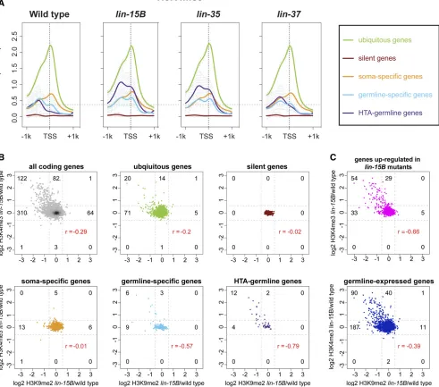
One of the major phenotypes of many synMuv B mutants, including lin-15B mutants, is the ectopic expression in so-matic cells of genes whose expression is normally restricted to the germline (Wanget al.2005; Petrellaet al.2011; Wu et al.2012). We investigated if genes that have an H3K9me2 promoter peak in wild-type L1s are enriched for genes that are specifically expressed in the germline. We analyzed four categories of expression: genes that are broadly expressed in all tissues (2576: ubiquitous), genes that are repressed in most tissues (415: silent), genes that are expressed specifi-cally in somatic tissues (1181: soma-specific), and genes that are expressed specifically in the germline (169: germline-specific). Genes with an H3K9me2 promoter peak in wild-type L1s are enriched for genes with germline-specific expression, but not for genes with ubiquitous, silent, or somatic expression (Figure 2B and Figure S5). These enrichments are mirrored when plotting H3K9me2 ChIP-seq signal around the TSS av-eraged over the genes in each expression category (Figure 2C). If H3K9me2 at germline gene promoters is correlated with synMuv B repression of germline gene expression in the soma, then we would predict that germline genes would lose H3K9me2 promoter peaks in synMuv B mutants. Indeed, in
lin-15B mutants, there were many fewer germline-specific
genes with an H3K9me2 promoter peak, and there was a large decrease in the signal of H3K9me2 at the TSS of germline-specific genes (Figure 2, B and C).lin-35andlin-37mutants resembled wild type in having genes with an H3K9me2 pro-moter peak enriched for germline-specific genes (Figure 2, B and C).
We also examined germline genes whose misregulation is correlated with the HTA phenotype (Petrellaet al.2011). HTA-germline targets are defined as genes normally expressed in the germline that are upregulated in arrestedlin-35mutant L1s at 26° and whose expression returns to near wild-type levels in HTA-suppressedlin-35;mes-4(RNAi)double mutant L1s at 26°(48: HTA-germline) (Petrellaet al.2011). Similar to what was seen with germline-specific genes, genes with an H3K9me2 promoter peak were enriched for HTA-germline genes in wild type,lin-35, andlin-37mutants, but this enrich-ment was much reduced inlin-15Bmutants (Figure 2, B and C). Together, these data reveal a striking loss of H3K9me2 at the promoters of germline-specific and HTA-germline genes in
lin-15Bmutants, but not inlin-35orlin-37mutants.
H3K9me2 promoter peaks are distributed along the whole length of autosomes
Previous work on H3K9me2 inC. elegansfocused on its dis-tribution in broad domains on autosomal arms and the role of
H3K9me2 in repressing repetitive sequences (Ikegamiet al. 2010; Liu et al.2011; Guoet al.2015; Zeller et al.2016). Little investigation has been done into what role the more narrowly focused H3K9me2 found at promoters may be serv-ing in gene regulation. InC. elegans, genes with expression that is higher in the germline than other tissues (germline-enriched genes) or with expression exclusive to the germline (germline-specific genes) show a biased localization to the centers of autosomes compared to the localization of all cod-ing genes (Figure S6). Therefore, if H3K9me2 promoter peaks are associated with regulation of germline gene expres-sion, we would predict that H3K9me2 promoter peaks would also be found in the center regions of chromosomes and not be biased toward arm localization. We compared the distri-butions along autosomes of genes with H3K9me2 in their gene body vs. at their promoter. In wild type, genes with H3K9me2 in their gene body demonstrated the previously reported pattern of H3K9me2 enrichment on autosomal arms compared to centers (Figure 3, A and B). For genes with an H3K9me2 gene body peak, all mutants showed the same autosomal arm bias as seen in wild type (Figure 3, A and B and Figure S7). In contrast, genes with an H3K9me2 pro-moter peak in wild type were more evenly distributed across autosomes, with weak or no depletion from autosomal cen-ters (Figure 3, A and B). Notably, lin-15Bmutants showed reduction of H3K9me2 promoter peaks in the center of all autosomes (Figure 3, A and B), suggesting that LIN-15B is needed for H3K9me2 localization at gene promoters in auto-somal centers where germline genes are enriched.lin-35 mu-tants showed a distribution of genes with H3K9me2 at their promoter similar to wild type (Figure S7). lin-37 mutants were intermediate betweenlin-15Bandlin-35mutants (Fig-ure S7). H3K9me2 promoter peaks in chromosome centers in wild type represent a pattern not previously described for H3K9me2 inC. elegansand place H3K9me2 promoter peaks in mainly euchromatic regions where they may affect coding gene expression. Additionally, the loss of H3K9me2 from pro-moters in autosomal centers inlin-15Bmutants suggests that
LIN-15Bplays a specific role in directing H3K9me2 to areas of
the genome where there are fewer repeats and more coding genes, especially germline genes.
Loss of H3K9me2 in mutants is associated with increased H3K4me3 on germline genes
Trimethylation of histone H3 on lysine 4 (H3K4me3) and lysine 36 (H3K36me3) are correlated with active gene ex-pression (Liuet al.2011; Hoet al.2014; Evanset al.2016). Thus, we expected to see increases in H3K4me3 and H3K36me3 on germline genes in synMuv B mutants. Indeed, synMuv B mutants displayed increases in H3K4me3 and H3K36me3 on germline-expressed, germline-specific, and HTA-germline genes but not on other categories of genes (Figure 4, Figure S8, and Figure S9). A total of 64% of genes (130 of 204,P-value,1310228, hypergeometric test) with
at least a 1.5-fold increase in H3K4me3 at their promoter in
expressed (Figure 4, B and C). Increased levels of H3K36me3 and, especially, H3K4me3 on germline-specific genes were observed at 20°and 26°in bothlin-15Bandlin-35mutants, but only at 26°inlin-37mutants (Figure S8 and Figure S9). This is consistent with previous data showing that misexpres-sion of germline genes inlin-37mutants is more sensitive to temperature than in lin-15B and lin-35 mutants (Petrella et al. 2011). HTA-germline genes showed larger increases in H3K4me3 and H3K36me3 than germline-specific genes. This is expected as HTA-germline genes were defined partly by requiring these genes to be upregulated inlin-35mutants (Petrellaet al.2011), while not all germline-specific genes are upregulated in synMuv B mutants. The increased levels of both H3K4me3 and H3K36me3 on germline-specific and HTA-germline genes in mutants is consistent with these genes being expressed at higher levels, most likely in a larger population of cells [i.e., somatic cells in addition to the two primordial germ cells (PGCs)] in these mutants.
We investigated if there is a correlation between loss from promoters of the repressive H3K9me2 chromatin modification and acquisition of H3K4me3, which is associated with gene activation. To compare those marks at promoters, we calcu-lated the log2 fold change of the signal of each modification in
lin-15B mutant/wild type within 250 bp upstream and downstream of the TSS. A higher histone modification signal inlin-15Bmutants than wild type would result in a positive log2 fold change; a lower histone modification signal in lin-15Bmutants than wild type would result in a negative log2 fold change. In lin-15Bmutants, 25% of all genes (122 of 448) that had at least a 1.5-fold reduction of H3K9me2 pro-moter signal also had at least a 1.5-fold increase of H3K4me3 promoter signal (Figure 4B). Strikingly, 40% of germline-specific (6 of 15) and 75% of HTA-germline genes (12 of 16) that had reduced H3K9me2 promoter signal also had increased H3K4me3 promoter signal (Figure 4B). We investigated if more of the 122 genes that showed reduced H3K9me2 and increased H3K4me3 promoter signal in lin-15B mutants had an indication of being germline-expressed and regulated by synMuv B mutants. We found that 74% (90 of 122,P-value, 1310227) of those genes have evidence of being
germline-expressed (Reinke et al. 2004; Wang et al.2009) and 44% (54 of 122,P-value,1310231) are upregulated inlin-15B
mutants (Petrella et al. 2011). The same analysis of genes that have a concurrent loss of H3K9me2 and gain of H3K4me3 inlin-35andlin-37mutants compared to wild type showed similar but muted trends as observed inlin-15B mu-tants (Figure S10). However, unlike inlin-15Bmutants, there was a subset of genes that in lin-35 and lin-37mutants dis-played increased H3K4me3 promoter signal without reduced
H3K9me2 promoter signal (Figure S10). Because we did not observe H3K9me2 at the promoter of these genes in wild type, we surmise that repression of this subset of genes in wild type does not depend on H3K9me2 at their promoter. Altogether, the germline genes that have enrichment of H3K9me2 at their pro-moter in wild type lose that enrichment when upregulated in any of the three mutants.
Global loss of H3K9me2 leads to phenotypes similar to lin-15B and DREAM complex mutants
To investigate if loss of H3K9me2 promoter localization plays an important role inlin-15Bmutant phenotypes, we analyzed mutants for the histone methyltransferases (HMTs) respon-sible for H3K9 methylation. Loss of these HMTs leads to a global loss of all H3K9 methylation (Towbin et al. 2012; Garrigues et al. 2015), which may phenocopylin-15B mu-tants. H3K9 methylation inC. elegansembryos is catalyzed by two HMTs,MET-2andSET-25, which primarily catalyze H3K9me1/2 and H3K9me3, respectively (Towbin et al. 2012). If loss of H3K9 methylation is associated with ectopic germline gene expression and the HTA phenotype, we would expect thatmet-2andset-25mutants would show these phe-notypes.set-25single mutants, which lose H3K9me3, showed neither an HTA phenotype nor an ectopic germline gene ex-pression phenotype, as assessed by staining for the germline-specific protein PGL-1 (Figure 5, A and B). Therefore, H3K9me3 does not appear to be important for repression of germline genes in the soma. In contrast, met-2 single mu-tants, which lose 80–90% of H3K9me2 and 70% of H3K9me3 (Towbinet al.2012), displayed80% larval arrest around the L3 stage at 26°, but no larval arrest at 24°(Figure 5A). Thus,met-2mutants show an HTA phenotype similar to but weaker thanlin-15Bmutants (Figure 5A; Petrellaet al. 2011). We also observed ectopic expression ofPGL-1inmet-2
mutants at 26°similar tolin-15B mutants, with thePGL-1
protein being primarily cytoplasmic and diffuse in intestinal cells (Figure 5B). To test if the remaining 10–20% of H3K9me2 catalyzed by SET-25 in met-2 mutants (Towbin et al. 2012) partially represses germline gene expression in somatic cells, we analyzed met-2 set-25 double mutants, which have been shown to completely lack H3K9 methyl-ation during embryonic stages (Towbin et al. 2012; Garrigueset al.2015). Consistent with residualSET-25 -me-diated H3K9me2 serving a role in repression of germline genes in somatic cells,met-2 set-25double mutants showed significantly enhanced larval arrest at 26°when compared to
met-2single mutants (Figure 5A). Similar tolin-15B,lin-35, and lin-37 mutants, met-2 set-25 double mutants did not show increased larval arrest at 24°. EctopicPGL-1inmet-2
set-25double mutants at 26°was similar to that seen inmet-2
single mutants (Figure 5B). Altogether, our results show that a global loss of H3K9me2 phenocopies both the HTA and ectopic germline gene expression seen in synMuv B mutants. One of the known proteins that binds to methylated H3K9 to create a repressive chromatin environment is HP1 (Couteau et al.2002; Nestorovet al.2013; Garrigueset al.2015). In
C. elegans there are two HP1 homologs,HPL-1and HPL-2.
hpl-2is a synMuv B gene.hpl-2mutants display a variety of phenotypes including HTA and ectopic germline gene expres-sion in the soma (Figure 5) (Couteau et al. 2002; Petrella et al.2011), whilehpl-1mutants generally lack observable phenotypes (Schott et al. 2006). Therefore, we compared genes with H3K9me2 promoter peaks with previously
published data on genes bound by HPL-2 in embryos (Garrigueset al.2015). We confirmed that most of the genes with an H3K9me2 promoter peak in our L1 wild-type samples also have such a peak in wild-type embryos (Figure S11A). We found that genes with an H3K9me2 promoter peak in wild type that is lost inlin-15Bmutants are enriched for pro-moter-boundHPL-2(Figure S11). Additionally, the 122 genes that have decreased H3K9me2 and increased H3K4me3 in
lin-15Bmutants as compared to wild type are enriched for
promoter-boundHPL-2(Figure S11). These data suggest that
HPL-2 binding may contribute to regulation of germline genes that are repressed in somatic cells through an H3K9me2 promoter peak. However, we noted differences in the pattern of PGL-1accumulation in the soma of hpl-2
mutants compared to eitherlin-15Bormet-2 set-25mutants; 75% (15/20) ofhpl-2mutant L1s displayed intestinalPGL-1
staining that was perinuclear and punctate, reminiscent of
PGL-1 staining in the germline (Figure 5B) (Petrella et al.
2011; Wuet al.2012). In contrast, none of thelin-15B,met-2, ormet-2 set-25mutants analyzed (n = 19–20) displayed that pattern of intestinal staining (Figure 5B), unlike the previously published analysis ofmet-2(Wuet al.2012). These differences in the pattern of ectopicPGL-1suggest that loss of H3K9me2 either at a subset of genes inlin-15Bmutants or globally in
met-2 set-25mutants is not equivalent to loss ofHPL-2.
Discussion
Repression of germline gene expression in the soma is vital, as loss of germline gene repression is a hallmark of various disease states including cancer. Investigating the changes to chromatin that occur when germline genes are misexpressed in the somatic cells of mutants is afirst step in understanding the mechanisms that repress germline genes to protect so-matic fates and development. Here, we investigated the changes to histone modifications that occur in a subset ofC. eleganssynMuv B mutants that misexpress germline genes in the soma. We defined a new localization pattern for the re-pressive histone modification H3K9me2 in wild type, at the promoter of coding genes; unlike the previously described broad domains of H3K9me2, promoter peaks of H3K9me2
are not enriched on autosomal arms (Liu et al. 2011; Garrigueset al.2015; Evanset al.2016; Ahringer and Gasser 2018). Promoter enrichment of H3K9me2 in autosomal cen-ters provides a new regulatory role for H3K9me2, in addition to its well-described regulation of repetitive elements on au-tosomal arms. We also found that, in wild-type somatic cells, genes with an H3K9me2 promoter peak are enriched for genes expressed specifically in the germline and genes that are synMuv B targets. The localization of H3K9me2 to germ-line genes and synMuv B targets is disrupted strongly in lin-15B mutants and weakly in DREAM complex mutants. We additionally showed that loss of H3K9me2 but not H3K9me3 phenocopies synMuv B mutants. Our data implicate H3K9me2 promoter enrichment as an important aspect of repression of germline gene expression in somatic cells.
There is strong evidence that a memory of gene expression/ repression and associated chromatin modifications are trans-mitted from the parental germline to the developing embryo (Furuhashiet al.2010; Rechtsteineret al.2010; Zenket al. 2017; Tabuchi et al.2018). For example, genes that were expressed in the germline continue to be marked with MES-4-generated H3K36me3 in embryos, even in the ab-sence of ongoing transcription in embryos (Furuhashiet al.
2010; Rechtsteiner et al. 2010; Kreher et al. 2018). It is thought that H3K36me3 marks these genes for re-expression in the germline during postembryonic development. How then are germline genes repressed properly in somatic tissues when those tissues inherit germline genes with marks of active ex-pression that potentially set those genes up for re-exex-pression? Our data, along with other recent work, strongly implicate deposition of H3K9me2 at the proper time in development as necessary to create proper patterns of repressive chromatin in differentiating somatic cells. In C. elegans, H3K9me2 and H3K9me3 levels are very low in the nuclei of early stage em-bryos, and only start to accumulate when cells are transition-ing from early embryogenesis to midembryogenesis at about the 50-cell stage (Mutluet al.2018). This is in part driven by the nuclear import of an activeMET-2complex that catalyzes conversion of H3K9me1 to H3K9me2. The timing of MET-2
import just precedes the stage in embryogenesis when zygotic transcription is upregulated and when tissue-specific expres-sion patterns emerge (Spenceret al.2011; Levinet al.2012; Robertson and Lin 2015; Mutluet al.2018). Concurrent with
MET-2import and increased global H3K9 methylation is the creation of regions of compact chromatin within the nucleus (Mutluet al.2018). In support of the role of synMuv B proteins in the timing of chromatin compaction during embryogenesis, the formation of compact chromatin is delayed inlin-15B, and
lin-35mutants (Costelloet al.2019). Developmental chroma-tin compaction likely plays a role in lineage-specific gene re-pression and is proposed to be driven at least in part by H3K9 methylation. Our data suggest that loss of H3K9me2, either through loss of theMET-2andSET-25HMTs that catalyze the mark or through loss of proper localization of H3K9me2 to germline genes inlin-15Bmutants, leads to misexpression of germline genes in somatic cells. We hypothesize that specific localization of H3K9me2 to germline gene promoters facili-tated byLIN-15Bis an important aspect of resetting the chro-matin landscape of germline genes to prevent their expression in somatic lineages.
A striking aspect of ourfindings is the difference in changes to promoter-enriched H3K9me2 between lin-15B mutants and DREAM complex mutants. It was previously proposed, based on phenotype analysis, thatLIN-15Bis a member of the DREAM complex (Wu et al.2012). Our data indicate that, althoughLIN-15Bbinds to and represses many of the same genes as the DREAM complex, its molecular function at those genes is probably distinct. The proposed DNA-binding do-main ofLIN-15Bmay allow it to be independently recruited to similar targets as the DREAM complex, where the two may function together to repress genes. This scenario has impli-cations for regulation of gene expression in the germline as well as in the soma. Recent work from the Seydoux labora-tory has implicated the loss ofLIN-15Bprotein in the germ-line as important for germgerm-line development (Leeet al.2017). Maternally provided LIN-15B is normally removed from the PGCs, while DREAM components are not (Lee et al. 2017). Our work suggests that loss of LIN-15B from the PGCs may protect essential germline genes from being
H3K9 methylated and repressed in those cells. How the dif-ferent synMuv B complexes work together to fully repress germline genes in somatic cells is still an open question. The establishment of H3K9me2 may be an initiating step in germline gene repression, or may be one aspect of a series of redundant steps necessary to repress germline genes. Analy-sis of the order and dependency ofMET-2,LIN-15B, and the DREAM complex binding to germline genes is necessary to address these questions.
The work presented here focuses on a subset of germline genes that are regulated through the LIN-15B/H3K9me2/ DREAM complex pathway. Although this pathway may regu-late only a subset of genes in this way, the repercussions to development are clear: organisms defective in this regulation cannot thrive in the face of challenges (e.g., high tempera-ture) when somatic fates are compromised. Recent work in Drosophilaunderscores the importance of H3K9 methylation in repression of a subset of coding genes to maintain proper cell fate. In the Drosophilaovary, loss of H3K9me3 leads to upregulation of testis-specific transcripts, and changes the fate of ovarian germ cells, leading to sterility (Smolkoet al. 2018). As inC. elegans, prior investigations of H3K9 methyl-ation loss inDrosophilahad focused primarily on upregula-tion of repetitive elements (Ranganet al.2011; Wanget al. 2011; Guoet al.2015; Zelleret al.2016). However, it is clear that H3K9me2/3 loss leading to upregulation of small sets of coding genes in a tissue-specific manner can have profound effects on cell fate and function. As more studies investigate the roles of H3K9me2/3 in repression of coding genes, it seems likely that new pathways will be uncovered that are necessary to create different patterns of H3K9me2/3 in dif-ferent tissues for maintenance of proper cell fate.
with chromatin (Janicet al.2010; Petrellaet al.2011; Wuet al. 2012), it will be interesting to investigate if ectopic expression of germline genes in human somatic tumors is due to loss of these conserved complexes. Finally, not all germline genes, but only a specific subset, are ectopically expressed in these mod-els. Why only certain germline genes are vulnerable to misex-pression, if those genes are the same across species, and which cellular processes are disrupted as a result of germline gene misexpression singularly or as a group, are open questions. Further investigation could have broad implications for under-standing conserved basic chromatin mechanisms and thera-peutic targets for cancer treatment.
Acknowledgments
Many thanks to Anita Manogaran for comments and discus-sion of the manuscript. Some strains were provided by the Caenorhabditis Genetics Center (CGC), which is funded by National Institutes of Health (NIH) Office of Research In-frastructure Programs (P40 OD010440). This work used the Vincent J. Coates Genomics Sequencing Laboratory at Uni-versity of California (UC) Berkeley, supported by NIH S10 OD018174 Instrumentation Grant. This work was supported by a NIH grants R00GM98436 and R15GM122005 to L.N.P. and NIH grant R01GM34059 to S.S.
Literature Cited
Ahringer, J., and S. M. Gasser, 2018 Repressive chromatin in
Caenorhabditis elegans: establishment, composition, and func-tion. Genetics 208: 491–511. https://doi.org/10.1534/genet-ics.117.300386
Al-Amin, M., H. Min, Y. H. Shim, and I. Kawasaki, 2016 Somatically expressed germ-granule components, PGL-1 and PGL-3, repress programmed cell death inC. elegans. Sci. Rep. 6: 33884.https://doi.org/10.1038/srep33884
Blaschke, K., E. T. Ebata, M. M. Karimi, J. A. Zepeda-Martínez, P. Goyalet al., 2013 Vitamin C induces Tet-dependent DNA de-methylation and a blastocyst-like state in ES cells. Nature 500: 222–226.https://doi.org/10.1038/nature12362
Brenner, S., 1974 The genetics ofCaenorhabditis elegans. Genetics 77: 71–94.
Costello, M. E., and L. N. Petrella, 2019C. eleganssynMuv B pro-teins regulate spatial and temporal chromatin compaction dur-ing development. bioRxivhttps://doi.org/10.1101/538801 Coustham, V., C. Bedet, K. Monier, S. Schott, M. Karali et al.,
2006 The C. elegansHP1 homologue HPL-2 and the LIN-13 zincfinger protein form a complex implicated in vulval devel-opment. Dev. Biol. 297: 308–322. https://doi.org/10.1016/j.yd-bio.2006.04.474
Couteau, F., F. Guerry, F. Muller, and F. Palladino, 2002 A het-erochromatin protein 1 homologue in Caenorhabditis elegans
acts in germline and vulval development. EMBO Rep. 3: 235– 241.https://doi.org/10.1093/embo-reports/kvf051
Cui, M., E. B. Kim, and M. Han, 2006 Diverse chromatin remod-eling genes antagonize the Rb-Involved synMuv pathways inC. elegans. PLoS Genet. 2: e74.https://doi.org/10.1371/journal. pgen.0020074
Ebata, K. T., K. Mesh, S. Liu, M. Bilenky, A. Fekete et al., 2017 Vitamin C induces specific demethylation of H3K9me2
in mouse embryonic stem cells via Kdm3a/b. Epigenetics Chro-matin 10: 36.https://doi.org/10.1186/s13072-017-0143-3 Edgar, R., M. Domrachev, and A. E. Lash, 2002 Gene expression
omnibus: NCBI gene expression and hybridization array data repository. Nucleic Acids Res. 30: 207–210. https://doi.org/ 10.1093/nar/30.1.207
Egelhofer, T. A., A. Minoda, S. Klugman, K. Lee, P. Kolasinska-Zwierzet al., 2011 An assessment of histone-modification an-tibody quality. Nat. Struct. Mol. Biol. 18: 91–93. https://doi. org/10.1038/nsmb.1972
Evans, K. J., N. Huang, P. Stempor, M. A. Chesney, T. A. Downet al., 2016 StableCaenorhabditis eleganschromatin domains sepa-rate broadly expressed and developmentally regulated genes. Proc. Natl. Acad. Sci. USA 113: E7020–E7029. https://doi. org/10.1073/pnas.1608162113
Fay, D. S., and J. Yochem, 2007 The SynMuv genes of
Caenorhabditis elegansin vulval development and beyond. Dev. Biol. 306: 1–9.https://doi.org/10.1016/j.ydbio.2007.03.016 Fraser, R., and C. J. Lin, 2016 Epigenetic reprogramming of the
zygote in mice and men: on your marks, get set, go! Reproduc-tion 152: R211–R222.https://doi.org/10.1530/REP-16-0376 Furuhashi, H., T. Takasaki, A. Rechtsteiner, T. Li, H. Kimuraet al.,
2010 Trans-generational epigenetic regulation of C. elegans
primordial germ cells. Epigenetics Chromatin 3: 15. https:// doi.org/10.1186/1756-8935-3-15
Garrigues, J. M., S. Sidoli, B. A. Garcia, and S. Strome, 2015 Defining heterochromatin inC. elegansthrough genome-wide analysis of the heterochromatin protein 1 homolog HPL-2. Genome Res. 25: 76–88.https://doi.org/10.1101/gr.180489.114 Goetsch, P. D., J. M. Garrigues, and S. Strome, 2017 Loss of the
Caenorhabditis elegans pocket protein LIN-35 reveals MuvB’s innate function as the repressor of DREAM target genes. PLoS Genet. 13: e1007088.https://doi.org/10.1371/journal.pgen.1007088 Guo, Y., B. Yang, Y. Li, X. Xu, and E. M. Maine, 2015 Enrichment
of H3K9me2 on unsynapsed chromatin inCaenorhabditis elegans
does not target de novo sites. G3 (Bethesda) 5: 1865–1878. https://doi.org/10.1534/g3.115.019828
Harrison, M. M., C. J. Ceol, X. Lu, and H. R. Horvitz, 2006 Some
C. elegans class B synthetic multivulva proteins encode a con-served LIN-35 Rb-containing complex distinct from a NuRD-like complex. Proc. Natl. Acad. Sci. USA 103: 16782–16787.https:// doi.org/10.1073/pnas.0608461103
Ho, J. W. K., Y. L. Jung, T. Liu, B. H. Alver, S. Lee et al., 2014 Comparative analysis of metazoan chromatin organization. Nature 512: 449–452.https://doi.org/10.1038/nature13415 Ikegami, K., T. A. Egelhofer, S. Strome, and J. D. Lieb, 2010
Caenorhabditis eleganschromosome arms are anchored to the nuclear membrane via discontinuous association with LEM-2. Genome Biol. 11: R120. https://doi.org/10.1186/gb-2010-11-12-r120
Janic, A., L. Mendizabal, S. Llamazares, D. Rossell, and C. Gonzalez, 2010 Ectopic expression of germline genes drives malignant brain tumor growth in Drosophila. Science 330: 1824–1827. https://doi.org/10.1126/science.1195481
Kawasaki, I., Y. H. Shim, J. Kirchner, J. Kaminker, W. B. Woodet al., 1998 PGL-1, a predicted RNA-binding component of germ granules, is essential for fertility in C. elegans. Cell 94: 635– 645.https://doi.org/10.1016/S0092-8674(00)81605-0 Kent, W. J., A. S. Zweig, G. Barber, A. S. Hinrichs, and D. Karolchik,
2010 BigWig and BigBed: enabling browsing of large distrib-uted datasets. Bioinformatics 26: 2204–2207.https://doi.org/ 10.1093/bioinformatics/btq351
Kreher, J., T. Takasaki, C. Cockrum, S. Sidoli, B. A. Garcia et al., 2018 Distinct roles of two histone methyltransferases in trans-mitting H3K36me3-based epigenetic memory across genera-tions in Caenorhabditis elegans. Genetics 210: 969–982. https://doi.org/10.1534/genetics.118.301353
Langmead, B., C. Trapnell, M. Pop, and S. L. Salzberg, 2009 Ultrafast and memory-efficient alignment of short DNA sequences to the human genome. Genome Biol. 10: R25. https://doi.org/10.1186/gb-2009-10-3-r25
Latorre, I., M. A. Chesney, J. M. Garrigues, P. Stempor, A. Appert
et al., 2015 The DREAM complex promotes gene body H2A.Z for target repression. Genes Dev. 29: 495–500.https://doi.org/ 10.1101/gad.255810.114
Lee, C. S., T. Lu, and G. Seydoux, 2017 Nanos promotes epige-netic reprograming of the germline by down-regulation of the THAP transcription factor LIN-15B. eLife 6: pii: e30201.https:// doi.org/10.7554/eLife.30201
Levin, M., T. Hashimshony, F. Wagner, and I. Yanai, 2012 Developmental milestones punctuate gene expression in the Caenorhabditis embryo. Dev. Cell 22: 1101–1108. https://doi.org/10.1016/j.devcel.2012.04.004
Liu, T., A. Rechtsteiner, T. A. Egelhofer, A. Vielle, I. Latorreet al., 2011 Broad chromosomal domains of histone modification patterns in C. elegans. Genome Res. 21: 227–236. https://doi. org/10.1101/gr.115519.110
Maeda, I. D., D. Okamura, Y. Tokitake, M. Ikeda, H. Kawaguchi
et al., 2013 Max is a repressor of germ cell-related gene ex-pression in mouse embryonic stem cells. Nat. Commun. 4: 1754. https://doi.org/10.1038/ncomms2780
Meissner, B., A. Warner, K. Wong, N. Dube, A. Lorchet al., 2009 An integrated strategy to study muscle development and myofilament structure in Caenorhabditis elegans. PLoS Genet. 5: e1000537. https://doi.org/10.1371/journal.pgen.1000537
Meister, P., B. D. Towbin, B. L. Pike, A. Ponti, and S. M. Gasser, 2010 The spatial dynamics of tissue-specific promoters during
C. elegans development. Genes Dev. 24: 766–782. https://doi. org/10.1101/gad.559610
Morgan, H. D., F. Santos, K. Green, W. Dean, and W. Reik, 2005 Epigenetic reprogramming in mammals. Hum. Mol. Genet. 14: R47–R58.https://doi.org/10.1093/hmg/ddi114 Mutlu, B., H. M. Chen, J. J. Moresco, B. D. Orelo, B. Yanget al.,
2018 Regulated nuclear accumulation of a histone methyl-transferase times the onset of heterochromatin formation inC. elegans embryos. Sci. Adv. 4: eaat6224. https://doi.org/ 10.1126/sciadv.aat6224
Nestorov, P., M. Tardat, and A. H. Peters, 2013 H3K9/HP1 and polycomb: two key epigenetic silencing pathways for gene regulation and embryo development. Curr. Top. Dev. Biol. 104: 243–291. https://doi.org/10.1016/B978-0-12-416027-9.00008-5
Petrella, L. N., W. Wang, C. A. Spike, A. Rechtsteiner, V. Reinke
et al., 2011 synMuv B proteins antagonize germline fate in the intestine and ensure C. elegans survival. Development 138: 1069–1079.https://doi.org/10.1242/dev.059501
Rangan, P., C. D. Malone, C. Navarro, S. P. Newbold, P. S. Hayes
et al., 2011 piRNA production requires heterochromatin for-mation in Drosophila. Curr. Biol. 21: 1373–1379. https://doi. org/10.1016/j.cub.2011.06.057
R Core Team, 2018 R: A Language and Environment for Statisti-cal Computing. R Foundation for StatistiStatisti-cal Computing, Vienna. Rechtsteiner, A., S. Ercan, T. Takasaki, T. M. Phippen, T. A. Egelhofer et al., 2010 The histone H3K36 methyltransferase MES-4 acts epigenetically to transmit the memory of germline gene expression to progeny. PLoS Genet. 6: e1001091.https:// doi.org/10.1371/journal.pgen.1001091
Reinke, V., and A. D. Cutter, 2009 Germline expression infl u-ences operon organization in the Caenorhabditis elegans
genome. Genetics 181: 1219–1228. https://doi.org/ 10.1534/genetics.108.099283
Reinke, V., I. S. Gil, S. Ward, and K. Kazmer, 2004 Genome-wide germline-enriched and sex-biased expression profiles in
Caenorhabditis elegans. Development 131: 311–323. https:// doi.org/10.1242/dev.00914
Robertson, S., and R. Lin, 2015 The maternal-to-zygotic transition inC. elegans. Curr. Top. Dev. Biol. 113: 1–42.https://doi.org/ 10.1016/bs.ctdb.2015.06.001
Roy, P. J., J. M. Stuart, J. Lund, and S. K. Kim, 2002 Chromosomal clustering of muscle-expressed genes in Caenorhabditis elegans. Nature 418: 975–979. https:// doi.org/10.1038/nature01012
Schott, S., V. Coustham, T. Simonet, C. Bedet, and F. Palladino, 2006 Unique and redundant functions ofC. elegansHP1 pro-teins in post-embryonic development. Dev. Biol. 298: 176–187. https://doi.org/10.1016/j.ydbio.2006.06.039
Sekinaka, T., Y. Hayashi, T. Noce, H. Niwa, and Y. Matsui, 2016 Selective de-repression of germ cell-specific genes in mouse embryonicfibroblasts in a permissive epigenetic environ-ment. Sci. Rep. 6: 32932.https://doi.org/10.1038/srep32932 Smolko, A. E., L. Shapiro-Kulnane, and H. K. Salz, 2018 The
H3K9 methyltransferase SETDB1 maintains female identity in
Drosophilagerm cells. Nat. Commun. 9: 4155.https://doi.org/ 10.1038/s41467-018-06697-x
Song, J. S., W. E. Johnson, X. Zhu, X. Zhang, W. Li et al., 2007 Model-based analysis of two-color arrays (MA2C). Ge-nome Biol. 8: R178.https://doi.org/10.1186/gb-2007-8-8-r178 Spellman, P. T., and G. M. Rubin, 2002 Evidence for large do-mains of similarly expressed genes in theDrosophilagenome. J. Biol. 1: 5.https://doi.org/10.1186/1475-4924-1-5
Spencer, W. C., G. Zeller, J. D. Watson, S. R. Henz, K. L. Watkins
et al., 2011 A spatial and temporal map of C. elegans gene expression. Genome Res. 21: 325–341. https://doi.org/ 10.1101/gr.114595.110
Strome, S., and W. B. Wood, 1983 Generation of asymmetry and segregation of germ-line granules in earlyC. elegansembryos. Cell 35: 15–25.https://doi.org/10.1016/0092-8674(83)90203-9 Tabuchi, T. M., A. Rechtsteiner, T. E. Jeffers, T. A. Egelhofer, C. T.
Murphyet al., 2018 Caenorhabditis eleganssperm carry a his-tone-based epigenetic memory of both spermatogenesis and oo-genesis. Nat. Commun. 9: 4310. https://doi.org/10.1038/ s41467-018-06236-8
Towbin, B., C. González-Aguilera, R. Sack, D. Gaidatzis, V. Kalck
et al., 2012 Step-wise methylation of histone H3K9 positions heterochromatin at the nuclear periphery. Cell 150: 934–947. https://doi.org/10.1016/j.cell.2012.06.051
Unhavaithaya, Y., T. H. Shin, N. Miliaras, J. Lee, T. Oyamaet al., 2002 MEP-1 and a homolog of the NURD complex component Mi-2 act together to maintain germline-soma distinctions inC. elegans. Cell 111: 991–1002. https://doi.org/10.1016/S0092-8674(02)01202-3
Wang, D., S. Kennedy, D. Conte, J. K. Kim, H. W. Gabel et al., 2005 Somatic misexpression of germline P granules and en-hanced RNA interference in retinoblastoma pathway mutants. Nature 436: 593–597.https://doi.org/10.1038/nature04010 Wang, X., Y. Zhao, K. Wong, P. Ehlers, Y. Kohara et al.,
2009 Identification of genes expressed in the hermaphrodite germ line of C. elegansusing SAGE. BMC Genomics 10: 213. https://doi.org/10.1186/1471-2164-10-213
Wang, X., L. Pan, S. Wang, J. Zhou, W. McDowell et al., 2011 Histone H3K9 trimethylase Eggless controls germline stem cell maintenance and differentiation. PLoS Genet. 7: e1002426.https://doi.org/10.1371/journal.pgen.1002426 Whitehurst, A. W., 2014 Cause and consequence of cancer/testis
Wu, X., Z. Shi, M. Cui, M. Han, and G. Ruvkun, 2012 Repression of germline RNAi pathways in somatic cells by retinoblastoma pathway chromatin complexes. PLoS Genet. 8: e1002542. https://doi.org/10.1371/journal.pgen.1002542
Zeller, P., J. Padeken, R. van Schendel, V. Kalck, M. Tijsterman
et al., 2016 Histone H3K9 methylation is dispensable for
Caenorhabditis elegans development but suppresses RNA:DNA hybrid-associated repeat instability. Nat. Genet. 48: 1385– 1395.https://doi.org/10.1038/ng.3672
Zenk, F., E. Loeser, R. Schiavo, F. Kilpert, O. Bogdanovic et al., 2017 Inherited H3K27me3 restricts enhancer function during maternal-to-zygotic transition. Science 357: 212–216.https:// doi.org/10.1126/science.aam5339
Zhang, Y., T. Liu, C. A. Meyer, J. Eeckhoute, D. S. Johnsonet al., 2008 Model-based analysis of ChIP-Seq (MACS). Genome Biol. 9: R137.https://doi.org/10.1186/gb-2008-9-9-r137