PLASTICITY OF PRIMARY SENSORY
NEURONS AND THEIR CENTRAL
CONNECTIONS: NORMAL DEVELOPMENT
AND THE EFFECTS OF NEONATAL AXOTOMY
Simon Beggs
Thesis submitted for the degree of PhD at
University of London
Department of Anatomy and Developmental Biology
University College London
ProQuest Number: 10010145
All rights reserved
INFORMATION TO ALL USERS
The quality of this reproduction is dependent upon the quality of the copy submitted.
In the unlikely event that the author did not send a complete manuscript and there are missing pages, these will be noted. Also, if material had to be removed,
a note will indicate the deletion.
uest.
ProQuest 10010145
Published by ProQuest LLC(2016). Copyright of the Dissertation is held by the Author.
All rights reserved.
This work is protected against unauthorized copying under Title 17, United States Code. Microform Edition © ProQuest LLC.
ProQuest LLC
789 East Eisenhower Parkway P.O. Box 1346
,
, -, ,,
. r
/ „ , ) , ' . ( , - : ; . :, : ] , , . . . , \ : ; ( ' ^
Abstract
Neonatal peripheral sensory neurons undergo considerable developmental and injury
induced plasticity. This thesis investigates this plasticity and the mechanisms that underlie
it. The first part describes the results o f investigations into the central consequences of
neonatal peripheral nerve injury. While nerve section in the adult mammal results in the
death o f axotomised sensory neurons and a rearrangement o f their central connections, the
same procedure performed neonatally results in more rapid and extreme changes. In
Chapter 1 the issue o f axotomy-induced cell death is addressed. Using unbiased
stereological counting techniques a full quantitative measure o f the extent and time course
o f dorsal root ganglion neuronal death is presented and shows a critical period o f postnatal
plasticity exists during which massive cell loss rapidly occurs. In addition,
immunocytochemical methods were used to identify the relative loss o f identified sub
populations o f axotomised sensory neurons. The results indicate that all cell types are
equally susceptible to axotomy and that the surviving population retains a normal
phenotypic distribution. Chapter 2 describes a novel method o f revealing and quantifying
the collateral sprouting o f primary sensory afferents within the spinal cord following
axotomy. Using the tracer Dil, post-mortem labelling o f central terminal fields can be
achieved without the disadvantages o f transport delay and possible nerve damage that
occur with in vivo dye application. Chapter 3 provides evidence for a role o f BDNF in mediating the axotomy-induced sprouting response. Immunocytochemistry and northern
blot analysis show an upregulation o f BDNF mRNA and protein within the dorsal horn o f
the spinal cord following sciatic axotomy. Further evidence comes from BDNF knockout
mice that exhibit reduced sprouting. The second part o f the thesis investigates the normal
developmental plasticity o f primary sensory neurons in the postnatal period, namely the
refinement o f afferent connections within the dorsal horn o f the spinal cord. Initially
diffuse AB-flbre projections gradually withdraw from superficial laminae until the adult
pattern is achieved. In chapter 4, chronic NMDA receptor blockade from the day o f birth
is shovm to halt this normal developmental process. Control experiments confirm that
other aspects o f dorsal horn development are unaffected. It is therefore concluded that
postnatal reorganisation o f afferent terminals is an NMDA receptor-mediated activity-
dependent process and that peripheral afferent input is required to fine-tune central
This series o f experiments provides further understanding o f the postnatal development of
spinal cord connectivity. The issues o f trophic control o f sensory neuron survival and
growth are discussed with reference to target removal and mechanisms are proposed for
collateral sprouting that incorporate the normal developmental and axotomy-induced
Abbreviations
5HT 5-Hydroxy tryptamine
AMPA Amino-hydroxy-Methyl-isoxalone Propionic Acid BDNF Brain-Derived Neurotrophic Factor
BHRP p subunit o f cholera toxin conjugated to Horseradish Peroxidase CCK Choleocystokinin
CGRP Calcitonin Gene-Related Peptide CNS Central Nervous System
DAB Diamino Benzidine DRG Dorsal Root Ganglion
EPSC Excitatory Post Synaptic Current EPSP Excitatory Post Synaptic Potential FRAP Fluoride-Resistant Acid Phosphatase GABA Y Amino Butyric Acid
GAP-43 Growth-Associated Protein 43
GDNF Glial-cell Derived Neurotrophic Factor HRP Horseradish Peroxidase
IB4 Isolectin B4
LGN Lateral Geniculate Nucleus LTD Long-Term Depression LTP Long-Term Depression
MRNA Messenger Ribose Nucleic Acid NF-L Light chain Neurofilament NGF Nerve Growth Factor NGS Normal Goat Serum N K l Neurokinin-1
NMDA V-Methyl-D-Aspartate NMJ Neuromuscular Junction NPY Neuropeptide Y
NT3 Neurotrophin 3 NT4/5 Neurotrophin 4/5
ODC Ocular Dominance Colurrm PBS Phosphate Buffered Saline PKC Protein Kinase C
SG Substantia Gelatinosa IM P Thiamine Monophosphatase TTX Tetrodotoxin
VIP Vasoactive Intestinal Peptide V Rl Vanilloid Receptor 1
VR-Ll Vanilloid Receptor-Like 1
Contents
Introduction
Chapter 1:
Sensory neuron changes following neonatal axotomy
1: Introduction
1.1: Primary afferents exhibit physiological, biochemical and morphological heterogeneity
1.2: Peripheral innervation 1
1.3: Central innervation 10
1.4: Consequences of neonatal axotomy 11
2: Materials and Methods
2.1: Total cell counts 16
2.2: Identification of sub-populations 17
2.3: Estimate of the proportion of sciatic projecting neurons in L4/5 after
neonatal axotomy 20
2.4: Identification of sciatic projecting neuron sub-populations 20
3: Results
3.1: Neonatal sciatic axotomy causes rapid and extensive neuronal cell
death in the DRG within a critical postnatal period 22
3.2: Approximately half of the remaining L4 and L5 ganglia neurons arise
from the sciatic nerve 24
3.3: Neonatal axotomy does not result in a change in the relative
proportions of DRG sub-populations in the surviving L4/5 DRGs 26
3.4: The surviving axotomised DRG neurons are not N52, CGRP, or IB4
positive 27
3.5: Populations of both myelinated and unmyelinated neurons survive PO
axotomy 32
4: Discussion
4.1: Neurotrophin dependency of postnatal DRG neurons 37
4.2: Sub-populations of surviving axotomised DRG neurons 40
Chapter 2:
Collateral sprouting of primary afferents following neonatal
axotomy
1: Introduction
1.1: Peripheral nerve injury and sprouting 45
1.2: A-fibre terminal sprouting following adult nerve injury 46
1.3: Neonatal peripheral nerve injury and central innervation 47
1.4: GABA in the postnatal spinal cord 51
1.5: The effect of loss of afferent input on GABA expression 53
2: Materials and methods.
2.1: Peripheral nerve section 56
2.2: Preparation of Dil-eoated wire 56
2.3: Dorsal column labelling procedure 56
2.4: GABA immunoreactivity 56
3: Results
3.1: Dil application to the dorsal columns labels labels primary afferent
terminals within the dorsal horn grey matter 59
3.2: Denervation of saphenous territory and sciatic sprouting is revealed
by Dil 59
3.3: GABA immunoreactivity is reduced in the spinal cord of neonatally
axotomised rats 63
4. Discussion
65
Chapter 3:
BDNF expression following neonatal axotomy
1: Introduction
1.1: The neurotrophin family of neurotrophic factors 67
1.2: The BDNF gene 70
1.3: Expression of BDNF during development and in adult 70
1.4: Effects of BDNF on differentiation, survival and maturation of DRG
neurons 71
1.5: Retrograde and anterograde transport of BDNF 73
1.6: Paracrine and autocrine actions of BDNF 75
1.7: The BDNF receptor: TrkB 76
1.8: BDNF responses in sensory neurons and motoneurons to injury 79
1.9: BDNF knockouts 84
1.10: Trk-B Knockouts 86
1.11: Regulation of BDNF in vivo by physiological stimulation 87
1.12: Role of BDNF in hippocampal LTP and spinal cord central
sensitization 88
1.13: BDNF and central sprouting following nerve injury 90
2: Materials and methods
2.1: BDNF protein expression in the lumbar horn following neonatal
sciatic axotomy 93
2.2: BDNF mRNA expression in the lumbar horn following neonatal
sciatic axotomy 94
2.3: Primary afferent sprouting in BDNF transgenic mice 96
3: Results
3.3: BDNF mRNA expression is upregulated in axotomised animals 99 3.4: Neonatal saphenous axotomy results in collateral sprouting of intact
sciatic nerve central projections in wild-type mice 102
3.5: BDNF^ mice exhibit reduced collateral sprouting following PO
saphenous nerve axotomy 102
4: Discussion.
4.1: BDNF changes in DRG post-axotomy 105
4.2: Mechanisms of BDNF action: role of trkB 106
4.3: Roles of BDNF 107
4.4: Evidence from the visual system 117
4.5: GABA, BDNF and developmental plasticity 121
Chapter 4:
Postnatal development of spinal cord connectivity
1: Introduction
1.1: The role of sensory experience in determining patterns of
connectivity during development 126
1.2: The NMDA receptor and activity-dependent plasticity during
development 128
1.3: The use of MK-801 as a pharmacological blocker of NMDA receptors
in vivo 129
1.4: Development of primary afferent innervation of the mammalian
dorsal horn 132
1.5: Possible mechanisms for the postnatal withdrawal of A-fibres from
lamina II 132
2: Materials and methods
2.1: Elvax manufacture 138
2.2: Elvax implantation 138
2.3: Sciatic nerve labelling 138
2.4: TMB reaction for horseradish peroxidase 138
2.5: Size-frequency profiles for labelled DRG neurons 141
2.6: Developmental profile of A-fibre withdrawal from SG 141
2.7: Immunohistochemistry 141
2.8: Dil labelling of neonatal spinal cord 143
3: Results
3.1: A-fibres remain in superficial laminae after chronic MK-801
treatment 144
3.2: Chronic NMDA antagonism does not alter the cellular specificity of
BHRP 144
3.3: C-fibre terminal patterns are not altered following chronic MK801
treatment 147
4: Discussion
4.1: A-fibre withdrawal from SG is an NMDA-dependent phenomenon. 149
4.2: Mechanisms for functional alteration in synapse efficacy. 149
4.4: Mechanisms responsible for silent synapses. 152
4.5: Developmental regulation of NMDA receptors. 153
4.6: Synaptic stabilisation and elimination. 154
4.7 BDNF and synaptic stability. 156
Conclusions
159References
163Tables
1. Physiological classification of primary afferents. 2
2. Functional and morphological classification of primary afferents. 4
3. Reported DRG cell losses following neonatal peripheral injury. 14
4. Total cell numbers in L4 DRG following axotomy at PO, PIG and P21. 24
5. Proportion of flruorogold-labelled neurons following neonatal axotomy 24
6. Total number of neuronal profiles. 27
7. Percentages of fluorogold labelled neurons positively immunolabelled. 32
8. Neuronal deficits in BDNF transgenic mouse CNS. 85
9. Changes in calcium binding protein expression in BDNF CNS structures. 86
10. Neuronal deficits in trkB transgenic mouse CNS. 87
Figures
1. Central consequences of neonatal peripheral axotomy. xi
2. Laminar organisation of primary afferent innervation of the adult
dorsal horn. 3
3. DRG profile counts protocol. 18
4. Ganglionic distribution of sciatic projecting neurons. 22
5. PO, PIO and P21 axotomy cell survival graph. 23
6. DRG surviving neuron subtypes. 25
7. N52, CGRP and IB4 developmental profile graph. 28
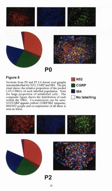
8. Immuno pie chart: PO and P2. 29
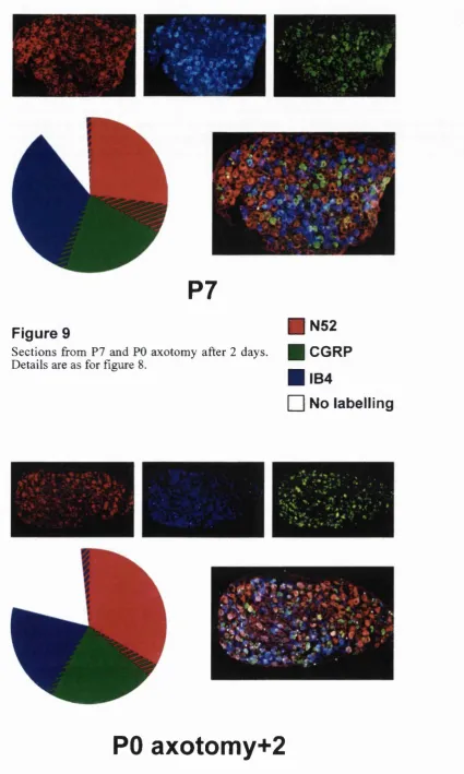
9. Immuno pie chart: P7 and PO ax+2. 30
10. Immuno pie chart: PO ax+7 and POax+adult. 31
11. N52 and CGRP - fluorogold labelling. 33
12. IB4 and peripherin - fluorogold labelling. 34
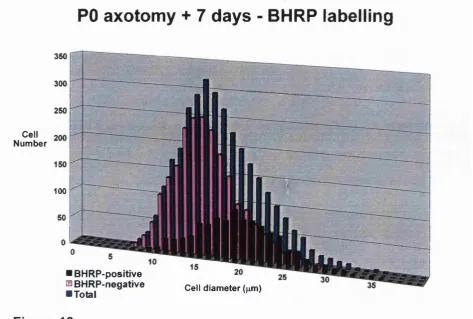
13. BHRP size-frequency graph. 35
14. WGAHRP size-frequency graph. 36
15. Phenotypic change in surviving DRG population. 44
16. Sciatic axotomy/Saphenous sprouting diagram. 49
17. Saphenous sprouting: Shortland and Fitzgerald 1994. 50
18. Dil and bulk-labelling comparison. 55
19. Dil labelling method 57
20. Dil results 60
25. Peripheral and central changes in BDNF following axotomy 80
26. BDNF plasmid schematic 95
27. Sciatic sprouting revealed by TMP depletion 98
28. BDNF immunoreactivity 100
29. BDNF mRNA expression 101
30. Axotomy-induced sprouting in wild-type mouse 103
31. Reduced sprouting following axotomy in BDNF^ mouse 104
32. Neuroprotective effects of BDNF 109
33. BDNF dependent changes in synaptic efficacy 112
34. Pre and postsynaptic effects of BDNF 113
35. Role of BDNF in inhibitory maturation 124
36. Visual system and whisker barrels 127
37. NMDA function 130
38. Neonatal afferent projections revealed by Dil 133
39. A-fibre withdrawal diagram 134
40. Developmental changes in the pattern of A-fibre labelling in the dorsal
horn 136
41. ELVAX implant method 139
42. ELVAX consequences 145
43. BHRP labelled neuron size-frequency graphs 146
44. NeuN, CGRP and IB4 following ELVAX implantation 148
45. Developmental change in dorsal horn silent synapses 151
Introduction
Many children are exposed to considerable pain in hospital as a result o f disease, surgery
or intensive care therapy (Owens 1984; Barker and Rutter 1995). In addition, chronic pain
can occur in young children as a result o f nerve damage or denervation syndromes
(Wilkins et al 1998; Anand 1998). There has been concern that intense noxious stimuli in
infancy, at a critical stage o f development, may have long-term consequences for the
developing CNS, particularly in the development o f pain states (Fitzgerald 1991;
Fitzgerald 1995). The differences between the sensory nervous system o f neonates and
adults, both spinal and supraspinal, in terms o f structural and functional connections,
transmitter/receptor expression and activity and local and descending modulation, indicate
that the relative responses to peripheral injury will he different. Clinical and laboratory
studies suggest that the newborn nervous system is subject to long lasting changes as a
result o f invasive procedures affecting peripheral tissue (Fitzgerald 1985; Shortland and
Fitzgerald 1994; Taddio et al 1997; Porter et al 1999; Anand et al 1999). Animal studies
have revealed that disruption o f the CNS, repetitive painful stimuli or prolonged tissue or
nerve damage, especially during a critical postnatal period, can produce alterations in
connectivity greater than those seen following similar disruptions to the adult CNS
(Fitzgerald 1985; Reynolds and Fitzgerald 1995). Do these changes in connectivity have
any hearing on long term changes in sensory experience and/or behaviour? Mechanical
allodynia and chronic pain have been reported to be greater in young rats compared to
adults following nerve injury (Chung et al 1995). Prolonged sensory disturbances and
altered pain perception manifests in children who have undergone early pain and trauma
(Porter et al 1999). The relatively mild surgery o f neonatal circumcision results in
increased pain behaviour in infants three months later (Taddio et al 1997). Similarly, early
intensive care leads to complex changes in pain perception and somatization that can be
separated from illness, social and family factors (Grunau 1994). Even the degree o f birth
trauma appears to be linked to the severity o f acute stress responses to painful stimulation
in infancy. Birth injury, although rare, occurring at a frequency o f 6-8/1000 births, most
commonly affects the brachial plexus and/or phrenic nerve. Such injuries have not been
The consequences o f neonatal invasive procedures are two-fold. Firstly, any
incision/damage will result in a local inflammatory response. Cutaneous innervation is
immature and the various sensory terminals are not fully differentiated. Wounding the
skin during a critical postnatal period results in a long lasting hyperinnervation o f the
wounded area o f skin, with associated mechanical hypersensitivity (Reynolds and
Fitzgerald 1995). Secondly, peripheral nerve damage has a dramatic effect upon the
sensory neurons innervating the periphery and the somatotopy o f their central projections
(Fitzgerald 1985: Shortland and Fitzgerald 1994).
The aim o f the series o f experiments presented here was to elucidate the central
consequences o f neonatal peripheral injury. The model used was sciatic nerve axotomy in
newborn rat pups. The development o f the rat pup does not mirror that o f the human
foetus, but there are sufficient similarities for a comparative developmental timetable to be
constructed. CNS development in the rat at the time o f birth corresponds to the human
foetus at 24 weeks o f gestation, and the first three postnatal weeks o f the rat’s life are
approximately equivalent to the third trimester o f gestation in the human (Fitzgerald et al
1987). This makes the neonatal rat model particularly suitable for the comparison o f
sensory pathway development with the pre-term infant (Fitzgerald 1991).
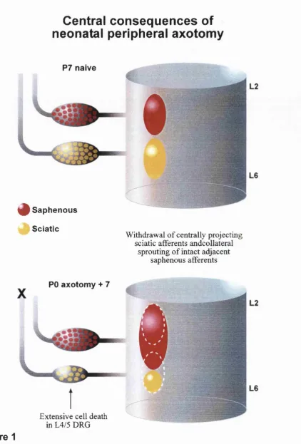
Two major consequences o f neonatal axotomy have been described: extensive death o f
axotomised DRG neurons with subsequent withdrawal o f their central projections (Himes
and Tessler 1989), and a collateral sprouting o f intact adjacent afferents into the
denervated central territory (Fitzgerald 1985; Shortland and Fitzgerald 1994) (Figure 1).
Although often described, quantitative studies o f the extent o f DRG cell death have
produced widely varying results and therefore a rigorous quantitative analysis was
performed (chapter 1). The collateral sprouting has also been widely described, and here a
novel method o f post-mortem labelling and quantification o f the sprouting response is
presented (chapter 2). To better understand the mechanisms underlying sprouting, I have
referred to other sensory systems that follow similar postnatal development and response
to injury. These have implicated the neurotrophin BDNF as a candidate for regulating
activity-dependent plasticity, both in normal development and following perturbations to
the system (Galuske et al 1992; Kang and Schuman 1995; Lu and Figurov 1997). Chapter
3 describes BDNF expression at the protein and mRNA level and shows a clear
Central consequences of
neonatal peripheral axotomy
P7 naive
^ S a p h e n o u s Sciatic
Withdrawal o f centrally projecting sciatic afferents andcollateral
sprouting o f intact adjacent saphenous afferents
PO axotomy + 7
Extensive cell death in L4/5 DRG
Figure 1
BDNF in axotomy-induced changes in connectivity within the spinal cord, but whether
that role is instructive or merely permissive is uncertain.
It is clear that considerable postnatal development occurs within the organisation o f the
spinal cord (Fitzgerald et al 1994). Perhaps an interaction with the tactile world is
necessary to establish the final discrete patterns o f connectivity within the spinal
somatosensory system in the same way that exposure to light is a prerequisite for honing
the connectivity required to establish stereoscopic vision (Katz and Shatz 1996). The
dynamics o f NMDA receptor activation have made it the source o f considerable interest in
studies o f activity-dependent plasticity (Fox and Zahs 1994). Chapter 4 describes the
consequences o f chronic NMDA receptor antagonism throughout the postnatal period
during which extensive reorganisation normally occurs. The results indicate that postnatal
changes in dorsal horn connectivity are activity-dependent and mediated by NMDA
Chapter 1
1: Introduction
1.1: Primary afferents exhibit physiological, biochemical and morphological
heterogeneity.
Primary sensory neurons within the DRG are a functionally and morphologically
heterogeneous population o f cells, exhibiting a wide range o f biochemical, anatomical and
physiological specificities. In order to elucidate the functional distinctions between the
various sub-populations they need to be clearly classified. Primary sensory neurons can
be distinguished in a variety o f ways: functionally specific peripheral endings, central
termination patterns, cell body size, axon diameter and degree o f myelination, membrane
properties and biochemical phenotype.
Physiological evidence for discrete populations o f primary afferents came from the work
o f Erlanger and Gasser in the 1930s (reviewed in Perl 1994). From measurements o f
conduction velocities within whole peripheral nerves they were able to reveal three
distinct populations o f nerve fibres separable by their action potential conduction
velocities. They subsequently defined these three groups as A, B and C fibres, with A
fibres having the fastest rates o f conduction and C the slowest. Since then the B-fibre
component has been shown to derive from preganglionic sympathetic fibres, leaving
primary afferents split into two groups, A and C. This nomenclature still persists in
defining fast and slow conducting nerve fibres and has been extended to classify the
ganglion cell bodies o f each type o f fibre. The total population o f neurons within a DRG
have been broadly split into two, large light and small dark cells, a distinction based upon
their relative expression o f neurofilament. These two groups possess A and C-fibres
respectively (Lawson 1979 and 1984). O f somatic primary afferents, approximately two-
thirds are small, unmyelinated C-fibre neurons (Willis and Coggeshall 1991). These are
almost exclusively nociceptive - a small proportion being purely thermoceptive - and are
responsive to mechanical, chemical and thermal stimuli, generally polymodally. Many of
these cell types are quiescent under normal conditions, particularly viscerally projecting
afferents, only active under pathological conditions, eg inflammation (McMahon and
Koltzenburg 1991). The rest o f the population o f the DRG are the large, myelinated A-
Chapter 1. Sensory neuron changes follow ing neonatal axotomy.
peripheral endings for transducing cutaneous mechanical stimuli, and the large Aa-fibres
that are proprioceptive, innervating muscle spindles and Golgi tendon organs (see table 1).
A x o n D ia m e te r
(|im)
C o n d u c tio n V e lo c ity
(m/s)
M y e lin a tio n
S o m a D ia m e te r
(H m )
F u n c tio n
A a
15-20 Fast (20-70) Myelinated Large (40-80) Proprioception
A P
10-15 Fast (12-55) Myelinated Medium/Large
(30-60)
Low threshold
Mechano-reception
AÔ 1-5 Medium (2-12) Thinly
Myelinated Small/Medium (15-50) High threshold Mechano-reception Thermo-reception Nociceptive
C <1 Slow (<2) Unmyelinated Small (10-25) Polymodal
Nociception
Table 1: Physiological classification of primary afferents.
The distribution o f different neuronal sub-types within the mammalian DRG does not
appear to exhibit any organisation in relation to function, morphology or phenotype,
unlike the situation in the chick (Honig 1982). Some evidence exists for a basic form of
somatotopy between separate peripheral nerves in an individual ganglion during
development (Wessels et al 1990) and in the adult (Puigdellivol-Sanchez et al 1998a),
otherwise the neuronal populations tend to be totally heterogeneous. The projections of
these afferents, however, are highly organised. Individual peripheral nerves innervate a
distinct region o f the skin that exhibits no overlap with adjacent nerves. These boundaries
are also respected by their central projections. Within the dorsal horn o f the spinal cord,
the primary afferent terminals are highly ordered in three dimensions. Peripheral
somatotopy is retained by the rostrocaudal and mediolateral boundaries with no overlap of
central projections o f adjacent peripheral nerves (Molander and Grant 1985, 1986; Swett
and W oolf 1985). In the adult, projections o f functionally distinct neuronal subtypes are
separated within the dorsoventral plane such that specific neuronal populations project to
distinct laminae o f the dorsal horn. Ap-fibres terminate in the deeper laminae o f the
dorsal horn, mainly restricted to lamina 111-V. Aô-fibres project to laminae 1 and V and C-
fibres are restricted to lamina 11 (Light and Perl 1979; Rivero-Melian and Grant 1990) (see
Laminar organisation of primary
afferent innervation of the aduit dorsal horn
m IV
Figure 2
Schem atic illu stratio n o f the central terminal distribution o f primary afferent populations.
A illustrates the segregation of myelinated and unmyelinated fibres within the dorsal horn. The largest diam eter A -afib res project to the motoneuron pools in the ventral horn. Ap-fibres, predominantly term inate in the deeper laminae o f the dorsal horn (III-V). AÔ-fibres project to lamina I and V, witii some exceptions (see text) and C-fibres exclusively terminate in lamina II.
B
CGRP p75
Ret TMP GFRa P2X3 IB4 VRl
Chapter 1. Sensory neuron changes follow ing neonatal axotomy. Fibre class Threshold Principal Transmitter Receptor expression Dorsal horn projection Physiology Sensation (norm al) Sensation (pathol.)
c
High Substance PCGRP EAA
N Kh2 N M DA AMPA mClu I lie N ociceptive Polymodal Silent N oxious Temp. hyperalgesia Cold allodynia
AP
Low EAA AMPA IIIIV
Mechano. ceptive
Tactile Mech.
allodynia
Table 2: Functional and morphological classification of primary afferents.
The classification o f cells as defined by morphology and physiology has been extended by
investigations o f chemical phenotype. The first example o f this came with the description
o f the peptide substance P within a population o f small sensory neurons and their central
terminals (Hokfelt et al 1975). This led to the sub-classification o f primary afferents into
peptidergic and non-peptidergic. Approximately half o f the C-fibre population o f adult
DRGs are peptidergic, most o f which express calcitonin gene-related peptide (CGRP)
(Averill et al 1995). Many other peptides, eg substance P, galanin, somatostatin, CCK,
VIP and NPY are also expressed by varying proportions o f these neurons (Verge et al
1995a; Alvares and Fitzgerald 1999). The non-peptidergic C-fibre population is defined
by its ability to bind the lectin IB4 (Alvarez et al 1991).
The phenotype is by no means fixed from the initial stages. For example, no neuropeptide
protein or RNA is detectable until target connection has been achieved, at E l 6/17 (Hall et
al 1997; Jackman and Fitzgerald 2000). This is due to the target-derived neurotrophic
requirement o f the innervating neurons to synthesise CGRP and substance P (Mulderry
1994). Even after this stage, it is clear that postnatal changes occur in the relative sizes o f
DRG sub-populations. The percentage o f cells expressing CGRP, substance P and
glutamate increases postnatally (Nitsos and Rees 1993). The expression o f other markers
eg somatostatin, does not begin until after birth (Bennett et al 1998a).
The entire population o f DRG neurons can be broadly divided into two by the relative
expression o f neurofilaments. In the adult animal, low molecular weight neurofilament
(NF-L) and the intermediate filament protein peripherin are expressed by two distinct
groups. Large/medium cells are almost exclusively immunoreactive for NF-L, whereas
peripherin is expressed by small cells, more than 50% o f which also express substance P
and/or CGRP (Goldstein et al 1991; Goldstein et al 1996). The cross-reactivity between
Chapter 1. Sensory neuron changes follow ing neonatal axotomy.
recordings from DRG in vitro show a correlation between neurofilament expression and DRG sub-type defined by conduction velocity and cross-sectional area: immunoreactivity
to RT-97 (phosphorlyated NF-L sub-unit) is detectable only in the A-fibre population, and
all RT-97 negative cell were C or A5 (Lawson and Waddell 1991). The relationship
between cell size and RT-97 immunoreactivity is also evident in late embryonic and
neonatal stages (Jackman and Fitzgerald 2000; Beland and Fitzgerald 2000). Analysis o f
mRNA expression in DRG neurons shows that the NF-L positive, peripherin negative
population also express RNAs for low, medium and high molecular weight
neurofilaments, whereas the peripherin positive population do not (Goldstein et al 1996).
Curiously, both cell types express peripherin mRNA. The absence o f peripherin
immunoreactivity in the large cells may be due to post-translational regulation or rapid
clearance o f the protein from the cell body and into the axons (Goldstein et al 1996). The
developmental regulation o f this expression shows that embryonically (E l 5/16) the DRG
appears as a homogeneous population o f cells all expressing peripherin and NF-L protein
and mRNA. This situation rapidly changes such that by E20 the two populations are
evident and by P2 the pattern o f expression seen in the adult is established (Goldstein et al
1996).
DRG neurons are supported throughout development by trophic support from one or more
o f the neurotrophins (reviewed in Davies 1994; Snider 1994). The expression of
individual neurotrophins and their receptors provides another means o f classification,
albeit providing less clear distinctions than those previously described. In the adult, 40%
o f sensory neurons, mainly unmyelinated C-fibres, express trkA (Averill et al 1995). The
IB4 population express G F R al and c-ret: the receptor complex for GDNF (Molliver et al
1997; Bennett et al 1998b). Approximately 35% o f the large A fibres express trkC
(McMahon et al 1994) and trkB is expressed by a proportion o f medium sized neurons, the
extent o f this population varying between studies from 5-40% (Kashiba et al 1995;
Karchewski et al 1999).
The small, unmyelinated population o f DRG neurons can be divided neurochemically into
two populations by their expression o f neuropeptides. The peptidergic population express
CGRP, amongst other peptides, and to a great extent co-express trkA, and are responsive
to NGF (Verge et al 1995b; Averill et al 1995; Michael et al 1997). A separate population
Chapter 1. Sensory neuron changes following neonatal axotomy.
1990). This IB4 population expresses the GDNF receptor complex components c-ret,
G FR a r and G FR a2 and responds to GDNF in vivo and in vitro (Molliver et al 1997; Bennett et al 1998b). The ontogeny o f this pattern o f expression is not achieved until the
second postnatal week. At E l 7, between 70-80% o f all DRG neurons express trkA and
are presumably dependent upon NGF for survival (Bennett et al 1996; Molliver et al
1997). Between E l 7 and P7, an upregulation o f ret occurs within a subset o f this
population and a corresponding down-regulation o f trkA. The outcome o f this phenotypic
change is a switch from NGF to GDNF for trophic support o f the IB4 population. The ret
expression persists until adulthood, when approximately 40% o f DRG neurons express
trkA, and the proportion that bind IB4 increases to around 40%, presumably this being the
same population o f small cells that downregulate trkA (Bennett et al 1996; Kashiba et al
1998). These two populations are also distinguishable by the distinct regions o f the
superficial dorsal horn in which their central projections terminate. The peptidergic
population terminate in lamina I and outer lamina II (IIo) whereas the IB 4/F R A P
population project to lamina Ilj (Silverman and Kruger 1990) (Figure 2 B ). These
distinctions between populations o f small sensory neurons clearly indicate possible
separate nociceptive roles and recent evidence suggests that there may indeed be
frmctional differences between them (Snider and McMahon 1998; Stucky and Lewin
1999). The P2X class o f ATP receptors consists o f 6 sub-types, all expressed in DRG
neurons (Bumstock and Wood 1996). However, only one, P2X], is restricted to small
sensory neurons, and this expression is coincident with the IB4 population (Bradbury et al
1998; Vulchanova et al 1998).
Recently, electrophysiological evidence has indicated that action potentials in the IB4
population are longer in duration than peptidergic nociceptive neurons (McCarthy and
Lawson 1997; Stucky and Lewin 1999). A possible mechanism underlying this is that
these neurons exhibit significantly greater TTX-resistant Na^ currents under voltage clamp
(Stucky and Lewin 1999). The question remains as to whether a functional difference
exists between these two populations in terms o f their nociceptive responsiveness. It is
clear that 40-50% o f cutaneous C-fibres respond to noxious heat (Koltzenburg et al 1997).
Both the IB4 and peptidergic populations exhibit responsiveness to heat but the latter elicit
far larger heat-activated currents than the IB4 population. Peptidergic neurons respond to
rapid increases in heat with a large burst o f action potentials, whereas IB4 cells fire only a
Chapter 1. Sensory neuron changes follow ing neonatal axotomy.
associated with a reduced ability to produce action potentials to heat-induced current
suggest that the IB4 population display poor heat sensitivity in vivo, indicating a greater role for the IB4 negative, peptidergic population in acute responses to heat. The majority
o f evidence that exists indicates the endogenous heat-gated ion channel to be the V Rl
capsaicin receptor (Caterina et al 1997; Tominaga et al 1998; Cesare et al 1999).
However, the patterns o f expression o f V R l in DRG neurons do not at first appear to fit
the physiological evidence proposed by Lewin et al. V R l protein and mRNA expressiom
has been shown to be restricted to small, unmyelinated nociceptive neurons, and within
that population is expressed by approximately 60% o f the IB4 population and 80-90% o f
the peptidergic population (Tominaga et al 1998; Michael and Priestley 1999; Guo et al
1999). If this proportion o f neurons express V R l, why are only 45% responsive to heat?
It is possible that the level o f V R l expression required by a neuron is greater than the
limits o f resolution o f in situ and immunohistochemical methods (see below). Alternatively, V R l may not be the only heat-activated ion channel. Heat sensitivity can
be exhibited by both small and large DRG cells, indicative o f the polymodal nature o f
many large myelinated cells. However, the response is quantitatively different. The small
nociceptive population o f heat sensitive DRG neurons has a threshold o f around 45°C, the
temperature at which heat starts to become noxious. Larger, predominantly
mechanoreceptive neurons have a higher threshold to heat, approximately 51 °C (Nagy and
Rang 1999). Only the former population o f cells is responsive to capsaicin, indicating
more than one heat-activated channel. This evidence fits neatly with the first homologue
to VRl so far discovered - V R L l, vanilloid receptor-like 1. This is expressed only by
large DRG neurons and its threshold to heat is 51°C. The possibility exists, and
circumstantial evidence suggests that further homologues underlie some o f the disparate
effects o f capsaicin (Szallasi and Blumberg 1999). It may well be that sub-population
specific expression o f one or more o f these homologues underlies the discrepancies
discussed.
The V R l quantification studies mentioned above are predominantly concerned with
expression in the soma o f DRG neurons. Clearly, heat detection is a peripheral function
and the assumption is that neurons expressing V R l protein in the perikarya o f the soma
also transport it axonally. Indeed, the Tominaga study does show central transport o f V Rl
Chapter 1. Sensory neuron changes follow ing neonatal axotomy.
projections - ie lamina I and IIo, there is a discrepancy o f V R l and IB4 co-expression,
with V Rl only located at the medial extent o f lamina Ilj. One interpretation is that this
may represent evidence for two neurochemically distinct sub-populations o f IB4 positive
neurons. It is clear that VRl endows thermal and chemical sensitivity upon neurons in
which it is expressed. The conventional polymodal nociceptor, however, is equally
responsive to noxious mechanical stimuli (Bessou and Perl 1969). Is it possible that two
discrete populations o f IB4 neurons with divergent modalities exist? Only investigation of
the mechanical responsiveness o f the two purported sub-populations will answer this.
However, the medio-lateral dissociation in the dorsal horn represents a topographical
distinction between proximal and distal peripheral innervation, and this result would
therefore indicate a greater thermal responsiveness of, for example, the distal regions of
the hindlimb, ie the plantar surface o f the paw. Furthermore, closer inspection o f fibres
within the DRG revealed some substance P positive fibres with no V R l labelling, as was
the case with a proportion o f IB4 fibres. Triple labelling o f cells also revealed a
population o f neurons expressing VRl but not substance P or IB4. A possibility here is
that the use o f substance P does not label all peptidergic cells, ie this population may
indeed express another peptide, eg CGRP. What is clear from previous studies is that
small cells within the DRG are a heterogeneous population. This heterogeneity is
manifest in a variety o f ways (Hunt et al 1992). Previous work on capsaicin sensitivity o f
DRG neurons has clearly shown that no sub-classification o f these small cells, be it
biochemical, anatomical or physiological, totally encompasses that population that
exhibits sensitivity to capsaicin (Holzer et al 1991). This is clearly also true for the
population expressing V Rl (Michael and Priestley 1999). It is possible that direct
correlation o f protein or mRNA expression and function is not appropriate. Clearly, a
critical level o f V R l expression will be necessary to give a cell capsaicin/heat
responsiveness and the method o f stimulation used to detect function may not always be
physiologically relevant ie heat or capsaicin applied to peripheral terminals or to
dissociated cells in culture. It is also not unreasonable to assume a gradual capsaicin
sensitivity across a population o f neurons expressing V R l to varying degrees. It has been
known for some time that capsaicin sensitivity does indeed vary between sensory neurons
(Seno and Dray 1991; Stucky et al 1998). An important contribution in understanding this
phenomenon has been the demonstration o f differential expression o f V R l mRNA within
defined sub-populations o f DRG neurons (Michael and Priestley 1999). It is clear that
Chapter 1. Sensory neuron changes following neonatal axotomy.
not all, IB4 cells show a relatively low level o f V R l mRNA expression, particularly low
among those that also co-express trkA. Furthermore, the somatostatin positive sub
population o f IB4 cells have uniformly low expression, consistent with studies showing
low capsaicin sensitivity o f these cells (Kashiba et al 1997). Levels o f V Rl were
generally higher in the substance P/CGRP population, but again, expression was variable.
Analysis o f adult DRG neurons following neonatal capsaicin treatment shows a greater
loss o f the CGRP population than IB4, consistent with these reported levels o f V Rl
expression (Torsney and Fitzgerald 2000). An interesting finding was a population of
very small cells, negative for trkA and IB4, but expressing ret, that had extremely high
levels o f V R l mRNA. This population was very small (-1% o f total) but the level o f V Rl
expression was such that they may have a unique function. If we assume the relative
levels o f mRNA expression within these groups to be comparable with protein expression
levels, then the physiological disparity between IB4 positive and negative cells in their
responsiveness to heat (Stucky and Lewin 1999) has a clear molecular basis. Furthermore,
the fact that only 45% o f small nociceptive neurons respond to noxious heat (Koltzenburg
et al 1997) is equally compatible.
It is important also not to consider these expression patterns as fixed, or to assume
expression o f V R l determines the physiological phenotype o f a given neuron. NGF
injections are capable o f producing a long-lasting hyperalgesia to heat in vivo (Lewin
1993). While the acute stage o f this hyperalgesia is almost certainly maintained by
cytokine release from mast cells (Lewin 1994), the longer-lasting secondary stage is
independent o f this as has recently been exhibited in DRG neurons in culture (Stucky and
Lewin 1999). The key to this may lie in the expression o f trkA in sub-populations of
small DRG neurons. The IB4 population responds physiologically less robustly to
noxious heat (Stucky and Lewin 1999), possibly due to lower levels o f V R l expression, as
detailed above. However, approximately 25% o f this IB4 binding population also express
trkA (Averill et al 1995; Molliver et al 1995; Michael and Priestley 1997) and this
population may be particularly sensitive to sensitization to NGF. Therefore NGF may
selectively increase the proportion o f the IB4 population that are responsive to heat. The
fact that GDNF has no effect indicates it is NGF specific. If this is the case then it
provides a novel mechanism for NGF induced sensitization by unmasking heat sensitivity
Chapter 1. Sensory neuron changes follow ing neonatal axotomy.
in DRG somata was as in the two previous studies, they found distinct expression o f V Rl
protein in fibres. V R l was shown to almost totally colocalise with P2X3 and IB4 in both
centrally and peripherally projecting fibres, but virtually no V R l was detectable in fibres
labelled with substance P or CGRP. The discrepancy here is unexplained as yet. One
possibility the authors propose is that ectopic V R l protein persists in the soma o f
peptidergic neurons and is not transported axonally. Alternatively, a further homologue o f
VRl may exist that is expressed selectively in fibres (cf the selective expression o f
neuropeptide Y receptors Y1 and Y2 on soma and fibres respectively (Hokfelt et al 1998).
1.2: Peripheral innervation.
Peripherally projecting axons first emerge from the rat and mouse lumbar DRG at E l 2
(Reynolds et al 1991; Mimics and Koerber 1995a). By E13, major nerves such as the
sciatic and saphenous have formed from the plexus. Although both A and C-fibres enter
the hindlimb at E l 3/14, the larger, RT97 positive A-fibres predominate, projecting more
distally and having a higher density o f skin terminals throughout embryonic life with the
smaller C-fibres slower to innervate the epidermis (Jackman and Fitzgerald 2000). Distal
regions are reached by E l 6.5 by which stage appropriate dermatomal boundaries have
been established (Mimics and Koerber 1995a). Peptidergic innervation o f the hindlimb
epidermis is not detectable until E l 9, when all regions simultaneously show expression,
indicating that the fibres are present but the onset o f peptide expression is delayed
(Jackman and Fitzgerald 2000).
The relative contributions o f C, A5 and Ap-fibre innervation to a given area o f skin are 70,
10 and 20% respectively, although this is variable. It is important to indicate that,
although each is deemed to have a specific function, they are differentially sensitive to
different stimulus modalities, rather than having all-or-nothing responses. Thus, under
normal conditions, all three are capable o f transducing innocuous information, although in
the rat very few C-fibres transmit innocuous stimuli, whereas noxious stimuli are
transmitted by C and Aô-fibres only. Following injury or inflammation, this situation can
be changed. It is also the case that a stimulus o f a single modality is unlikely to occur in
the normal situation. By investigating the receptive field properties o f mouse primary
afferents, and using conduction velocity measurements for identification, the relative
proportions o f receptive modalities have been elucidated (Koltzenburg et al 1997). In the
Chapter 1. Sensory neuron changes following neonatal axotomy.
adapting (RA) and slowly adapting (SA) afférents. The former are likely to be hair follicle
afferents and the latter arising from Merkel cells in touch domes. The Aô population of
myelinated afferents are mostly high threshold mechanoreceptors (66%), the remainder
comprising D-hair receptors. The unmyelinated component all exhibited high threshold
mechanoreceptive properties. These relative proportions are established by P I4, although
conduction velocities are lower, presumably as a consequence o f immature myelination
(Koltzenburg et al 1997).
Under normal physiological conditions, nociceptive C-fibres are not spontaneously active.
However, a small subset o f these, between 10-20% and mostly in joints and viscera,
remain inactive even with acute noxious stimulation and have been termed ‘silent
nociceptors’ (McMahon and Koltzenburg 1990; Koltzenburg 1995). In pathological
conditions, such as inflammation or tissue injury, these nociceptors become sensitized and
responsive to chemical mediators that are typically a product o f such states (Koltzenburg
1995; Dmitrieva and McMahon 1996). This recruitment is presumed to enhance the C-
fibre barrage to the dorsal horn and underlie aspects o f pain states associated with
injury/inflammation. This adaptivity o f primary afferent neurons and ability to selectively
modulate activity in the spinal cord is also an underlying feature o f the normal postnatal
changes that occur in connectivity o f the somatosensory system.
1.3: Central innervation.
Spinal cord differentiation proceeds along a ventrodorsal and rostrocaudal gradient, such
that ventral motoneurons are the first neuronal type to be generated, between E l 1-13, and
lamina II neurons the last, E l 4-16 (Redmond et al 1997). Within the dorsal root ganglia
(DRG), cells destined to be large, myelinated neurons are the first to be bom. O f the small
cells, those that are IB4 positive develop before those that will express peptides (Kitao et
al 1996). Central projections o f primary afferents from the DRG start to accumulate in the
dorsolateral Bundle o f His at E l 2, where they remain for 3 days. The large diameter
fibres enter at this time, projecting to the deeper laminae o f the dorsal horn (Reynolds et al
1991; Snider et al 1992; Mimics and Koeher 1995), although their arbors initially project
superficially (Fitzgerald et al 1994). The small diameter fibres enter shortly before birth at
E19 (Fitzgerald 1987; Mimics and Koerber 1995b; Jackman and Fitzgerald 2000). Unlike
Chapter 1. Sensory neuron changes follow ing neonatal axotomy.
The dorsal horn o f the spinal cord is innervated by primary afferents transducing a wide
variety o f sensory information from the periphery. The terminal arborizations of these
afferents are segregated between the laminae o f the dorsal horn based upon fibre type and
receptive field properties (Light and Perl 1979; Brown 1981). Unmyelinated C-fibres
terminate almost exclusively within lamina I and II. Within this population o f afferents
there is a distinction between peptidergic and non-peptidergic afferents which is discussed
in more detail later. Thinly myelinated Aô fibres consist o f two functionally different
groups. One, mainly nociceptive, terminate in laminae I and V, while the other, which
innervate hair follicles (D-hair afferents), project to laminae II and III. Large, myelinated,
Ap-fibres terminate in the deeper laminae o f the dorsal horn, typically III-V, the pattern of
which is dependent upon their specific phenotypes (Brovm 1981) (see figure 1).
The cellular organisation o f the dorsal horn itself is, however, far more complex. The
laminar organisation that is conventionally used for afferent arborizations is based upon
cytoarchitectural boundaries observed by Nissl staining (Rexed 1952; Molander et al
1984). These neurons subserve a variety o f functions, some are the cells o f origin o f
ascending tracts, and many are excitatory or inhibitory intemeurons (Brown 1981; Willis
and Coggeshall 1991) and importantly, the extent o f their dendritic processes is greater
than the laminar organisation o f their cell bodies suggest. In some cases, these cells are
restricted to specific laminar regions. For example, the dendritic trees o f lamina II islet
cells are contained within lamina I and II (Gobel 1982), and spinocervical tract cells,
found in deeper laminae, have dendritic projections that remain within the deeper laminae
(Brown 1981). The afferent innervation o f these two specific cell types is therefore likely
to be restricted to a particular subset.
This convenient laminar organisation is not necessarily true o f all dorsal horn neurons,
however. Many cells have dendrites that cross several laminar boundaries (Willis and
Coggeshall 1991) and are therefore capable o f receiving monosynaptic input from widely
divergent afferent types. A population o f neurons in laminae III and IV which express the
N K l receptor have dendrites that terminate in laminae I and II and are presumably targets
for unmyelinated afferents projecting to lamina II (Brown et al 1995; Mantyh et al 1995).
The dendrites o f these neurons can extend from lamina V up to lamina I and presumably
receive monosynaptic input from all classes o f afferent as well as from numerous dorsal
horn interneurons. These neurons have been shown to project to the spinothalamic tract
Chapter 1. Sensory neuron changes follow ing neonatal axotomy.
al 1997). They therefore relay nociceptive input, and yet their cell bodies reside in the
deeper laminae o f the dorsal horn, a region that receives low threshold mechanical
innervation from the periphery. Similarly, Lamina II neurons, normally considered to
relay noxious information from lamina II projecting afferents, extend dendrites into deeper
laminae where they make synaptic contacts with low threshold Ap-fibres (W oolf and
Fitzgerald 1983; Baba et al 1999). Clearly, inferring functional differences in sensory
processing merely from differences in laminar segregation o f primary afferent terminals is
too simplistic. Indeed, elegant experiments combining the specificity to myelinated fibres
o f cholera toxin conjugated HRP and N K l immunohistochemistry have revealed that N K l
labelled neurons do receive monosynaptic input from large calibre, myelinated afferents
(Naim et al 1998). It is important to stress, however, that the density o f synapses from
these afferents was far less than those o f substance P or CGRP containing afferents, such
that the latter, peptidergic C-fibres constitute a substantially greater input. In addition,
anatomical evidence suggests these synapses are often functionally 'silent' (Baba et al
1999). Activation o f such silent connections, and the establishment o f novel ones, are
thought to be critical mediators o f central changes underlying acute and chronic sensory
processing abnormalities following peripheral inflammation/injury (W oolf et al 1992). It
is these same mechanisms that are responsible for the extensive changes in connectivity
that occur during early postnatal development. The interaction between primary afferent
input and postsynaptic spinal cord cells is clearly more elaborate than a simple laminar
mechanism can elucidate. The modelling o f mono and polysynaptic connections within
the spinal cord is crucial for the development o f a system capable o f discerning the wide
range o f sensory modalities that constitute the tactile world.
1.4: Consequences of neonatal axotomy.
It is clear from numerous studies that peripheral nerve injury has far more dramatic
consequences if it occurs during a critical period o f neonatal plasticity. Most commonly
described has been the extensive death o f axotomised dorsal root ganglion cells that occurs
(Risling et al 1980; Aldskogius and Risling 1981; Bondok and Sansone 1984; Yip et al
1984; Schmalbruch 1987; Himes and Tessler 1989; Rich and Hollowell 1990; Cheema et
al 1984). In the adult, axotomy induced cell death is not initiated until approximately
Chapter 1. Sensory neuron changes following neonatal axotomy.
cells that die has been a matter o f some debate due to the counting methods employed in
various studies. This is discussed in more detail later. An initial consequence o f the cell
death is a transganglionic degeneration and subsequent withdrawal o f central terminals of
those cells (Aldskogius et al 1982; Bondok and Sansone 1984). This is followed by the
sprouting o f adjacent, intact axon collaterals into the denervated region (Fitzgerald 1985a;
Jacquin and Rhoades 1985; Fitzgerald et al 1990) (see chapter 2). Cell death, while
extensive, is not complete for all axotomised neurons, and consequently some survive
neonatal axotomy. These also undergo a sprouting response, similar to that seen in the
adult, such that they innervate inappropriate laminae o f the cord (Shortland and Fitzgerald
1994). The widespread death o f DRG neurons following axotomy and indeed the ability of
some to survive has been proposed to depend on their relative maturity and sensitivity to
trophic factors (White et al 1993). However, attempts to quantify the extent o f the
neuronal death that occurs have yielded widely varying results (Whiteside et al 1998).
Table 3 summarises these reports:
Yip et al (1984) 40-50% cell loss from L4/5 PI sciatic crush
Schmalbruch (1987) 50-90% loss from L4-L6 PO sciatic axotomy
Himes and Tessler (1989) 50% loss from L5 PO sciatic axotomy
Rich and Hollowell (1990) 32% loss from L4/5 PO sciatic axotomy
Nothias et al (1993) 50% loss from L4/5 PO sciatic axotomy
Cheema et al (1994) 75% loss from L5 PO sciatic axotomy
Table 3: Reported DRG ceil losses following neonatal peripheral injury.
The extreme variation between studies may be due to the use o f different or inappropriate
counting techniques (Coggeshall 1992). However, the studies are in agreement as to the
rapidity o f the onset o f cell death, all claiming the majority o f cells to have died within the
initial 24 hour period following injury. This has recently been supported by the detection
o f numerous apoptotic neuronal profiles within L4 and L5, 24 hours post neonatal sciatic
axotomy (Oliveira et al 1997; Sugomoto et al 1998; Whiteside et al 1998).
The aim o f this series o f experiments was to quantitatively describe the degree and time
course o f DRG cell death following neonatal axotomy o f the sciatic nerve using unbiased
Chapter 1. Sensory neuron changes follow ing neonatal axotomy.
clearly show that cell death is far more substantial following axotomy at early postnatal
ages. The extent o f this period o f higher susceptibility is unclear from those studies and
therefore axotomy was performed at a range o f ages to establish the duration o f this
critical period. Once the degree o f cell death was established, the question remained as to
whether a particular sub-population o f DRG neurons is particularly sensitive to axotomy,
as has been proposed for the longer term consequences o f adult axotomy (Coggeshall et al
1997). Immunohistochemical analysis o f axotomised DRGs was performed in order to
establish whether any particular neuronal phenotype bestowed a susceptibility to axotomy,
Chapter 1. Sensory neuron changes follow ing neonatal axotomy.
2: Materials and Methods
2.1: Total cell counts.
For all experiments Sprague Dawley rats o f both sexes were used. The age between birth
and 24 hours old was defined as PO.
Sciatic nerve axotomy was performed at three ages; PO, FIG and P21.
Surgical procedure
PO rat pups were anaesthetised by hypothermia. Under aseptic conditions, an incision was
made in the left thigh and the sciatic nerve exposed above the popliteal fossa. The nerve
was ligated with 7/0 suture and cut distal to the ligation. The wound was sealed using
Vetbond adhesive and the pups warmed and returned to their mother.
PIO and P21 rats were anaesthetised by halothane inhalation (4% induction, 2%
maintenance). The sciatic nerve was exposed as described above, ligated and sectioned.
Muscle layer and skin were sutured using 5/0 sutures. Rats were allowed to recover and
returned to their mother.
For all three ages, animals were divided into two groups (n=4 for each). One group was
left for 2 days and the other for 7 days after the sciatic nerve axotomy. At these times,
animals were terminally anaesthetised with 200mg/ml pentobarbitone and perfused
transcardially with 0.9% saline followed by a 3% paraformaldehyde, 3% glutaraldehyde,
0.1% picric acid fixative. L4 and L5 lumbar ganglia were identified by tracing the sciatic
nerve, removed and post-fixed in fixative solution.
The ganglia were dehydrated in ascending concentrations o f ethanol, embedded in a
mixture o f epon and araldite and serially sectioned at a thickness o f 4pm. Sections were
counterstained in Toluidine blue (0.33% in 0.1 M sodium cacodylate buffer).
Counting paradigm
Estimates o f total cell numbers were made using a physical dissector paradigm
(Coggeshall 1992; Pover et al 1993). Neuron numbers (N) are calculated by multiplying
the numbers o f neurons per unit volume (Ny) by the total volume o f the ganglion (Vref):
N = N y X Vref
Ny is calculated using five pairs o f adjacent sections per ganglion such that the five pairs
are equally distributed throughout the ganglion. The first o f each pair is deemed the
reference section and the second the look-up section. Each section is photographed and
neuronal nuclear profiles subsequently drawn. The profiles for the reference and look-up
Chapter 1. Sensory neuron changes follow ing neonatal axotomy.
profile is seen in the reference but not the look-up section. The area o f each section is
determined and multiplied by the thickness to give the volume. The number o f tops per
unit volume (Nv) can then be calculated. Vref is calculated by taking approximately ten
evenly spaced sections throughout the ganglion and multiplying mean ganglion area by the
total number o f sections and the thickness.
Cell counts were made in collaboration with Professor Richard Coggeshall.
2.2: Identification of sub-populations.
Sciatic nerve section was performed on a different litter o f PO rats as described above and
the pups returned to their mother. Pups were taken 2 days (n=4), 7 days (n=4) following
axotomy and a further group left to reach adulthood (n=4). Additionally, 2 day and 7 day
old naive pups were used as controls (n=4 for each). Rats were terminally anaesthetised as
above and perfused with 0.9% saline followed by 4% paraformaldehyde fixative. L4 and
L5 lumbar DRGs were identified, removed and post-fixed in a 30% sucrose/fixative
solution overnight at 4°C.
Individual DRGs were mounted in Tissue Tek and frozen for cryostat sectioning. 10pm
serial sections were cut and collected directly onto gelatinised slides. Sections were
allowed to dry at room temperature overnight before immunohistochemical labelling.
Immunohistochemistry 1.
Sections were immunolabelled with the following antibodies:
Anti-N52 (NF200 clone N52 mouse monoclonal IgG Sigma)
Anti-CGRP (rabbit polyclonal IgG Chemicon)
IB4 (Biotinylated lectin Sigma)
O Slides were washed 3x10 minutes in 0.1 M PBS.
0 Blocking stage:
20% NGS, 0.2% triton in 0.1 M PBS
1 hour at room temperature
© Primary antibody:
aN 52 (1:1000)
aCG RP (1:2000) In 4% NGS, 0.2% triton in 0.1 M PBS
1B4 (lOpg/ml)
Cell profile counts method
1. L4 and L5 DRGs taken from each rat. 2. Each ganglion serially sectioned at 10pm.
3. Five sections, evenly distributed throughout the entire ganglion, used for profile counts (red outline). Sections were therefore far enough apart wi&in the ganglion to ensure that no single cell was counted more than once.
4. For each individual animal, L4 and L5 numbers were pooled, ie profile counts made from ten sections per animal.
R at1 R at 2
L4 , L 6 )
R at 3 R at 4
5. No. o f positive profiles in L4+L5 ^ |qq = Total no. o f profiles in L4+L5
6. P I + P2 + P3 + P4 _= Final value (%) (where appropriate)
Figure 3
Summary o f the cell profile analysis used to determine DRG neuron sub-population proportions. This method was employed for all comparative studies described (optical dissector technique was used for absolute quantification). Mean o f percentages was only used to obtain a figure for the sciatic component o f L4/5 where the percentage o f total profiles (as revealed by neutral red counterstain) that were labelled with fluorogold was measured (n=6). In all other cases, the percentage values for each animal were recorded individually to allow further statistical analysis.