_____________________________________________________________________________________________________
*Corresponding author: E-mail: [email protected];
(Past name: British Journal of Medicine and Medical Research, Past ISSN: 2231-0614, NLM ID: 101570965)
The Expression of Programmed Death Ligand-1
within Stages of Oral Squamous Cell Carcinoma:
Immunohistochemical Analysis
Moomal Aslam Khan
1*, Serajuddaula Syed
1, Noor Ul Wahab
2,
Saima Akram Butt
3and Jabbar Ahmed Qureshi
41
Department of Pathology, Ziauddin University, 4/8 Shahra e Galib, Clifton Block 6, Karachi, Pakistan. 2
Department of Oral and Maxillofacial Surgery, Ziauddin College of Dentistry, Pakistan.
3Department of Oral Pathology, Ziauddin College of Dentistry, Pakistan.
4Department of Pharmacology, Ziauddin Medical University, Pakistan.
Authors’ contributions
This work was carried out in collaboration among all authors. All authors read and approved the final manuscript.
Article Information
DOI: 10.9734/JAMMR/2020/v32i230368 Editor(s): (1) Dr. Thomas I. Nathaniel, University of South Carolina, School of Medicine-Greenville, Greenville, USA. Reviewers: (1)Heba Gamal Abd El-Aziz Nasr, Al-Azhar University, Egypt.
(2)Arthur Chuemere, University of Port Harcourt, Nigeria. Complete Peer review History:http://www.sdiarticle4.com/review-history/55165
Received 17 December 2019 Accepted 26 February 2020 Published 28 February 2020
ABSTRACT
Background: Oral squamous cell carcinoma is prevalent in South Asian countries with rising cases of its incidence and mortality. Despite advancements in treatment, survival and recurrence rates are poor. Immunotherapy is a novel therapeutic modality in immunooncology. Immune checkpoint proteins are under investigation for clinical implications amongst which Programmed Death Ligand-1 has shown valuable results in certain malignancies.
Aims: To determine the immunohistochemical expression of Programmed Death Ligand-1(PD-L1) in oral squamous cell carcinoma and to find an association of Programmed Death Ligand-1 with stage and clinicopathological parameters of oral squamous cell carcinoma.
Study Design: Cross-sectional study.
Place and Duration of Study: Ziauddin Medical University, Karachi, 1 Year duration during 2018-2019.
Methods: A total number of 140 biopsy confirmed cases of oral squamous cell carcinoma were recruited in the study. Immunohistochemical expression of Programmed Death Ligand-1 was
evaluated and associated with the clinicopathological parameters of oral squamous cell carcinoma (OSCC). The data was statistically analyzed through Descriptive statistics and Chi square test by using SPSS v.20.
Results: Out of 140 participants, 74% were males (n=103) and 26% were females (n=37). Programmed Death Ligand-1 positivity was observed in 62.1% of cases (n=87). The Mean age of the participants was 48.91 ± 11.7 years. The most common site of cancer involvement was buccal mucosa and majority of participants were habitual of consuming chewable products i.e. Pan, Gutka and betel nut (89; 64%). Stage III and IV tumours comprised a major portion of cases in our study. (52; 37%), (56; 40%). A statistically significant p-value was noted for the association of Programmed Death Ligand-1 with stage II and IV tumours. (P-values: 0.029, 0.001)The association of Programmed Death Ligand-1 with other variables such as age, gender, ethnicity, sites or habits was not statistically significant.
Conclusion: This study concludes that the statistical significance of Programmed Death Ligand-1 expression with tumour stage is suggestive of worsening prognosis and might have detrimental effects as tumour progresses in advanced stage. Programmed Death Ligand-1 positivity in patients having oral squamous cell carcinoma could be useful in future research in the light of cancer immunotherapy which has shown success in oncology.
Keywords: Programmed death ligand -1(PD-L1); squamous cell carcinoma; immunotherapy; immune checkpoint protein.
1. INTRODUCTION
Melanesia and South East Asian countries have observed a dramatic increase in the prevalence of oral squamous cell carcinoma (OSCC), ranking it as 16th most common cancer worldwide as per GLOBOCON 2018 [1] Pakistan has a reported prevalence of 10% and oral cancers are the 2nd most prevalent malignancy in this country with 18,881 new cases reported, in both sexes and all age groups in the year 2018 [1].
Oral cancer is present amongst 3rd to 7thdecade age groups and most diagnosed cases are of the male population as compared to the female gender [2,3]. A multitude of causative factors result in carcinogenesis and development of this tumour [4]. These factors maybe Extrinsic, such as alcohol consumption or use of tobacco and addictive chewable products, or Intrinsic, which mainly involve genetic elements, viral infections, nutritional deficiencies and most importantly deficiencies in the immune system that result in immune suppression and cancer development [4]. The habits of using tobacco products and chewable items such as Gutka, Paan, chalia are common in the population subset of Pakistan.
Oral cancers begin as premalignant lesions and over a period of time result in bleeding, painful ulcers appearing as exophytic growth with life threatening conditions [5,6,7]. Oral inspection and radiological techniques aid in diagnosis, however, histopathological examination remains the gold standard for diagnostic purpose [8]. Oral
cancer bears poor prognosis, having a 5-year survival rate of 60% according to literature. Almost two-thirds of oral tumours are associated with regional or distant metastasis i.e. in stages III and IV respectively. Metastasis of OSCC usually involves ipsilateral cervical lymph nodes and distant metastases to lungs and esophagus [9]. Tumour grade and stage serve as tools to determine prognosis. Regardless of myriads of treatment modalities, the overall survival and prognosis remains to be poor up to date. Around 30% of affected individuals die because of recurrence and distant metastatic spread.
Advances in treatment have recently focused on the use of immune checkpoint inhibition to treat cancers. Programmed death ligand 1(PD-L1) has yielded promising results in management of various carcinomas including Non-small cell lung carcinomas, Melanoma and those of the head and neck [10,11,12-18].
PD-L1 is an immune checkpoint protein which negatively modulates immune cell responses by involving the PD-1 receptor which is present on T lymphocytes. The PD-L1 overexpressing tumour cells result in exhaustion of T cells which enable tumour growth, proliferation and escape from immune surveillance mechanism. PD-L1 has been linked with poor prognosis in some studies and its expression level has been reported to be higher with advanced stage cancers [19,20,21].
novelty of this biomarker to usefulness in the field of cancer immunotherapy. Studies are rapidly being undertaken to explore the expression of PD-L1 in oral malignancies and have presented a series of diversified results warranting further research in this domain. In such phase of ongoing researches we aimed to determine the relation of PD-L1 expression with the stages of oral cancer along with clinical parameters.
2. METHODS
This was a cross sectional study which was carried out at Ziauddin hospital during the years 2018 to 2019. A total number of 140 cases of OSCC were recruited in the study by purposive sampling technique. The clinical parameters and demographic characteristics of patients diagnosed with OSCC were collected through a questionnaire. The diagnosis of cases was based on clinical and histopathological evaluation. Patients diagnosed with OSCC, irrespective of gender, age and ethnicity were included in the study and patients with other malignancies and tumours with a metastatic spread to oral sub sites were excluded.
Following this, laboratory procedures were carried out. Paraffin- embedded formalin-fixed biopsy blocks were selected. Around 4 µm tissue sections were cut and stained with Hematoxylin and Eosin (H & E) for observation through light microscope. Tumour staging was done by applying TNM staging system given by AJCC 7th Edition [22].
2.1 Immunohistochemical Analysis
Monoclonal antibody for PD-L1 (Cell marque Clone ZR3) was applied for immune-histochemistry as per the instructions given by the manufacturer. (Cell Marque, catalog No.
438R-25). Evaluation of PD-L1 staining was
done via light microscopy and this was followed by scoring system mentioned in similar studies. A four tiered Grading method was adopted and different scores were labelled for the stained cancer cells percentage. Score 0 was labelled as no staining with 0- <5% of tumour cell percentage. Score +1 was labelled as weak staining with ≥5%- ≤30% tumour cell percentage. Likewise, scores +2 and +3 were labelled as moderate and strong staining with tumour cell percentage of ≥31%- ≤60%, and ≥61%- ≤100% respectively. PD-L1 positive expression was defined as at least 5% of cancer cells showing membranous staining at any of the mentioned
intensities. The cut off figure of 5% has been applied in the clinical trials involving Head and cancers [23,24]. SPSS version 20 was used for statistical evaluation.
The percentage of viable tumour cells that showed complete, circumferential or partial linear plasma membrane staining at any intensity were considered for scoring. Immune cells, normal cells, necrotic cells and cytoplasmic staining were excluded. Squamous lung tissue was taken as a positive control. 4X objective magnification was used to examine tumour zones on the slides and 10-40x magnification was used to score viable tumour cells in the entire specimen.
2.2 Statistical Analysis
Quantitative data were expressed as mean and standard deviation, whereas, qualitative data were expressed as frequency and percentages. The association of PD-L1 with all variables was determined by applying the Chi square test. P value of less than 0.05 was taken as significant. The study was carried out after the approval of Ethics Review Committee (ERC).
3. RESULTS
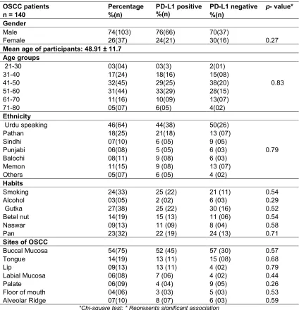
Out of 140 participants, around 74% were men (n=103) in contrast to women (26%, n=37). PD-L1 positivity was found in 62.1% of samples (n=87) and the major portion of PD-L1 positive cases belonged to males. (76%, n=66). The mean age of participants was calculated to be 48.91 ± 11.7 years. The clinical and demographic details of all participants and association with PD-L1 is shown in Table 1. The majority of cases showed moderate staining with a score of +3 for Tumour proportion Score (TPS) (Tables 2 & 3).
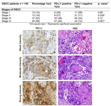
The expression of PD-L1 via immune-histochemistry according to various intensities in tissue specimens of OSCC is shown in Fig. 1 and Control Tissue in Fig. 2.
Table 1. The association of PD-L1 with clinicopathological parameters of all study participants
OSCC patients n = 140
Percentage %(n)
PD-L1 positive %(n)
PD-L1 negative %(n)
p- value*
Gender
Male 74(103) 76(66) 70(37)
0.27
Female 26(37) 24(21) 30(16)
Mean age of participants: 48.91 ± 11.7 Age groups
21-30 03(04) 03(3) 2(01)
0.83
31-40 17(24) 18(16) 15(08)
41-50 32(45) 29(25) 38(20)
51-60 31(44) 33(29) 28(15)
61-70 11(16) 10(09) 13(07)
71-80 05(07) 6(05) 4(02)
Ethnicity
Urdu speaking 46(64) 44(38) 50(26)
0.79
Pathan 18(25) 21(18) 13 (07)
Sindhi 07(10) 6 (05) 9 (05)
Punjabi 06(08) 5 (05) 6 (03)
Balochi 08(11) 9 (08) 6 (03)
Memon 11(15) 9 (08) 13 (07)
Others 05(07) 6 (05) 4 (02)
Habits
Smoking 24(33) 25 (22) 21 (11) 0.54
Alcohol 03(05) 2 (02) 6 (03) 0.29
Gutka 27(38) 25 (22) 30 (16) 0.52
Betel nut 14(19) 15 (13) 11 (06) 0.54
Naswar 09(13) 11 (09) 8 (04) 0.58
Pan 23(32) 22 (19) 24 (13) 0.71
Sites of OSCC
Buccal Mucosa 54(75) 52 (45) 57 (30) 0.57
Tongue 14(19) 13 (11) 15 (08) 0.68
Lip 09(13) 13 (11) 4 (02) 0.79
Labial Mucosa 06(08) 7 (06) 4 (02) 0.44
Palate 06(09) 4 (04) 9 (05) 0.26
Floor of mouth 04(06) 3 (03) 5 (03) 0.53
Alveolar Ridge 07(10) 8 (07) 6 (03) 0.59
*Chi-square test; * Represents significant association
According to TNM staging, in our study stage III and IV formed a major portion of oral cancer cases (52; 37%), (56; 40%). This was followed by stage II oral cancers (18; 13%) and stage I (14; 10%) respectively as shown in Table 4.
Statistical analysis was done to find an association of PD-L1 with all the parameters studied in our research. A significant p value was observed for PD-L1 in relation to stage II and IV tumours (Table 4). There was no statistically significant p value reported for the association of PD-L1 with the rest of the parameters such as
gender, age, ethnical background, anatomical sub sites and habits (Table 1).
4. DISCUSSION
have not been explored in this population subset as yet. Moreover, the low socio economic status has compromised the health care system which could enable easy access and treatment for a multitude of cancer types prevalent in the region. This has resulted in poor prognosis and reduced survival rates in patient population.
Table 2. Distribution of cases according to staining intensity of PD-L1
Staining intensity Distribution % (n)
No staining 37% (53)
Weak staining 13%(18)
Moderate staining 26%(36)
Strong staining 24%(33)
Table 3. Distribution of case according to scoring of PD-L1
TPS scores Distribution %(n)
0 0 - <5% 37%(53)
+1 ≥ 5% - ≤30% 21%(29)
+2 ≥ 31% - ≤60% 19%(26)
+3 ≥61% - 100% 23%(32)
In the current study we observed 61.2% PD-L1 positive cases of OSCC. This value of positive PD-L1 percentage falls within the range of 46-87%, which is the range mentioned in the results of related studies [9]. Such a range of variable percentage of PD-L1 positivity is probably due to using different cut off points, considering cytoplasmic and/or membranous staining for scoring or probably the use of different immune assays for PD-L1 with different antibody detection methods [9].
Literature has mentioned studies that have adopted varying cut off points for PD-L1 scoring as well as interpreting cytoplasmic and
membranous staining collectively or
independently for scoring purpose [27-29]. In our study, positivity of PD-L1 was defined by taking on a 5% cut off value exhibiting exclusive membranous staining which is in accordance with the physiological function of PD-L. Also, majority of researches as well as clinical trials have undertaken membranous positivity of PD-L1 with a 5% cut off point for proportion of stained tumour cells.
As per our findings, the majority of participants were men which is in line with most international studies that have reported that oral cancer is predominantly diagnosed in males [30-32]. Male preponderance is likely due to exposure of males
to etiological factors like regular usage of tobacco products and alcohol intake [33]. The age of most participants in our study was between to 5th to 7th decades. These findings are consistent with findings in other studies [34,35]. Variable lifestyle in all ages, poor oral hygiene and habits play a key role in development of OSCC in South Asian countries [36]. In the current research, PD-L1 positivity was mostly seen in male gender (n-66) than in females. (n=21).This agrees with Lin et al that has reported higher expression of PD-L1 in males. In contrast, some studies have also reported increased expression of PD-L1 in female gender, whereas, few studies have not reported any significance of PD-L1 linked with gender [37-40]. Although predominance in males and early onset of cancer was observed, we did not find a statistical significance of PD-L1 expression with age and gender which is in agreement with some studies [37,38].
Ethnicity of a population carries a diversified genetic lineage and cultural influence. In Pakistan, most oral malignancies are in south of Karachi in which multiple ethnicities reside such as Urdu speaking community, natives like Sindhis, Punjabis, Balochis and Pathans etc. All these ethnicities are habitual of different types of smoking and chewable habits [3]. Major portion of the study participants belong to Urdu speaking background. This finding is reinforced by other regional studies [41,32,42-44]. Most participants use chewable products and tobacco as this habit has been an element of the Indo Pak culture [3,45].
As far as significance of PD-L1 with ethnicity is concerned, we did not find a significant statistical link of PD-L1 expression with this parameter. This is in parallel with studies showing little or no evidence of the importance of PD-L1 with racial predisposition or ethnic consideration in study participants. These studies included Asiatic population, Caucasians and African Americans as their study participants [46,47].
Table 4. Association of PD-L1 with different stages of Oral squamous cell carcinoma
OSCC patients n = 140 Percentage %(n) PD-L1 positive %(n)
PD-L1 negative %(n)
p- value*
Stages of OSCC
Stage I 10 (14) 9 (08) 11 (06) 0.68
Stage II 13 (18) 8 (07) 21 (11) 0.029*
Stage III 37 (52) 32 (28) 45 (24) 0.12
Stage IV 40 (56) 51 (44) 23 (12) 0.001*
*Chi-square test; * Represents significant association
Fig. 1. Photomicrograph ( B,D,F) showing H & E stained sections of OSCC Tissue;40x Magnification and PD-L1 Immuno stained sections (A,C,E) exhibiting (A) Weak, (C) Moderate and (E) Strong immune reactivity; 40 x Magnification, showing Strong membranous staining
(arrows)
The most common site of oral cavity affected by oral malignancies in Indo- Pak region is the buccal mucosa which is similarly reported in our study [48]. Products consisting of tobacco and other additives are placed in the buccal vestibule eliciting a carcinogenic effect. Troeltzsch et al. reported high PD-L1 expression in structures connected to tongue or mandible as compared with soft palate or Maxillary structures. On the contrary, Satgunaseelan reported increased PD-L1 expression in cancer originating in bucco lingual region than from the floor of mouth or gingival structures. Our study, showed no statistical link of PD-L1 in relation with anatomical locations affected by OSCC, which is in agreement with a study conducted in Japan [50]. However, we noted most cases of oral tumours involving the buccal mucosa which is attributed to the chewable habits prevalent in our society.
Tumour stage is an essential tool to determine prognosis of cancers [51]. The stages I to IV are according to the worsening prognosis and severity of cancer. Early detection of tumours is effective for improved survival therefore early stage cancers confer better outcomes [52].
Our study reports higher prevalence of stage III & IV tumours which is in line with local studies that have reported a higher prevalence of advanced stage of oral cancers [3,31,32,21].
Some studies also report early stage presentation of oral cancers i.e. stage I and stage II [32,53].
In this study we found a significant p value for the association of PD-L1 with stage II and stage IV tumours. The significant association of PD-L1 with late stage agrees with previous studies, suggesting that tumour aggressiveness is linked to tumour immune resistance [24,40]. However, some previous studies also reported no prognostic significance of PD-L1 with oral cancers [40].
We believe that the significant association of PD-L1 with stage II tumours in our study is probably due to limited number of stage II cases of OSCC in our sample set.
Advanced stage of presentation is due to lack of awareness and illiteracy amongst the people who are socio economically poor and with compromised living standards with lack of professional care set ups for routine checkups and early diagnosis.
Prolonged delays in presentation of oral cancers is associated with late stage disease [54]. It would therefore, be wise to inform the general public about tumour symptoms which would enable earlier visits to health care institutions [55].
Extensive research needs to be done on PD-L1 as it is essential in the area of cancer immune therapeutics [51]. Treatment approach by incorporating immune checkpoint inhibitors could be a new turn for oral cancer management in economically compromised countries. This, to our information is the initial study carried out in the sub group of Pakistani population to find an association of PD-L1 with the clinicopathological characteristics of oral squamous cell carcinoma.
Although we observed positive findings, a few limitations and recommendations of the study have been reported. Elaborated research stressing the mechanistic role of PD-L1 consisting of a larger sample size and inclusion of a control group would have provided an opportunity to determine and compare the role of PD-L1 in progression of a dysplastic lesion to carcinoma, Studies to explore the value of PD-L1 in oral pre-cancerous lesions could also give a better understanding of PD-L1 in this cancer. Limited budget prevented us from utilizing other immune checkpoint proteins in immune assay along with PD-L1 for analysis and comparison of results in our study. Moreover, inclusion of blood samples might as well be helpful in comparing the data with regards to PD-L1 evaluation in peripheral blood and tissue specimens.
5. CONCLUSION
To conclude our findings, we support the literature stating that oral cancer is present in individuals who consume tobacco and chewable items and that buccal mucosa is the most frequent site involved in this tumour in the Pakistani population subset.
prevailing disease of concern in South Asian countries including Pakistan.
CONSENT
Informed and written Consent was obtained preceding the selection of cases.
ETHICAL APPROVAL
The study was carried out after the approval of Ethics Review Committee (ERC).
COMPETING INTERESTS
Authors have declared that no competing interests exist.
REFERENCES
1. Bray F, Ferlay J, Soerjomataram I, Siegel RL, Torre LA, Jemal A. J. Cacjfc. Global cancer statistics: Globocan estimates of incidence and mortality worldwide for 36 cancers in 185 countries. 2018;68(6):394-424.
2. Manoharan S, Karthikeyan S, Essa MM, Manimaran A, Selvasundram R. An overview of oral carcinogenesis. Inter-national Journal of Nutrition, Pharmaco-logy, Neurological Diseases. 2016;6(2): 51.
3. Alamgir MM, Jamal Q, Mirza T. J. Pjoms. Conventional clinical and prognostic variables in 150 oral squamous cell carcinoma cases from the indigenous population of Karachi. 2016;32(3):672. 4. AN KNaK. Insight in to squamous cell
carcinoma of head and neck region: Current Statistics. J Dent App. 2015; 2(6):232-9.
5. Sarode SC, Sarode GS, Tupkari JV. Oral potentially malignant disorders: A proposal for terminology and definition with review of literature. Journal of oral and maxillofacial pathology: JOMFP. 2014; 18(Suppl 1):S77.
6. Neville BW, Day TA. Oral cancer and precancerous lesions. CA: A Cancer Journal for Clinicians. 2002;52(4):195-215. 7. Singh MP, Kumar V, Agarwal A, Kumar R,
Bhatt M, Misra S. Clinico-epidemiological study of oral squamous cell carcinoma: A tertiary care centre study in North India. Journal of Oral Biology and Craniofacial Research. 2016;6(1):32-5.
8. Carreras-Torras C, Gay-Escoda C. Techniques for early diagnosis of oral squamous cell carcinoma: Systematic review. Medicina Oral, Patologia Oral Y Cirugia Bucal. 2015;20(3):e305.
9. Straub M, Drecoll E, Pfarr N, Weichert W, Langer R, Hapfelmeier A, et al. CD274/PD-L1 gene amplification and PD-CD274/PD-L1 protein expression are common events in squamous cell carcinoma of the oral cavity. Oncotarget. 2016;7(11):12024-34.
10. Fehrenbacher L, Spira A, Ballinger M, Kowanetz M, Vansteenkiste J, Mazieres J, et al. Atezolizumab versus docetaxel for patients with previously treated non-small-cell lung cancer (POPLAR): A multicentre, open-label, phase 2 randomised controlled trial. The Lancet. 2016;387(10030):1837-46.
11. Addeo R, Caraglia M, Iuliano G. Pembrolizumab: The value of PDL1 biomarker in head and neck cancer. Taylor & Francis; 2016.
12. Powles T, Eder JP, Fine GD, Braiteh FS, Loriot Y, Cruz C, et al. MPDL3280A (anti-PD-L1) treatment leads to clinical activity in metastatic bladder cancer. Nature. 2014; 515(7528):558.
13. Spranger S, Spaapen RM, Zha Y, Williams J, Meng Y, Ha TT, et al. Up-regulation of PD-L1, IDO and Tregs in the melanoma tumor microenvironment is driven by CD8+ T cells. Science Translational Medicine. 2013;5(200):200ra116-200ra116.
14. Li Y, Li F, Jiang F, Lv X, Zhang R, Lu A, et al. A mini-review for cancer immuno-therapy: Molecular understanding of PD-1/PD-L1 pathway & translational blockade of immune checkpoints. International Journal of Molecular Sciences. 2016; 17(7):1151.
15. Reck M, Rodríguez-Abreu D, Robinson AG, Hui R, Csőszi T, Fülöp A, et al. Pembrolizumab versus chemotherapy for PD-L1–positive non–small-cell lung cancer. New England Journal of Medicine. 2016;375(19):1823-33.
16. McCall NS, Dicker AP, Lu B. Beyond concurrent chemoradiation: The emerging role of PD-1/PD-L1 inhibitors in stage III lung cancer. Clinical Cancer Research. 2018;24(6):1271-6.
subgroup analysis of a randomized clinical trial. JAMA Oncology. 2016;2(9):1179-86. 18. Hamanishi J, Mandai M, Ikeda T, Minami
M, Kawaguchi A, Murayama T, et al. Safety and antitumor activity of anti–PD-1 antibody, nivolumab, in patients with platinum-resistant ovarian cancer. Journal of Clinical Oncology. 2015;33(34):4015-22. 19. Maruse Y, Kawano S, Jinno T, Matsubara R, Goto Y, Kaneko N, et al. Significant association of increased PD-L1 and PD-1 expression with nodal metastasis and a poor prognosis in oral squamous cell carcinoma. International Journal of Oral and Maxillofacial Surgery; 2018.
20. Stasikowska-Kanicka O, Wągrowska-Danilewicz M, Wągrowska-Danilewicz MJP, Research O. Immunohistochemical analysis of Foxp3+, CD4+, CD8+ cell infiltrates and PD-L1 in Oral Squamous Cell Carcinoma. 2018;24(3):497-505.
21. Chen XJ, Tan YQ, Zhang N, He MJ, Zhou GJPR. Practice expression of programmed cell death-ligand 1 in oral squamous cell carcinoma and oral leukoplakia is associated with disease progress and CD8+ tumor-infiltrating lymphocytes. 2019; 215(6):152418.
22. Edge SB, Compton CC. The American Joint Committee on Cancer: The 7th edition of the AJCC cancer staging manual and the future of TNM. Annals of Surgical Oncology. 2010;17(6):1471-4.
23. Lenouvel D, Gonzalez-Moles MA, Talbaoui A, Ramos-Garcia P, Gonzalez-Ruiz L, Ruiz-Avila I, et al. An update of knowledge on PD-L1 in head and neck cancers: Physiologic, prognostic and therapeutic perspectives. Oral Diseases; 2019.
24. Straub M, Drecoll E, Pfarr N, Weichert W, Langer R, Hapfelmeier A, et al. CD274/PD-L1 gene amplification and PD-CD274/PD-L1 protein expression are common events in squamous cell carcinoma of the oral cavity. 2016;7(11):12024.
25. Satgunaseelan L, Gupta R, Madore J, Chia N, Lum T, Palme CE, et al. Programmed cell death-ligand 1 expression in oral squamous cell carcinoma is associated with an inflammatory phenotype. Pathology. 2016;48(6):574-80.
26. Wang Q, Liu F, Liu L. Prognostic significance of PD-L1 in solid tumor: An updated meta-analysis. Medicine. 2017; 96(18):e6369.
27. Mattox AK, Lee J, Westra WH, Pierce RH, Ghossein R, Faquin WC, et al. PD-1
expression in head and neck squamous cell carcinomas derives primarily from functionally anergic CD4+ TILs in the presence of PD-L1+ TAMs. 2017; 77(22):6365-74.
28. Chen N, Fang W, Zhan J, Hong S, Tang Y, Kang S, et al. Upregulation of PD-L1 by EGFR activation mediates the immune escape in EGFR-driven NSCLC: Implica-tion for opImplica-tional immune targeted therapy for NSCLC patients with EGFR mutation. 2015;10(6):910-23.
29. Hanna G, Woo S-B, Li Y, Barletta J, Hammerman P, Lorch JJIjoo, et al. Tumor PD-L1 expression is associated with improved survival and lower recurrence risk in young women with oral cavity squamous cell carcinoma. 2018;47(5):568-77.
30. Zini A, Czerninski R, Sgan‐Cohen HD. Oral cancer over four decades: Epidemiology, trends, histology and survival by anatomical sites. Journal of Oral Pathology & Medicine. 2010;39(4):299-305.
31. Saleem MW, Baig FA, Memon ZJAJoM, Health. Immunohistochemical expression of epstein-bar virus in biopsies bearing oral squamous cell carcinoma. 2018;1-7. 32. Zafar M, Hadi NI, Baig S, Zehra NJBJMR.
Association between interleukin 6 gene polymorphism and human papilloma virus infection in oral squamous cell carcinoma patients. 2015;10(6):1-9.
33. García-Martín JM, Varela-Centelles P, González M, Seoane-Romero JM, Seoane J, García-Pola MJ. Epidemiology of oral cancer. Oral Cancer Detection: Springer. 2019;81-93.
34. Alamgir MM, Jamal Q, Mirza T. Conventional clinical and prognostic variables in 150 oral squamous cell carcinoma cases from the indigenous population of Karachi. Pakistan Journal of Medical Sciences. 2016;32(3):672.
35. Manoharan S, Karthikeyan S, Essa MM, Manimaran A, Selvasundram RJIJoN, Pharmacology, neurological diseases. An Overview of Oral Carcinogenesis. 2016; 6(2):51.
36. Udeabor SE, Rana M, Wegener G, Gellrich N-C, Eckardt AM. Squamous cell carcinoma of the oral cavity and the oropharynx in patients less than 40 years of age: A 20-year analysis. Head & Neck Oncology. 2012;4(1):28.
expression correlates with metastasis and poor prognosis in oral squamous cell carcinoma. 2015;10(11):e0142656.
38. Satgunaseelan L, Gupta R, Madore J, Chia N, Lum T, Palme CE, et al. Programmed cell death-ligand 1 expression in oral squamous cell carcinoma is associated with an inflammatory phenotype. 2016; 48(6):574-80.
39. Straub M, Drecoll E, Pfarr N, Weichert W, Langer R, Hapfelmeier A, et al. CD274/L1 gene amplification and
PD-L1 protein expression are common events in squamous cell carcinoma of the oral cavity. Oncotarget. 2016;7(11): 12024.
40. Troeltzsch M, Woodlock T, Pianka A, Otto S, Troeltzsch M, Ehrenfeld M, et al. Is there evidence for the presence and relevance of the PD-1/PD-L1 pathway in oral squamous cell carcinoma? Hints from an immunohistochemical study. 2017; 75(5):969-77.
41. Mehdi RF, Sheikh F, Khan R, Fawad B, Haq Auja Pjocp A. Survivin Promoter Polymorphism (-31 C/G): A Genetic Risk Factor for Oral Cancer. 2019;20(4):1289-93.
42. Khyani IAM, Qureshi MA, Farooq MU, Mirza T. Molecular diagnosis of oral pre-malignant lesions & oral squamous cell carcinoma in saliva-a breakthrough in pakistan. Inter J Endorsing Health Sci Res. 2014;2:108-16.
43. Younus S, Hadi NI, Ahmed F, Mohammad H. Role of chewing habits and cigarette smoking in differentiation of oral squamous cell carcinoma; 2017.
44. Ali J, Sabiha B, Jan HU, Haider SA, Khan AA, Ali SS. Genetic etiology of oral cancer. Oral Oncology. 2017;70:23-8.
45. Bhurgri Y, Bhurgri A, Usman A, Pervez S, Kayani N, Bashir I, et al. Epidemiological
review of head and neck cancers in Karachi. 2006;7(2):195.
46. Peng L, Wu Y-LJJotd. Immunotherapy in the Asiatic population: Any differences from Caucasian population? 2018; 10(Suppl 13):S1482.
47. Xian P, Ge D, Wu VJ, Patel A, Tang WW, Wu X, et al. PD-L1 instead of PD-1 status is associated with the clinical features in human primary prostate tumors. 2019; 7(3):159.
48. Naseer R, Naz I, Mahmood MK. Frequency of delayed diagnosis of oral squamous cell carcinoma in Pakistan. Asian Pacific Journal of Cancer Prevention: APJCP. 2016;17(11):5037. 49. Kouketsu A, Sato I, Oikawa M, Shimizu Y,
Saito H, Takahashi T, et al. Expression of immunoregulatory molecules PD-L1 and PD-1 in oral cancer and precancerous lesions: A cohort study of Japanese patients. Journal of Cranio-Maxillofacial Surgery; 2017.
50. Wong T, Wiesenfeld D.J. Adj. Oral Cancer. 2018;63:S91-S9.
51. van der Schroeff MP, de Jong RJBJOo. Staging and prognosis in head and neck cancer. 2009;45(4-5):356-60.
52. Han S, Chen Y, Ge X, Zhang M, Wang J, Zhao Q, et al. Epidemiology and cost analysis for patients with oral cancer in a University Hospital in China. 2010;10(1): 196.
53. Brouha XD, Tromp DM, Hordijk GJ, Winnubst JA, de Leeuw JRJJH, Sciences NJft, et al. Oral and pharyngeal cancer: Analysis of patient delay at different tumor stages. 2005;27(11):939-45.
54. Hassona Y, Sawair F, Matarweh D, Abdalhamid A, Thweib D, Scully CJJoCE. Oral cancer early detection: What do patients need to know? 2018;33(4):865- 9.
_________________________________________________________________________________
© 2020 Khan et al.; This is an Open Access article distributed under the terms of the Creative Commons Attribution License (http://creativecommons.org/licenses/by/4.0), which permits unrestricted use, distribution, and reproduction in any medium, provided the original work is properly cited.
Peer-review history: