OPTIMAX
#
2014
Radiation dose and image quality
optimisation in medical imaging
L i s b o n , P o r t u g a l
OPTIMAX 2014 – radiation dose and image
quality optimisation in medical imaging
L i s b o n , P o r t u g a l
Edited by Peter Hogg
1, and Luis Lança
2 1) Professor of Radiography, University of Salford, Manchester, UKP U B L I S H I N G I N F O R M A T I O N
Open Source Publisher
Attribution-NonCommercial-ShareAlike CC BY-NC-SA
ISBN 978-1-907842-60-3
A C K N O W L E D G E M E N T S
Siemens Healthcare Portugal For the loan of essential equipment
Sumed Medical
For the loan of essential equipment
Coimbra Health School
For the loan of paediatric phantom
SAMS Hospital (Lisbon)
A C K N O W L E D G E M E N T S
We should like to thank the following people:
Carla Lança
Lecturer in Orthoptics
Orthoptic Department – Lisbon School of Health Technology, Polytechnic Institute of Lisbon
Lisbon, Portugal
For performing and analysing the visual acuity tests
Leslie Robinson Senior Lecturer
School of Health Sciences University of Salford
Manchester, United Kingdom
For delivering essential lectures on team theory and project management
Maria da Luz Antunes Head Librarian
Lisbon School of Health Technology, Polytechnic Institute of Lisbon Lisbon, Portugal
Editor advisory
Miguel Mendes Wok Design Designer
Foreword ... 6
Introduction ... 7
Iterative Reconstruction in CT ... 9
Review Article – An evaluation of SAFIRE’s potential to reduce the dose received by paediatric patients undergoing CT: a narrative review 9
Experimental article – Maintaining image quality for paediatric chest CTs while lowering dose: FBP versus SAFIRE 15
Review article – The impact of Sinogram-Affirmed
Iterative Reconstruction on patient dose and image
quality compared to filtered back projection: a
narrative review 21
Research article – A comparison of Sinogram
Affirmed Iterative Reconstruction and filtered back projection on image quality and dose reduction in
paediatric head CT: a phantom study 27
Cobb Angle ... 37
Review article - Optimisation of exposure parameters for spinal curvature measurements in paediatric radiography 37
Research article - Optimisation of paediatrics computed radiography for full spine curvature measurements using a phantom: a pilot study 43
Foreign bodies in orbits ... 53
Review article - X Radiation dose implications in screening patients with ferromagnetic IOFBs prior to MRI: A literary review 53
Experimental article - A balance between image quality and effective dose in orbital X-ray screening for ferromagnetic IOFBs: A Pilot Study 59
Pressure mapping ... 69
Review article - The effects of clinical support
surfaces on pressure as a risk factor in the
development of Pressure Ulcers, from a radiographical perspective - A narrative literature
review. 69
Experimental article - An experimental study to compare the interface pressure and experience of healthy participants when lying still for 20 minutes in a supine position on two different imaging surfaces 75
Paediatric pelvis - Cu filtration ... 81
Review article – A narrative review on the reduction of effective dose to a paediatric patient by using
different combinations of kVp, mAs and additional
filtration whilst maintaining image quality 81
Experimental article - Reducing effective dose to a paediatric phantom by using different combinations
of kVp, mAs and additional filtration whilst
Following the successful OPTIMAX summer school held in Salford, 2013, we organised another OPTIMAX summer school in Lisbon during August, 2014. Sixty six people par-ticipated, comprising PhD, MSc and BSc students as well as tutors from the 5 European partners. Professional mix was drawn from engineering, medical physics / physics, radiog-raphy and occupational therapy. The summer school was hosted by the Lisbon School of Health Technology, Polytech-nic Institute of Lisbon, Portugal. It was funded by Erasmus, aside one additional student who was funded by Nuffield. The summer school comprised of lectures and group work in which experimental research projects were conducted in six teams. Team project focus varied, with two concentrating on iterative reconstruction (CT), one into interface pressure
mapping (between human body and imaging couch) whilst the remaining three focused to determining ways to reduce dose whilst preserving image quality for different projection radiography procedures. The summer school culminated in a conference, in which each team presented two oral papers. One paper reviewed the literature on their area of interest, whilst the other considered their experimental findings. The oral papers were also presented in written format, in journal article style, and after editing they have been included within this book. At the time of editing this book, several of the experimental papers had been submitted to conferences and some lecturers have commenced development work in order to make them fit for submission to journals.
Foreword
OPTIMAX 2014 Steering Committee
• Lança L, Lisbon School of Health Technology, Polytechnic Institute of Lisbon, Por-tugal
• Buissink C, Department of Medical Imaging and Radiation Therapy, Hanze Uni-versity of Applied Sciences, Groningen, The Netherlands
• Jorge J, Haute École de Santé Vaud – Filière TRM, University of Applied Sciences and Arts of Western Switzerland, Lausanne, Switzerland
• Sanderud A, Department of Life Sciences and Health, Oslo and Akershus University College of Applied Sciences, Oslo, Norway
Introduction
Medical imaging is a powerful diagnostic tool. Conse-quently, the number of medical images taken has increased vastly over the past few decades. The most common medical imaging techniques use X-radiation as the primary inves-tigative tool. The main limitation of using X-radiation is associated with the risk of developing cancers. Alongside this, technology has advanced and more centres now use CT scanners; these can incur significant radiation burdens com -pared with traditional X-ray imaging systems. The net effect is that the population radiation burden is rising steadily. Risk arising from X-radiation for diagnostic medical purposes needs minimising and one way to achieve this is through reducing radiation dose whilst optimising image quality.
All ages are affected by risk from X-radiation however the increasing population age highlights the elderly as a new group that may require consideration. Of greatest concern are paediatric patients: firstly they are more sensitive to radi -ation; secondly their younger age means that the potential detriment to this group is greater.
Containment of radiation exposure falls to a number of professionals within medical fields, from those who request imaging to those who produce the image. These staff are supported in their radiation protection role by engineers, physicists and technicians. It is important to realise that radiation protection is currently a major European focus of interest and minimum competence levels in radiation protection for radiographers have been defined through the integrated activities of the EU consortium called MEDRA-PET. The outcomes of this project have been used by the European Federation of Radiographer Societies to describe the European Qualifications Framework levels for radiog -raphers in radiation protection. Though variations exist between European countries radiographers and nuclear medicine technologists are normally the professional groups who are responsible for exposing screening populations
and patients to X-radiation. As part of their training they learn fundamental principles of radiation protection and theoretical and practical approaches to dose minimisation. However dose minimisation is complex – it is not simply about reducing X-radiation without taking into account major contextual factors. These factors relate to the real world of clinical imaging and include the need to measure clinical image quality and lesion visibility when applying X-radiation dose reduction strategies. This requires the use of validated psychological and physics techniques to measure clinical image quality and lesion perceptibility.
The OPTIMAX summer school allowed students and tutors to experience new ways of optimising dose and image quality. The summer school has radiation dose limitation and image quality as core themes and it draws on exper-tise in radiography, radiobiology, psychology and medical physics. The target groups for OPTIMAX include under- and post-graduate students of diagnostic radiography, nuclear medicine technology, biomedical science, engineering and physics. Indirect target groups include qualified staff, mainly physicists and radiographers.
I
tEratIvE
r
EconstructIon
In
ct
Review Article – An evaluation of SAFIRE’s potential to reduce the dose
received by paediatric patients undergoing CT: a narrative review
Synnøve Borge
a, Nina Campbell
b, Ana Gomes
c, Aysha M. Raszkowski
a, Jan Willem Rook
d,
Audun Sanderud
a, Anique Vallinga
d, Audrey Vouillamoz
e, Carst Buissink
da) Department of Health, Radiography, Oslo and Akershus University College of Applied Sciences, Norway
b) School of Health Sciences, University of Salford, United Kingdom c) Lisbon School of Health Technology (ESTeSL), Portugal
d) Department of Medical Imaging and Radiation Therapy, Hanze University of Applied Sciences, Groningen, The Netherlands e) Haute École de Santé Vaud – Filière TRM, University of Applied Sciences and Arts of Western Switzerland, Lausanne, Switzerland
K E Y W O R D S
SAFIRE
Iterative Reconstruction CT Paediatric
Patients Chest
Radiation risk Dose reduction
A B S T R A C T
Introduction: The purpose of this review is to gather and analyse current research publications to evaluate Sinogram-Affirmed Iterative Reconstruction (SAFIRE). The aim of this review is to investigate whether this algorithm is capable of reducing the dose delivered during CT imaging while maintaining image quality. Recent research shows that children have a greater risk per unit dose due to increased radiosensitivity and longer life expectancies, which means it is particularly important to reduce the radiation dose received by children.
Discussion: Recent publications suggest that SAFIRE is capable of reducing image noise in CT images, thereby enabling the potential to reduce dose. Some publications suggest a decrease in dose, by up to 64% compared to filtered back projection, can be accomplished without a change in image quality. However, literature suggests that using a higher SAFIRE strength may alter the image texture, creating an overly ‘smoothed’ image that lacks contrast. Some literature reports SAFIRE gives decreased low contrast detectability as well as spatial resolution. Publications tend to agree that SAFIRE strength three is optimal for an acceptable level of visual image quality, but more research is required. The importance of creating a balance between dose reduction and image quality is stressed. In this literature review most of the publications were completed using adults or phantoms, and a distinct lack of literature for paediatric patients is noted.
Conclusion: It is necessary to find an optimal way to balance dose reduction and image quality. More research relating to SAFIRE and paediatric patients is required to fully investigate dose reduction potential in this population, for a range of different SAFIRE strengths.
I N T R O D U C T I O N
Computed tomography (CT) is valuable for diagnostic insight. However, X-ray images taken during CT examina-tions expose the patient to a high dose of radiation, which has the potential to cause cancer.
Recent US cancer risk projections estimate 1 cancer per
1000 brain CT scans for patients under 5 years of age1; it
is therefore understandable that radiation dose has been a longstanding concern for paediatric patients, particularly when multiple scans are required.
Reconstruction (SAFIRE), developed by Siemens, is one of the newest available iterative algorithms. Based on its noise reduction capabilities, it is believed that this algorithm may have the potential to significantly reduce dose in children undergoing CT scans without sacrificing image quality.
This review attempts discusses whether SAFIRE is suita-ble for dose reduction in patients undergoing CT. Our focus is to analyse whether dose can be reduced for paediatric patients whilst maintaining an image quality that is accept-able for diagnosis.
Literature search and review strategy
Literature searching was conducted on several comput-erised databases (ScienceDirect, PubMed), online journals and publishers were also utilised, such as AJR Online and Springer.
As SAFIRE is relatively new, published articles available are limited. English articles from all years of publishing were included in this literature review; dates ranged from 2012 to 2014. Keywords used whilst searching for literary references were: SAFIRE, paediatric, CT. Due to a small number of arti-cles, research focussing on SAFIRE being used for adults was also considered for this review article.
Articles were excluded on the basis of not being related to: SAFIRE CT reconstruction, CT exposure for paediatric patients and the related risks, comparisons of SAFIRE with standard FBP. Most articles related to angiography were also excluded due to the use of high-contrast dyes. Ultimately, 21 articles were selected for inclusion in this review article.
D I S C U S S I O N
CT for paediatric patients
Use of CT has increased in recent years and, according to studies by Shah and Brenner et al.2-3, in 2007 there were 62
million CT examinations taken in the USA; 7 million of which were children. This is a concern for paediatric patients and, unfortunately, this number is steadily increasing each year.
The risk per unit dose for paediatric patients is greater than for adults, and it is a concern that some institutions do not lower the exposure for younger patients4.
There are two reasons why children have a higher risk of developing cancer due to radiation exposure. Firstly, the
life expectancy is longer than in adults. Secondly, children have rapidly dividing cells which makes them more sensitive to radiation2.
The radiosensitivity of children has been subject to debate and it is currently estimated that for 25% of cancer types, children are more susceptible than adults, and for 20% of tumour types the data is inconclusive5. It has been
esti-mated that a one year old child is as much as ten times more susceptible to radiation-induced cancer than an adult4.
In recent years, based on the steadily increasing use of CT, more attention has been focussed on trying to reduce patient dose. Frush and McCollough et al.6-7 describe
differ-ent strategies that are currdiffer-ently used or have been proposed to solve this problem, such as the use of different modalities or a reduction in acquisition parameters. Another possibil-ity is the use of an iterative reconstruction (IR) algorithm instead of FBP.
FBP and IR
CT image reconstruction was a longstanding issue of debate, mostly because the images created with the original technique (back projection) did not have sufficient quality. Back projection created projections of the object from many angles and the end result was a blurry image. FBP was a development of back projection, which additionally filtered all of the raw data to minimize artefacts and give better overall image quality8. FBP is currently the most used reconstruction technique. Unfortunately, this algorithm has a trade-off between dose and image quality, which limits by how much the dose can be reduced9.
IR techniques generate images using several iterative steps to create images that are more precise10, meaning dose can be lowered. IR was used in first generation CTs, but despite it’s potential for dose reduction, it was dismissed due to too much data and too little computer power available11.
In the past few years new and improved IR algorithms have emerged. Unfortunately there are concerns among some radiologists that IR creates a ‘smeared’ effect12, which in
turn could mean that pathology could go unnoticed. Equally there is a perception that there is not yet an IR technique which produces better visual (clinical) image quality than FBP for a lower dose13. However it is well known that
phys-ical measures of image quality (e.g., noise and CNR) improve when using IR.
in Image Space, 2009). A study by Hu et al. noted that IRIS gave the possibility of 40% dose reduction while maintaining image quality. Unfortunately, the time needed to reconstruct the images was 4-5 times longer than for FBP14, making IRIS difficult to use clinically.
SAFIRE
The Sinogram-Affirmed Iterative Reconstruction algo -rithm is a new technique developed by Siemens, and it has been widely considered innovative in comparison to its predecessors15. SAFIRE uses both projection space data and
image space data to reconstruct images quickly with high spatial resolution. There are currently five different strengths of SAFIRE that can be utilised, with SAFIRE 5 being the highest16.
One of the earliest publications (Schulz et al.17) relating to SAFIRE tested all five of the strengths, for soft and hard kernels, for CT slices of 1 mm and 3 mm. It was suggested that SAFIRE performed best in the bony kernel and that SAFIRE 5 had the greatest noise reduction potential, with noise being reduced by 15-85%. A similar result was found in the publication of Wang et al.9, who also found that noise
was reduced in SAFIRE images. Furthermore, they compared full-dose FBP images with half-dose SAFIRE 3 images and concluded the noise to be of the same level, suggesting that SAFIRE had a great potential for dose reduction whilst main-taining image quality.
This potential was furthered by a study conducted by Kalmar et al. who investigated the use of SAFIRE for thoracic and abdominal CTs for standard dose FBP and reduced dose IR18. Subjectively, both images received approximately the same image quality ratings. It was evaluated that an average dose reduction of 64% and 58% was achieved for thoracic and abdominal CTs respectively. Similarly, Baker et al.19
found that SAFIRE created images with less noise at 70% and 50% dose when compared to FBP at 100%. It was concluded that SAFIRE could create images with higher CNR at lower doses. However, for low-dose and low-contrast images, objects were invisible. Thus, Baker et al. emphasised a need for finding a balance between dose and image quality.
On the other hand, a study by Bratanova et al. concluded that low contrast detectability was higher with SAFIRE than FBP, and it increased with SAFIRE strength. They used two phantoms (low and high contrast), where one part simulated lesion-free background and with another part simulating hypodense lesions. The results showed that not only did SAFIRE produce images with lower noise, but also equal spatial resolution compared to FBP20.
The diagnostic accuracy of SAFIRE was also studied by Moscariello et al. This study used full dose FBP and half dose SAFIRE images. Half dose images were created using 50% of the raw data containing the full dose projections. The results stated that SAFIRE had higher CNR and lower noise. At the same time, image quality was equal or better than that of FBP. This allows for a possible dose reduction of 50% (FBP: 6.4 ± 4.3 mSv and SAFIRE: 3.2 ± 2.1 mSv)21.
A phantom-based study by Ghetti et al.15 suggested that
the quality-dose balance recommended by Baker19 could be
achieved by utilising the different strengths of SAFIRE. A noise-power-spectrum (NPS) showed that SAFIRE 4 and SAFIRE 5 performed better at lower frequencies, and con-cluded that this could be the compromise for dose and image quality in SAFIRE. Furthermore, a study conducted by Yang et al.22 tested all strengths of SAFIRE for low dose lung CT
images and determined that a higher strength did not nec-essarily mean a greater image quality, even though the noise decreased. This was because the higher strengths altered the texture pattern of the image and resulted in unfamiliar image impressions, such as blotchy artefacts in sharp transi-tion zones due to excessive smoothing. Yang et al. concluded that SAFIRE 3 was optimal for the lung. Similarly, a second study23 investigated SAFIRE 3 and SAFIRE 4 for lung CTs for
patients with mean BMI of 22.7 and 25.8 kg/m2 respectively,
and found that all 120 datasets were feasible for analysis. Diagnostic image quality was assessed as 100% and 98% for SAFIRE at 100 kVp and 80 kVp, an improvement from the 96% and 88% assessed for FBP. Subsequently, doses of 0.7 mSv and 0.4 mSv were deemed acceptable for diagnostic imaging with the use of SAFIRE, further suggesting that indi-vidually selecting the strength level for IR is advantageous.
Following this, another experiment performed by Lee et al. tested to see if an ultra low dose CT (ULDCT) of 0.3 mSv reconstructed using SAFIRE was achievable. This was compared with a reduced dose CT (RDCT) of 2.9 mSv. It was found that 91.9% of ULDCT and 100% of RDCT were considered sufficient for diagnosis24. However, patients with
a BMI of less than 25 had a greater success rate of 95% for SAFIRE, thus agreeing with the previous study. From the results of the previous study, completed on adults, it could be implied that SAFIRE’s ability is greatly affected by selecting the relevant strength depending on patient size or BMI. This could concern paediatric patients due to their smaller sizes and smaller BMI. However, studies assessing SAFIRE’s use with children for a range of BMIs or sizes are limited.
Han et al. studied the use of SAFIRE for cardiac CT datasets, for patients with a mean age of 4.1 years25. They
kVp if under 65 kg, 100 kVp if above. Their results were in agreement with previous studies; there was little to no dif-ference in diagnostic quality of FBP versus SAFIRE images, and IR images had a decrease in noise and an increase of SNR and CNR. Effective dose and SAFIRE strength used was not reported.
The strength of SAFIRE frequently goes unmentioned in articles, and so it is challenging to predict which strength might be the most efficient for younger patients. Based on aforementioned studies it might be concluded that SAFIRE 2 or SAFIRE 3 would be best, depending on patient size. Kim et al.26 investigated SAFIRE strengths 2, 3 and 4 specifically,
for paediatric abdominal CT patients with a range of kVp and mAs. SAFIRE 3 was concluded as optimal for subjective image quality but SAFIRE 4 was optimal for objective image quality, thus agreeing with previously mentioned studies relating to the lung. It is suggested that the mid-strengths (2, 3 or 4) tend to rate higher for physical and visual image quality measures, due to SAFIRE 5 becoming too blurred.
According to the study the possibility of 64.2% dose reduc-tion exists for paediatric abdominal patients.
C O N C L U S I O N
It can be concluded that SAFIRE can significantly reduce noise in images in comparison to FBP. Multiple articles have stated the potential of SAFIRE for significant dose reduction, with most implying a capability of reducing dose by around 50%.
Overall, SAFIRE does have the potential to significantly reduce dose delivered to paediatric patients undergoing CT. However, more research is required to study the extent at which the dose can be reduced, as articles relating to paedi-atric patients are limited. It is also imperative to further test different strengths of SAFIRE, particularly at lower levels of kVp and mAs, for paediatric patients in order to test the effects on image quality and dose reduction.
1. Pearce MS, Salotti JA, Little MP, McHugh K, Lee C, Kim KP, et al. Radiation exposure from CT scans in childhood and sub-sequent risk of leukaemia and brain tumours: a retrospective cohort study. Lancet. 2012;380(9840):499-505.
2. Shah NB, Platt SL. ALARA: is there a cause for alarm? Reducing radiation risks from computed tomography scanning in chil-dren. Curr Opin Pediatr. 2008;20(3):243-7.
3. Brenner DJ, Hall EJ. Computed tomography: an increasing source of radiation exposure. New Eng J Med. 2007;357(22):2277-84.
4. Brenner DJ, Elliston CD, Hall EJ, Berdon WE. Estimated risks of radiation-induced fatal cancer from pediatric CT. AJR Am J Roentgenol. 2001;176(2):289-96.
5. Rehani MM. Are children more sensitive to radiation than adults? Eur Soc Radiol News. 2013 [cited 2014 Aug 11];(Sep). Available from: http://www.myesr.org/html/img/pool/Radi-ation_Protection_ESR_Work_Sept_2013.pdf
6. Frush DP. Pediatric CT: practical approach to diminish the radi-ation dose. Pediatr Radiol. 2002;32(10):714-7.
7. McCollough CH, Primak AN, Braun N, Kofler J, Yu L, Christ-ner J. Strategies for reducing radiation dose in CT. Radiol Clin North Am. 2009;47(1):27-40.
8. Smith SW. Special imaging techniques. In Smith SW, editor. The scientist and engineer’s guide to digital signal processing [Inter-net]. San Diego: California Technical Publishing; 1997. chap. 25. Available from: http://www.dspguide.com/ch25.htm
9. Wang H, Tan B, Zhao B, Liang C, Xu Z. Raw-data-based iterative reconstruction versus filtered back projection: image quality of low-dose chest computed tomography examinations in 87 patients. Clin Imaging. 2013;37(6):1024-32.
10. Beister M, Kolditz D, Kalender WA. Iterative reconstruction methods in X-ray CT. Phys Medica. 2012;28(2):94-108.
11. Hsieh J, Nett B, Yu Z, Sauer K, Thibault JB, Bouman CA. Recent advances in CT image reconstruction. Curr Radiol Rep. 2013;1(1):39-51.
12. Harrison L. Iterative reconstruction equal to filtered back pro-jection. Medscape Med News [Internet]. 2014 May 9 [cited 2014 Aug 10]. Available from: http://www.medscape.com/viewarti-cle/824933
13. Schindera ST, Odedra D, Raza SA, Kim TK, Jang HJ, Szucs-Far-kas Z, et al. Iterative reconstruction algorithm for CT: can radiation dose be decreased while low-contrast detectability is preserved? Radiology. 2013;269(2):511-8.
14. Hu XH, Ding XF, Wu RZ, Zhang MM. Radiation dose of non-enhanced chest CT can be reduced 40% by using iterative reconstruction in image space. Clin Radiol. 2011;66(11):1023-9.
15. Ghetti C, Palleri F, Serreli G, Ortenzia O, Ruffini L. Phys-ical characterization of a new CT iterative reconstruction method operating in sinogram space. J Appl Clin Med Phys. 2013;14(4):4347.
16. Grant K, Raupach R. SAFIRE: Sinogram Affirmed Iterative Reconstruction [Internet]. 2012 [cited 2014 Aug 11]. Available from: https://www.healthcare.siemens.com/computed-tomog-raphy/options-upgrades/clinical-applications/safire
17. Schulz B, Beeres M, Bodelle B, Bauer R, Al-Butmeh F, Thal-hammer A, et al. Performance of iterative image reconstruction in CT of the paranasal sinuses: a phantom study. AJNR Am J Neuroradiol. 2013;34(5):1072-6.
18. Kalmar PI, Quehenberger F, Steiner J, Lutfi A, Bohlsen D, Talakic E, et al. The impact of iterative reconstruction on image quality and radiation dose in thoracic and abdominal CT. Eur J Radiol. 2014;83(8):1416-20.
19. Baker ME, Dong F, Primak A, Obuchowski NA, Einstein D, Gandhi N, et al. Contrast-to-noise ratio and low-contrast object resolution on full- and low-dose MDCT: SAFIRE versus filtered back projection in a low-contrast object phantom and in the liver. AJR Am J Roentgenol. 2012;199(1):8-18.
20. Von Falck C, Bratanova V, Rodt T, Meyer B, Waldeck S, Wacker F, et al. Influence of sinogram affirmed iterative reconstruction of CT data on image noise characteristics and low-contrast
detect-ability: an objective approach. PLoS One. 2013;8(2):e56875.
21. Moscariello A, Takx RA, Shoepf UJ, Renker M, Zwerner PL, O’Brien TX, et al. Coronary CT angiography: image quality, diagnostic accuracy, and potential for radiation dose reduction using a novel iterative image reconstruction technique: com-parison with traditional filtered back projection. Eur Radiol. 2011;21(10):2130-8.
22. Yang WJ, Yan FH, Liu B, Pang LF, Hou L, Zhang H, et al. Can sinogram-affirmed iterative (SAFIRE) reconstruction improve imaging quality on low-dose lung CT screening compared with traditional filtered back projection (FBP) reconstruction? J Comput Assist Tomogr. 2013;37(2):301-5.
23. Baumueller S, Winklehner A, Karlo C, Goetti R, Flohr T, Russi EW, et al. Low-dose CT of the lung: potential value of iterative reconstructions. Eur Radiol. 2012;22(12):2597-606.
24. Lee SW, Kim Y, Shim SS, Lee JK, Lee SJ, Ryu YJ, et al. Image quality assessment of ultra low-dose chest CT using sinogram-affirmed iterative reconstruction. Eur Radiol. 2014;24(4):817-26.
25. Han BK, Grant KL, Garberich R, Sedlmair M, Lindberg J, Lesser JR. Assessment of an iterative reconstruction algorithm (SAFIRE) on image quality in pediatric cardiac CT datasets. J Cardiovasc Comput Tomogr. 2012;6(3):200-4.
Experimental article – Maintaining image quality for paediatric
chest CTs while lowering dose: FBP versus SAFIRE
Synnøve Borgea, Nina Campbell
b, Ana Gomes
c, Aysha M. Raszkowski
a, Jan Willem
Rook
d, Audun Sanderud
a, Anique Vallinga
d, Audrey Vouillamoz
e, Carst Buissink
da) Department of Health, Radiography, Oslo and Akershus University College of Applied Sciences, Norway
b) School of Health Sciences, University of Salford, United Kingdom c) Lisbon School of Health Technology (ESTeSL), Portugal
d) Department of Medical Imaging and Radiation Therapy, Hanze University of Applied Sciences, Groningen, The Netherlands e) Haute École de Santé Vaud – Filière TRM, University of Applied Sciences and Arts of Western Switzerland, Lausanne, Switzerland
S A F I R E
FBP CT
Paediatric patients Chest
Image quality Dose reduction
A B S T R A C T
Objectives: Children have a greater risk from radiation, per unit dose, due to increased radiosensitivity and longer life expectancies. It is of paramount importance to reduce the radiation dose received by children. This research concerns chest CT examinations on paediatric patients. The purpose of this study was to compare the image quality and the dose received from imaging with images reconstructed with filtered back projection (FBP) and five strengths of Sinogram-Affirmed Iterative Reconstruction (SAFIRE). Methods: Using a multi-slice CT scanner, six series of images were taken of a paediatric phantom. Two kVp values (80 and 110), 3 mAs values (25, 50 and 100) and 2 slice thicknesses (1 mm and 3 mm) were used. All images were reconstructed with FBP and five strengths of SAFIRE. Ten observers evaluated visual image quality. Dose was measured using CT-Expo.
Results: FBP required a higher dose than all SAFIRE strengths to obtain the same image quality for sharpness and noise. For sharpness and contrast image quality ratings of 4, FBP required doses of 6.4 and 6.8 mSv respectively. SAFIRE 5 required doses of 3.4 and 4.3 mSv respectively. Clinical acceptance rate was improved by the higher voltage (110 kV) for all images in comparison to 80 kV, which required a higher dose for acceptable image quality. 3 mm images were typically better quality than 1 mm images. Conclusion: SAFIRE 5 was optimal for dose reduction and image quality.
I N T R O D U C T I O N
Chest CTs are one of the most commonly used diagnostic imaging techniques for paediatric patients1. Unfortunately,
radiation dose delivered during a CT examination is con-cerning, particularly for children, who have a greater risk per unit dose2.
The radiosensitivity of children has been subject to debate and it is currently estimated that for 25% of cancer types, children are more susceptible than adults, and for 20% of tumour types the data is inconclusive3. It has been
estimated that a one year old child is as much as ten times more susceptible to cancer than an adult2. It is therefore
understandable that radiation dose has been a longstanding concern for paediatric patients, particularly when multiple
scans are required.
Various imaging techniques can be used to reduce the radiation dose. Sinogram Affirmed Iterative Reconstruction (SAFIRE), developed by Siemens, is one of the possible new techniques. It is an alternative to conventional filtered back projection (FBP) and has been demonstrated to have signifi -cant dose reduction potential for adults. It also has the ability to decrease noise in the images4. Images are reconstructed
using two correction loops; one occurs in image space to reduce noise, and one utilises sinogram data to correct imperfections5.
visual image quality compared to FBP while reducing dose6-8.
This study aims to assess the dose reduction potential of SAFIRE for paediatric chest CTs, as compared to FBP, while maintaining image quality. To quantify image quality, contrast, sharpness, clinical acceptance and image noise were analysed.
M A T E R I A L S A N D M E T H O D S
CT protocol
An ATOM® Dosimetry Verification Phantom, modelled on the body of a 5 year old patient, was used for CT imaging9.
Thorax dimensions were 14 x 17 cm and lung inserts with spherical targets were utilised. Images were taken with a Siemens SOMATOM® Perspective 128 multi-slice CT (MSCT)
scanner at 110 kV and 80 kV, with mAs values of 25, 50 and 100. The images were reconstructed with a slice thickness of 1 mm and 3 mm for each mAs value. We chose to use images reconstructed with a soft kernel, as this kernel can increase the low-contrast detectability. FBP images were reconstructed using the B31s kernel, and SAFIRE images using the I31s filter10.
SAFIRE has five strengths, with SAFIRE 5 being the smoothest. The number of interactions is not dependable on the strength chosen. Each strength has different levels of noise reduction and can create different textures5. To ensure that SAFIRE’s potential was fully tested, all five strengths were used for reconstruction in this research. Images were also reconstructed using FBP for comparison purposes. This yielded 72 images for analysis in total.
The imaging and reconstruction processes were performed by Siemens, who then provided the images for analysis.
Visual analysis
Ten observers were chosen to review the images, nine of which were graduate or qualified radiographers with varying levels of experience. One observer was a medical physicist.
Images were rated visually based on sharpness, con-trast and noise. Each image was reviewed individually and the observers rated contrast and sharpness on a 5 point Likert scale (1 = poor, 2 = fair, 3 = moderate, 4 = good, 5 = extreme). Noise was rated on a 3 point scale (1 = noise affects the interpretation, 2 = acceptable noise, 3 = very little noise). Observers were also asked whether they deemed the image clinically acceptable for diagnostic purposes. Images were displayed using ViewDex, on a 30”
monitor with a resolution of 1440 x 900, for CT images with a matrix of 512 x 512.
Each image was randomised and rated individually; they were not compared with each other. This was done in hopes of achieving a quality score for each image while minimising bias. The observers rated each image twice, in separate sessions.
Dose analysis
Dose was measured using CT-Expo v2.3.1, using a ‘child’ age group and a scan range of 22 to 44 cm. The scanner model was input as Siemens and scanner as ‘perspective series’. The mode was ‘spiral mode’. Dose was calculated for each combination of mAs and kV.
Parameters were entered into CT-Expo (kV, mAs, slice thickness) and effective dose (mSv), organ dose, CT dose index (CTDI) and dose length product (DLP) were calculated using the ICRP 103 method.
Statistical analysis data
Statistical analysis was performed using SPSS. Differ-ences between techniques in sharpness and contrast were analysed by means of linear regression analysis. A p-value of less than 0.05 was considered to be statistically significant. The B coefficients (as shown in Tables 1 and 2) were used to create formulae for calculating dose for specific image qualities and visa versa. The equation used for calculation dose was as follows:
Dose = [IQ - (B1×Model1 ) - (B2×Model2 )
- (B3×Model3) - (B4×Model4 )
- (B5×Model5 ) - constant] / BDose
Where model1 was SAFIRE 1, model2 was SAFIRE 2 etc, and B1 was the B coefficient corresponding to SAFIRE 1 etc. Dose could be calculated using image quality.
Image Quality (IQ) = (Dose×BDose )+ (B1×Model1 )
+(B2×Model2 )+ (B3×Model3 )
+ (B4×Model4 )+ (B5×Model5 ) + constant
R E S U L T S
Image sharpness and contrast
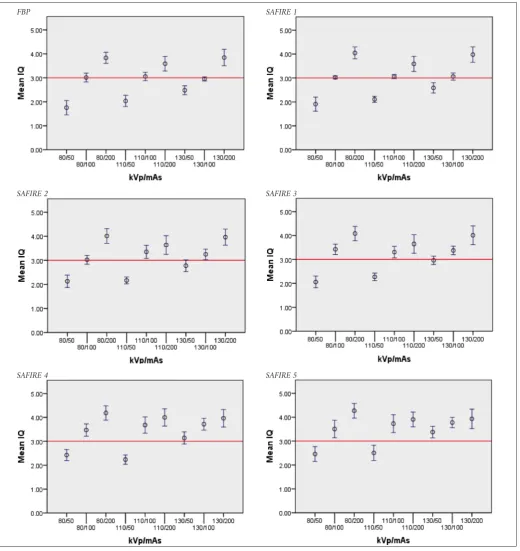
Linear regression was calculated with respect to both sharpness and contrast. The outcomes of the linear regres-sion are shown in Figures 1 and 2.
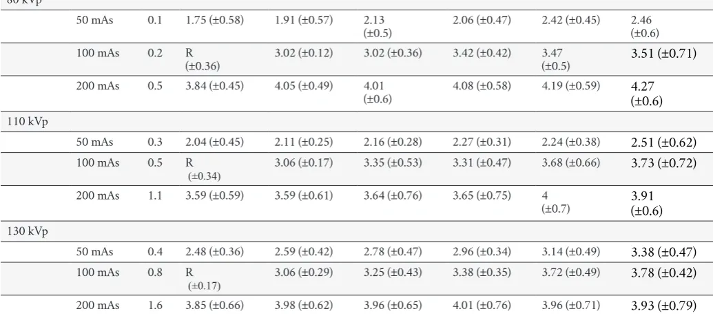
An equation to calculate the effect of the dose on image quality for FBP- and all of the SAFIRE-reconstructions was formed using the B-value coefficients. This correlation is shown by Figures 1 and 2. It can be seen that FBP always required a higher dose to achieve the same image quality as SAFIRE. Relating to contrast, an image quality score of 4 required a dose of 3.4 mSv for SAFIRE 5 reconstruction. FBP required a dose of 6.8 mSv. For sharpness-related image quality, a rating of 4 using SAFIRE 5 required 4.4 mSv whereas FBP needed 6.4 mSv.
Table 3 shows the dose reduction potential while
maintaining an image quality of 4 for all reconstruction techniques.
Image noise
The visual ratings regarding image noise are shown in Table 4. Percentages include noise ratings of average and less than average (scores of 2 and 3). The table shows that for 80 kV and 1.2 mSv, 100% of observers evaluated SAFIRE 5 images to have acceptable noise levels. In comparison, only 55% of observers rated FBP reconstructed images as accept-able. It can also be seen that slice thickness generally affects the amount of noise in the images. When comparing FBP images with 80 kV and 2.4 mSv, the acceptance level raised by 45% between 1 and 3 mm.
[image:18.595.101.558.66.437.2]110 kV greatly improves noise ratings in comparison to 80 kV. The difference between FBP and all strengths of SAFIRE is almost non-existent at this voltage. All 3 mm images were rated to have acceptable noise by at least 90% of observers.
Table 1: Statistical significance of SAFIRE for sharpness
Figure 1: linear regression for sharpness for SAFIRE and FBP.
Clinical acceptability
The visual ratings regarding the clinical acceptability are shown in table 5.
It appears that SAFIRE and FBP are equally accepted at
110 kV with doses of 3 and 6 mSv. For 80 kV, SAFIRE con-sistently has a higher percentage of acceptance than FBP, particularly for the higher doses. Higher strengths of SAFIRE are also generally more clinically acceptable for lower doses, but SAFIRE strengths 2, 3 and 4 also receive good scores for slightly higher doses.
[image:19.595.36.553.329.432.2]Table 3: Dose reduction (mSv) for SAFIRE strengths compared with FBP
Table 4: Percentage of observers who scored noise as acceptable or better
D I S C U S S I O N
It is suggested that SAFIRE 5 can provide a dose reduc-tion of 50% in comparison to FBP, as rated according to image contrast. For image sharpness, the dose reduction is smaller but still significant and is approximately 30%.
Literature suggests that SAFIRE strengths 3 and 4 are best for image quality11-13. Our results suggest that SAFIRE 5 is optimal
for dose reduction while maintaining image quality. The reason for SAFIRE 5 being optimal could be due to the phantom being child-sized. Research is limited regarding all SAFIRE strengths for paediatric patients and so it is difficult to compare.
Dose reductions for all SAFIRE strengths are shown in Table 3. It shows that dose can be reduced by 0.9 to 3.4 mSv, depending on the strength of SAFIRE used. SAFIRE 5 always has the greatest dose reduction, and also has the best rated clinical acceptance for almost all doses.
Previous studies suggested a potential dose reduction from 15 to 85% (14), depending on parameters, patient size
and SAFIRE strength. However, most studies tended to fall in the region of around 50%6,12-13,15-17. Our estimated dose
reduction for SAFIRE 5 is approximately 50%, which agrees with other studies.
A linear model was used for data analysis due to meas-ured dose values suggesting a linear trend. For 80 kV it was found that the effective doses were 2.4, 1.2 and 0.6 mSv for 100, 50 and 25 mAs respectively; this shows that although the overall trend might not be linear, for the window of data we were considering it was almost perfectly linear. This trend continued up to our highest measured dose value of 6.1 mSv. In reality, the dose and image quality relationship is not linear, it is asymptotic.
Clinical acceptability was higher for 3 mm image slices than for 1 mm slices, and increased as the SAFIRE strength increased. The higher voltage also had better acceptability overall. 3 mm images contain more data than 1 mm images which allows for greater noise reduction during reconstruc-tion. This might not be true for a clinical CT scan because there is a possibility that pathologies and anatomical struc-tures might be overlooked.
For Tables 4 and 5, percentages that end with 5, for example 85% and 95%, suggest that one or more observers rated images differently during the test and re-test. This could be due to user error or could be a sign of decreased intra-observer reliability. Observers may have rated images differently the second time due to being more acquainted with the image rating procedure. Further research is sug-gested to investigate this phenomenon.
Noise decreased with increasing dose, and there was less noise in the 110 kV datasets than in the 80 kV images. All of the SAFIRE strengths had acceptable levels of noise for doses of 2.4 mSv and above. For the lower doses at 80 kV, the higher strengths of SAFIRE performed better. SAFIRE 5 received a
90% acceptable noise rating for every dose except 0.6 mSv at 80 kV with 1 mm thickness. SAFIRE 4 was also suitable for most doses, and received a percentage score of 90 and above, excluding 0.6 mSv at 80 kV for 1 mm and 3 mm thicknesses.
Objective data was analysed and then disregarded, based on the fact that results were inconsistent. This is potentially due to the field of view in the received images differing. Changes in the field of view led to the ROI moving and changing in size. This lead to different amount of pixels being included which caused anomalous data.
Visual noise rating was evaluated using a three point scale. Linear regression is invalid for a three point scale, meaning that it could not be used during the analysis of signal to noise ratio. It is expected that dose reduction could be calculated if the linear regression was used.
Further research could utilise more observers, or observ-ers with a higher level of experience. Also, real clinical images could be utilised instead of a phantom; lack of anatomical structures makes the evaluation of the images less realistic.
C O N C L U S I O N
Dose reduction increased with higher SAFIRE strengths. SAFIRE 5 was optimal and estimated to have dose reduc-tions of approximately 30% and 50% relating to sharpness and contrast image quality respectively. SAFIRE was most effective for dose reduction at lower kV.
A C K N O W L E D G E M E N T S
The authors wish to thank Erasmus for funding this project and Siemens for providing image data. We also wish to thank W. Schaake, R. Visser and P. Hogg for helping analyse our data. Finally, we would like to thank the observ-ers who took the time to participate in this research.
1. Pearce MS, Salotti JA, Little MP, McHugh K, Lee C, Kim KP, et al. Radiation exposure from CT scans in childhood and sub-sequent risk of leukaemia and brain tumours: a retrospective cohort study. Lancet. 2012;380(9840):499-505.
2. Brenner D, Elliston C, Hall E, Berdon W. Estimated risks of radiation-induced fatal cancer from pediatric CT. AJR Am J Roentgenol. 2001;176(2):289-96.
3. Rehani MM. Are children more sensitive to radiation than adults? Eur Soc Radiol News [Internet]. 2013 [cited 2014 Aug 11];(Sep). Available from: http://www.myesr.org/html/img/ pool/Radiation_Protection_ESR_Work_Sept_2013.pdf
4. Lee SW, Kim Y, Shim SS, Lee JK, Lee SJ, Ryu YJ, et al. Image quality assessment of ultra low-dose chest CT using sinogram-affirmed iterative reconstruction. Eur Radiol.
2014;24(4):817-26.
5. Grant K, Raupach R. SAFIRE: Sinogram Affirmed Iterative Reconstruction [Internet]. Siemens; 2012 [cited 2014 Aug 11]. Available from: https://www.healthcare.siemens.com/comput-ed-tomography/options-upgrades/clinical-applications/safire
6. Moscariello A, Takx RA, Shoepf UJ, Renker M, Zwerner PL, et al. Coronary CT angiography: image quality, diagnos-tic accuracy, and potential for radiation dose reduction using a novel iterative image reconstruction technique - compar-ison with traditional filtered back projection. Eur Radiol. 2011;21(10):2130-8.
7. Wang H, Tan B, Zhao B, Liang C, Xu Z. Raw-data-based itera-tive reconstruction versus filtered back projection: image quality of low-dose chest computed tomography examinations in 87 patients. Clin Imaging. 2013;37(6):1024-32.
8. Beister M, Kolditz D, Kalender WA. Iterative reconstruction methods in X-ray CT. Phys Med. 2012;28(2):94-108.
9. CIRS. ATOM dosimetry verification phantoms [Inter-net]. Meditron; 2013 [cited 2014 Aug 14]. Available from: http://www.meditron.ch/medical-imaging/index.php/ quality-assurance/phantoms/anthropomorphic-phantoms/ product/60-atom-dosimetry-verification-phantoms
10. Bredenhöller C, Feuerlein U. SOMATOM sensation 10/16 spplication guide: protocols, principles, helpful hints [Internet]. Siemens; 2005. Available from: http://www.healthcare.siemens. com/siemens_hwem-hwem_ssxa_websites-context-root/ wcm/idc/siemens_hwem-hwem_ssxa_websites-context-root/ wcm/idc/groups/public/@global/@imaging/@ct/documents/ download/mdaw/mje1/~edisp/applicationguide_sensa-tion16_01-00209688.pdf
11. Kim JH, Kim MJ, Kim HY, Lee MJ. Radiation dose reduction and image quality in pediatric abdominal CT with kVp and mAs modulation and an iterative reconstruction technique. Clin Imaging. 2014;38(5):710-4.
12. Yang WJ, Yan FH, Liu B, Pang LF, Hou L, Zhang H, et al. Can sinogram-affirmed iterative (SAFIRE) reconstruction improve imaging quality on low-dose lung CT screening compared with traditional filtered back projection (FBP) reconstruction? J Comput Assist Tomogr. 2013;37(2):301-5.
13. Baumueller S, Winklehner A, Karlo C, Goetti R, Flohr T, Russi EW, et al. Low-dose CT of the lung: potential value of iterative reconstructions. Eur Radiol. 2012;22(12):2597-606.
14. Schulz B, Beeres M, Bodelle B, Bauer R, Al-Butmeh F, Thal-hammer A, et al. Performance of iterative image reconstruction in CT of the paranasal sinuses: a phantom study. AJNR Am J Neuroradiol. 2013;34(5):1072-6.
15. Schindera ST, Odedra D, Raza SA, Kim TK, Jang HJ, Szucs-Farkas Z, et al. Iterative reconstruction algorithm for CT: can radiation dose be decreased while low-contrast detectability is preserved? Radiology. 2013;269(2):511-8.
16. Kalmar PI, Quehenberger F, Steiner J, Lutfi A, Bohlsen D, Talakic E, et al. The impact of iterative reconstruction on image quality and radiation dose in thoracic and abdominal CT. Eur J Radiol. 2014;83(8):1416-20.
Review article – The impact of Sinogram-Affirmed Iterative
Reconstruction on patient dose and image quality compared
to filtered back projection: a narrative review
Abdulfatah Ahmed
a, André Garcia
b, Astrid Bakker
c, David Tomkinson
a, Julie Salamin
d, René de Lange
c,
Sergey A. Buyvidovich
e, Tina Sohrabi
e, Alexandre Dominguez
d, Cosmin Campeanu
d, Paul Plasman
ca) School of Health Sciences, University of Salford, Manchester, United Kingdom b) Lisbon School of Health Technology (ESTeSL), Polytechnic Institute of Lisbon, Portugal
c) Department of Medical Imaging and Radiation Therapy, Hanze University of Applied Sciences, Groningen, The Netherlands d) Haute École de Santé Vaud – Filière TRM, University of Applied Sciences and Arts of Western Switzerland, Lausanne, Switzerland e) Department of Life Sciences and Health, Radiography, Oslo and Akershus University College of Applied Sciences, Oslo, Norway
K E Y W O R D S
Comparison
Filtered back projection Sinogram-affirmed iterative reconstruction
Dose reduction Paediatric CT
Computed tomography Image quality
A B S T R A C T
Objective: Summarize all relevant findings in published literature regarding the potential dose reduction related to image quality using Sinogram-Affirmed Iterative Reconstruction (SAFIRE) compared to Filtered Back Projection (FBP).
Background: Computed Tomography (CT) is one of the most used radiographic modalities in clinical practice providing high spatial and contrast resolution. However it also delivers a relatively high radiation dose to the patient. Reconstructing raw-data using Iterative Reconstruction (IR) algorithms has the potential to iteratively reduce image noise while maintaining or improving image quality of low dose standard FBP reconstructions. Nevertheless, long reconstruction times made IR unpractical for clinical use until recently.
Siemens Medical developed a new IR algorithm called SAFIRE, which uses up to 5 different strength levels, and poses an alternative to the conventional IR with a significant reconstruction time reduction. Methods: MEDLINE, ScienceDirect and CINAHL databases were used for gathering literature. Eleven articles were included in this review (from 2012 to July 2014).
Discussion: This narrative review summarizes the results of eleven articles (using studies on both patients and phantoms) and describes SAFIRE strengths for noise reduction in low dose acquisitions while providing acceptable image quality.
Conclusion: Even though the results differ slightly, the literature gathered for this review suggests that the dose in current CT protocols can be reduced at least 50% while maintaining or improving image quality. There is however a lack of literature concerning paediatric population (with increased radiation sensitivity). Further studies should also assess the impact of SAFIRE on diagnostic accuracy.
I N T R O D U C T I O N
CT is one of the most used radiographic modalities in clinical practice but it also comes with a significant radia-tion dose to patients. Consequently, this research focused on dose reduction, particularly for paediatric examina-tions. These patients are more susceptible to long-term effects of radiation exposure, with higher potential for an increased lifetime risk of malignancy. Filtered back projection (FBP) is the standard reconstruction algorithm.
However IT developments in recent years permit itera-tive image reconstruction (IR) to become compatible with routine clinical practice.
in several validation loops. After the first correction loop, the result is compared with the original raw-data and an updated image is generated for the next iteration leading to further noise reduction. Where IR only uses a single correc-tion loop, SAFIRE uses up to 5 correccorrec-tion loops to further decrease image noise1. The level of noise reduction and noise
texture varies with SAFIRE strength for each reconstruction. SAFIRE strength does not translate the number of iterations and does not affect reconstruction time2.
The purpose of this review article is to summarize the current research comparing SAFIRE and FBP. It inves-tigates image quality and the potential of dose reduction provided by SAFIRE, compared to FBP. Data from articles are discussed bearing in mind SAFIRE’s potential for dose reduction while maintaining diagnostic image quality.
D A T A S O U R C E S A N D S E A R C H E S
MEDLINE, ScienceDirect and CINAHL data bases were searched, using the following key words: comparison, filtered back projection, sinogram-affirmed iterative reconstruction, dose reduction, paediatric CT, computed tomography, image quality. The research equation was: (Computed tomography AND sinogram-affirmed iterative reconstruction AND radi -ation dose AND image quality AND filtered back projection) NOT (contrast media). We excluded articles concerning previ-ous generation iterative reconstruction algorithms and articles focusing on cardiac CT on obese patients because of the dif-ference of size between head examination and those patients.
Eleven articles were included in our review article, dating from 2012 to 2014, for examinations of chest, abdomen, head and cardiac on anthropomorphic phantoms and adult or pae-diatric patients.
M A T E R I A L S A N D M E T H O D S
Patients/phantoms
Data came from CT scans performed on patients and phan-toms. Patients were mainly adults but some studies focused on paediatric protocols. Scans were performed on physical and anthropomorphic phantoms (chest, head). One study used data from both patients and phantom scans for comparison.
Paediatric vs adult protocols
Three articles focused on paediatric examinations,
“paediatric” denomination including children from 0 to 18 years old. Two explored cardiac CT and one abdomen. What mainly differs from adult studies were tube voltage (70 to 100-120 kVp) and tube current (lower mAs). Both were gen-erally adapted to weight, size and age.
Data acquisition
Since all of the data-sets acquired in these studies had to be reconstructed with the SAFIRE algorithm, almost all exams were performed on the dual-source CT scanner Somatom Definition Flash from Siemens. Filtered back projections were sometimes acquired on other Siemens equipments.
The range of tube voltage explored was usually 100kVp and 120 kVp, sometimes also 80 kVp for ultra low doses. Tube current was variable, either fixed (at 25, 50 and 100 mAs or percentage reduction) or automatically modulated.
Images reconstruction
Acquisitions were reconstructed with FBP and SAFIRE. For SAFIRE, either all strengths (S1-S5) were explored or median strength like strengths S2 to S4 or S3 (recommended by manufacturer)3.
Usually images were reconstructed with a medium smooth kernel or smooth and sharp kernels to compare changes in image quality.
Image quality analysis
For the physics analysis of image quality, noise and Sig-nal-to-Noise Ratio (SNR) were the main criteria calculated. Contrast and Contrast-to-Noise Ratio (CNR) were less often measured. Only one study on phantoms went further by examining the Noise Power Spectrum (NPS), the spatial resolution, the linearity and accuracy of CT numbers.
For visual analysis, in most of the articles, the images were analysed by at least two radiologists with 3 years experience or more in a specific radiological field. Further details about the method of image analysis were often not provided. Visual criteria generally considered image noise (e.g., graininess), quality of contour delineation (i.e., sharpness) and general impression (i.e., overall image quality). Han et al. (2012)4 referred to European Image Quality Assessment (i.e., sharp-ness, noise, noise texture, diagnostic confidence).
details needed (e.g., level 1: lack of vessel wall definition due to marked motion artefact, poor vessel opacification, promi -nent structural discontinuity, or high image noise rendering the segment non-diagnostic).
R E S U L T S
Chest/thorax
Christe et al. (2013)6 conclude that while using SAFIRE
instead of FBP it was possible to achieve a dose reduction of 30, 52 and 80% for bone, soft tissue and air, respectively. Image quality was verified objectively using signal, noise and contrast measurements. With the same radiation dose, an average of 34% more CNR was achieved by changing respec-tively from FBP to SAFIRE. For the same CNR, an average of 59% dose reduction was produced for SAFIRE. The visual classification was given by two radiologists. For the same visual image quality, the dose could be reduced by 25% using SAFIRE. This study only used SAFIRE S3.
Wang et al. (2013)7 explored SAFIRE strengths S2-S4 after
excluding the extremes (S1 and S5), as they were considered to be, respectively, too “noisy” and too “smooth”. The results of this study suggests there was no significant difference in the objective noise and SNR on mediastinal images between full-dose (FD) images reconstructed with FBP and half-dose images reconstructed with SAFIRE. But, on lung images, noise was significantly lower and SNR was significantly higher in half-dose images reconstructed with SAFIRE. Subjective image noise was similar on mediastinal and lung images with half-dose SAFIRE and full-dose FBP reconstruction.
Amongst all strengths, SAFIRE S3 had the best results for physics and visual image quality. Authors conclude that, compared to full-dose CT images reconstructed with the con-ventional FBP algorithm, SAFIRE with three iterations could provide similar or better image quality at 50% less dose.
Ghetti et al. (2013)10 explored image quality using 3
phan-toms. Noise was analysed on images reconstructed with all 5 SAFIRE strengths and a conventional medium-smooth kernel. Additionally, on images with strength SAFIRE S3, different kernels were selected to evaluate a possible differ-ence in noise reduction due to the filter applied. For the same dose, noise reduction of iterative reconstruction increases with the SAFIRE strength applied in a proportional way.
CT number accuracy and linearity were verified to assess SAFIRE reconstructions influences on them. The different
SAFIRE strengths did not change mean CT values and showed no considerable differences from values obtained with FBP.
Images were reconstructed with three different levels of SAFIRE strength (S1, S3, S5) and FBP at 3 different dose levels. CNR was measured for all images. CNR is always greater for SAFIRE and it increases with the strength of SAFIRE applied. But there is no evidence of a significant difference between the different filters in the SAFIRE outcomes. The spatial resolution was measured through different modules with two dose levels (at 120 kVp). Image texture changes increased with SAFIRE strength, resulting in an overall image quality improvement. Detail edge is sharper with less background noise using SAFIRE.
Abdomen
Greffier et al. (2013)8 analysed the data from 10 patients who had a normal dose abdominal CT and who then under-went a second CT scan examination. The first sequence was acquired with 30% less mAs than the original CT and the second acquisition with 70% less mAs. The raw-data of the two scans was reconstructed with FBP and SAFIRE (S1-S5) and medium kernel.
Physics analysis concluded there was no significant dif -ference in the measured signal when using FBP and SAFIRE. Noise significantly decreased (11% between FBP and SAFIRE 1) with SNR and CNR increase after each iteration. Good image quality was obtained with 30% less dose by using SAFIRE S2. Furthermore by using S5, it was possible to achieve up to 70% dose reduction while still maintaining image quality.
In the work of Kim et al. (2014)9, a first group of paedi
-atric abdominal patients was scanned with kVp and mAs modulation. Raw-data was reconstructed using SAFIRE (S2-S4). A second group of patients underwent the same exam in emergency room on a CT scanner with only mAs modula-tion and the raw-data was reconstructed with FBP. Physics and visual analysis of image quality showed that SAFIRE was able to achieve an average 64.2% in dose reduction compared to the control group with FBP. The objective image noise of the SAFIRE S2 and S3 was comparable to that of the control group. For visual image quality analysis, SAFIRE S2 and S3 showed better image quality than the control group in terms of diagnostic acceptability. Moreover, strength S3 scored better in terms of subjective image quality compared to S2.
Head
Schulz et al. (2013)1 worked on data from a phantom
image was reconstructed using two different kernels with FBP and SAFIRE (S1-S5) algorithms. Image noise was evaluated and showed that compared to FBP, all iterative reconstruction techniques reduced the noise by 15%-85% depending on the iterative strength, rendering kernel, and dose parameters. Visual image quality was evalu-ated on images acquired at tube currents of 100% (FBP), 50% (SAFIRE), and 25% (SAFIRE). Visual evaluation of the images suggested that FBP images at full dose were preferred to 50% dose SAFIRE reconstruction. Their con-clusion was that SAFIRE has a potential in CTs exam since even slight increase in iteration can yield important noise reduction.
Corcuera-Solano et al. (2014)11 aimed to assess dose
reduction for patients in the neurosurgical intensive care unit who undergo multiple head CT scans. While maintain-ing similar image quality and SNR levels, ultra-low-dose CT (ULDCT) reconstructed with SAFIRE represented a 68% lower CTDIvol compared to standard-dose CT (SDCT) with FBP technique in the same patients. SAFIRE recon-struction low-dose CT (LDCT) offered higher image quality than FBP standard-dose CT with no differences in SNR at a 24% lower CTDIvol. Compared with LDCT, ULDCT had significantly lower SNR but demonstrated clinically satisfactory measures of image quality. In visual analyses, there were no major differences in quality between ULDCT and SDCT.
Korn et al. (2013)12 described an increase of 47% in
CNR when using SAFIRE reconstruction instead of FBP in reduced-dose examination, because the degradation of image quality at lower dose was more than compensated by SAFIRE. Through objective measurements of image sharp-ness, they found that it was similar for FBP and SAFIRE reconstructions. Compared with FBP standard-dose (320 mAs) reconstructions, low-dose (255 mAs) SAFIRE recon-structions also allowed for an improvement in visual grading of noise as well as overall image quality.
Authors concluded that with 20% dose reduction, recon-struction of head CT by SAFIRE provides above standard objective and subjective image quality.
Cardiac
Han et al. (2012)4 evaluated the impact of SAFIRE on
image quality in paediatric cardiac CT datasets. From a visual point of view, no change was observed in spatial resolution, sharpness improved in 9% of cases, image noise in 63% cases and noise texture in 85% cases when using SAFIRE. The diagnostic confidence was similar in both groups. The improvement and reduction of noise was similar for helical and axial acquisition techniques. Visual image quality anal-ysis resulted on a lower contrast from 1% for SAFIRE but clinically not significant, noise decreased (34%) and CNR (41%) and SNR (56%) increased with SAFIRE.
Wang et al. (2013)5 analysed images from patients and
phantoms. Data from dual source equipment was recon-structed using FBP and data from single source was reconstructed with SAFIRE and FBP, to assess image quality with only half dose. Images from the phantom suggested that noise proportionally decreased as current increased. No significant difference in SNR and noise was found between full-dose FBP and half-dose SAFIRE neither for phantom nor patients. Similar visual results between full-dose FBP and half-dose SAFIRE were performed in visualising coronary segments. For half-dose FBP, significantly fewer segments were visible. It suggested that with an estimated dose reduc-tion of 50%, there was no significant difference in noise, SNR and overall image quality with SAFIRE reconstruction com-pared to full-dose standard protocol reconstructed with FBP.
Nie et al. (2014)3 evaluated the impact of SAFIRE on
image quality for a tube voltage of 70 kVp. The mean scores of visual analysis were significantly higher with SAFIRE algorithm than with FBP algorithm regarding to graininess, sharpness and overall image quality. Noise was lower and SNR and CNR significantly higher with SAFIRE. Radiolo -gists evaluated the diagnostic accuracy. SAFIRE scored better than FBP algorithm but no significant difference in diagnos -tic accuracy between FBP and SAFIRE was found (p > 0.05).
Authors Part of body
examinated SAFIRE Strength
Dose reduction Image quality results
Christe et al.
(2013)6 Chest S3
80% at same noise 45% at same SNR 59% at same CNR 25% at same subjective IQ
-44 % noise, +36 % SNR, +34 % CNR with SAFIRE Better subjective IQ for SAFIRE with same dose
Wang et al.
(2013)7 Chest (low dose) S3 similar IQ with FBP 100% dose and SAFIRE 50% dose Full-dose FBP noise comparable to half-dose SAFIRESubjective IQ evaluation in noise, SNR and lesion detection comparable with full-dose FBP or half-dose SAFIRE
Ghetti et al.
(2013)10 Chest,Water, Catphan 600 and
3D phantom
S1-S5 S1,S3,S5
Unique dose of 13.4 mGy tested for noise
Doses tested for CNR : 20.2, 13.4 and 6.7 mGy
Up to 60% noise reduction with SAFIRE 5 for 2mm slices with same dose
Noise decreases and CNR increases when SAFIRE strength rises
Greffier et al.
(2013)8 Abdomen S1-S5 Dose reduced at 30% and 70% from full dose SNR and CNR improved with the increase in SAFIRE levels
Kim et
al.(2014)9 Abdomen (paediatric) S2,S3,S4 64.2% average dose reduction for similar image quality with SAFIRE Noise decreases and IQ increases with SAFIRE strengths No significant difference between SAFIRE S4 and FBP
Schulz et al.
(2013)1 Head:paranasal sinuses
S1-S5 100% FBP, 50% SAFIRE, 25%
SAFIRE Image noise always greater with FBPWith 25% dose, mean noise reduction 47.5% for 3mm and 49.4% for 1mm slices with SAFIRE
Best IQ with 100% dose level with FBP
Corcuera-Solano et al. (2014)11
Head S3 ULDCT 68% dose reduction
LDCT 24% dose reduction Image quality similar with full dose FBP and LDCT reconstructed with SAFIRE S3
Korn et al.
(2013)12 Head S3 20% dose reduction + 48% SNR, + 47% CNR with SAFIRE for same doseSimilar sharpness IQ SAFIRE scored better than FBP
Han et al.
(2012)4 Cardiac - - - 34% noise, + 56% SNR, + 41% CNR using SAFIRE vs FBP using the same dose Wang et al.
(2013)5 CardiacWater phantom
- Simulating a 50% radiation dose
reduction No significant noise and SNR difference and equivalent image quality between full dose FBP and half dose SAFIRE
Nie et al.
(2014)3 Cardiac S3 Same dose 70 kVp Significantly lower image noiseSignificantly higher SNR and CNR for SAFIRE Higher scores for subjective IQ with same dose
D I S C U S S I O N
Although specific values differ from one study to another, all studies concluded that SAFIRE allows for a significant dose reduction, while maintaining adequate image quality. Nevertheless some limitations were identified.
The studies included in this review used different param-eters to measure image quality. There was no standard way in how both physical and visual image quality was meas-ured. Different sizes of ROI’s and different Likert scales were used. Furthermore, not all articles assessed both physical and visual image qualities.
The studies assessing visual image quality only used two radiologists as observers. In order to reduce observer bias, a larger group is needed. Monitor characteristics and display parameters were completely missing as well as the visual acuity performance of the observers.
The images were only classified according to their diag -nostic or visual quality, but not their diag-nostic accuracy. More studies must be done regarding if SAFIRE provides better diagnostic accuracy than FBP.
[image:26.595.41.561.94.551.2]In some studies the image sets were acquired using dif-ferent equipment for FBP and SAFIRE reconstructions.