comm
en
t
re
v
ie
w
s
re
ports
de
p
o
si
te
d r
e
se
a
rch
refer
e
e
d
re
sear
ch
interacti
o
ns
inf
o
rmation
Evolution of selenium utilization traits
Héctor Romero
*†
, Yan Zhang
‡
, Vadim N Gladyshev
‡
and Gustavo Salinas
§
Addresses: *Laboratorio de Organización y Evolución del Genoma, Dpto. de Biología Celular y Molecular, Instituto de Biología, Facultad de
Ciencias, Iguá 4225, Montevideo, CP 11400, Uruguay. †Escuela Universitaria de Tecnología Médica, Facultad de Medicina, Piso 3 Hospital de
Clínicas, Avda. Italia s/n, Montevideo, CP 11600, Uruguay. ‡Department of Biochemistry, University of Nebraska, Lincoln, NE 68588-0664,
USA. §Cátedra de Inmunología, Facultad de Química/Ciencias, Instituto de Higiene, Avda. A. Navarro 3051, Montevideo, CP 11600, Uruguay.
Correspondence: Gustavo Salinas. E-mail: [email protected]
© 2005 Romero et al.; licensee BioMed Central Ltd.
This is an Open Access article distributed under the terms of the Creative Commons Attribution License (http://creativecommons.org/licenses/by/2.0), which permits unrestricted use, distribution, and reproduction in any medium, provided the original work is properly cited.
Evolution of selenium utilization traits
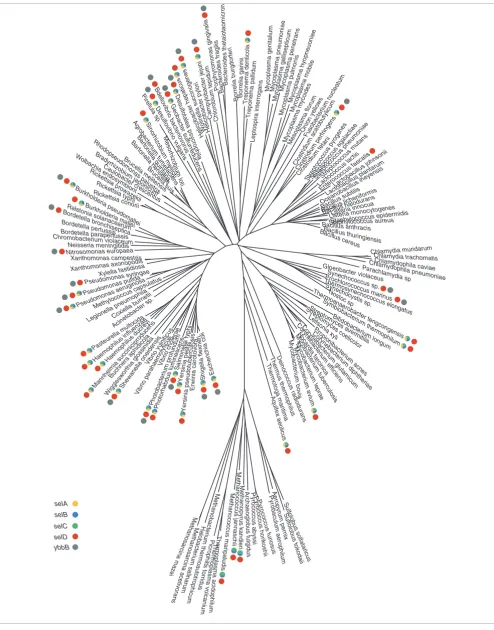
<p>Completely sequenced genomes were analyzed for occurrence of <it>SelA</it>, <it>B</it>, <it>C</it>, <it>D </it>and <it>ybbB </ it>genes. <it>SelB </it>and <it>SelC </it>were found to be signatures for the Sec decoding trait, while <it>SelD </it>defines the overall selenium utilization.</p>
Abstract
Background: The essential trace element selenium is used in a wide variety of biological processes. Selenocysteine (Sec), the 21st amino acid, is co-translationally incorporated into a
restricted set of proteins. It is encoded by an UGA codon with the help of tRNASec (SelC),
Sec-specific elongation factor (SelB) and a cis-acting mRNA structure (SECIS element). In addition, Sec synthase (SelA) and selenophosphate synthetase (SelD) are involved in the biosynthesis of Sec on
the tRNASec. Selenium is also found in the form of 2-selenouridine, a modified base present in the
wobble position of certain tRNAs, whose synthesis is catalyzed by YbbB using selenophosphate as a precursor.
Results: We analyzed completely sequenced genomes for occurrence of the selA, B, C, D and ybbB genes. We found that selB and selC are gene signatures for the Sec-decoding trait. However, selD is also present in organisms that do not utilize Sec, and shows association with either selA, B, C and/ or ybbB. Thus, selD defines the overall selenium utilization. A global species map of Sec-decoding and 2-selenouridine synthesis traits is provided based on the presence/absence pattern of selenium-utilization genes. The phylogenies of these genes were inferred and compared to organismal phylogenies, which identified horizontal gene transfer (HGT) events involving both traits.
Conclusion: These results provide evidence for the ancient origin of these traits, their independent maintenance, and a highly dynamic evolutionary process that can be explained as the result of speciation, differential gene loss and HGT. The latter demonstrated that the loss of these traits is not irreversible as previously thought.
Background
Selenium (Se) is an essential trace element for numerous organisms that belong to the three domains of life. The most relevant biological form of selenium is the rare amino acid selenocysteine (Sec), the selenium analog of cysteine (Cys). Sec is co-translationally incorporated into protein [1-3]. In
functionally characterized selenoproteins, Sec is the catalytic group in the active site and is directly involved in redox catal-ysis. It is thought that Sec confers a functional advantage over cysteine at these active sites, increasing the catalytic effi-ciency of the enzymes [4]. Despite this selective advantage, the set of selenoproteins in any given organism is small [5,6]. Published: 27 July 2005
Genome Biology 2005, 6:R66 (doi:10.1186/gb-2005-6-8-r66)
Received: 20 April 2005 Revised: 7 June 2005 Accepted: 27 June 2005 The electronic version of this article is the complete one and can be
Sec is inserted into selenoproteins at in-frame UGA codons
(usually termination codons) by tRNASec (SelC) [2,7].
Inter-pretation of UGA as Sec requires translational reprogram-ming, which is provided by the Sec insertion sequence (SECIS) element, a cis-acting stem-loop structure present in the selenoprotein mRNA [2]. The decoding of Sec in bacteria also involves a Sec-specific elongation factor (SelB) which
binds GTP, the SECIS element and the tRNASec [8,9]. In
eukaryotes, this function is carried out by two proteins: EF-Sec and SECIS-binding protein (SBP2). EF-EF-Sec is a EF- Sec-spe-cific elongation factor, distantly related to bacterial SelB, that
binds GTP, tRNASec and SBP2; this latter protein, in turn,
binds the SECIS element [10]. Sec synthesis is the other part of the metabolic pathway required for biosynthesis of
seleno-proteins. It takes place on tRNASec, which is first
aminoa-cylated with serine (by a canonical seryl-tRNA synthetase) and then modified to selenocysteinyl-tRNA, in the reaction that uses selenophosphate as the selenium donor [9]. In the Bacteria, this reaction is catalyzed by Sec synthase (SelA). The functional equivalent of SelA in Archaea and Eukarya has not
been described. A phosphoseryl-tRNASec kinase (PSTK) has
been recently identified only in eukaryotic and archaeal Sec-incorporating organisms [11]. It has been suggested that this protein can play a role in Sec biosynthesis and/or regulation. The synthesis of selenophosphate is catalyzed by selenophos-phate synthetase (SelD) from ATP and selenide in both prokaryotes and eukaryotes.
Selenophosphate has also been described as a precursor for the last step of the synthesis of the modified base 5-methyl-aminomethyl-2-selenouridine in the wobble position of the anticodons of Lys, Glu and Gln tRNAs [12], and this reaction was reported to be catalyzed by YbbB in Escherichia coli [13]. The function of this modified base is not known.
Thus, considerable efforts in recent years have been made to elucidate molecular details of Sec decoding in the three domains of life. In addition, the selenoproteome of several species has been the subject of intensive research [5,6,14-16]. Despite this progress, fundamental issues relating to the evo-lution of Sec utilization remain unclear. On the basis of the complexity and similarity of the Sec-insertion mechanisms in
different organisms, it has been proposed that the Sec-decod-ing trait arose once, before the division of the three domains of life, and was subsequently lost in some lineages. It is also thought that once an organism has lost the Sec-insertion sys-tem, it cannot re-emerge. Whether the Sec biosynthesis and insertion pathway evolved before the fixation of the genetic code or whether this was a late addition is not known [17-19].
Here we provide a map of Sec-incorporating and selenourid-ine-utilizing organisms within the tree of life, based on the analysis of completely sequenced genomes. From phyloge-netic analysis of all components of the Sec-decoding machin-ery, we present clear evidence for the loss of the trait in many lineages at different taxonomic levels, and examples of acqui-sition of the trait by horizontal gene transfer (HGT). In addi-tion, we describe and explain the maintenance of selenophosphate synthetase in non-Sec-incorporating organ-isms, and use this information to define a selenouridine-utili-zation trait as well as a general selenium-utiliselenouridine-utili-zation trait. We find that the 2-selenouridine pathway can also be acquired by HGT. These data suggest that the loss of selenium utilization is not irreversible.
Results
A map of selenium utilization within the tree of life
Figure 1 displays a phylogenetic tree, based on rRNA, of the 155 species whose entire genomes have been sequenced (see Materials and methods for the rationale behind the use of rRNA and other alternatives). The criteria for the occurrence of the Sec-decoding trait included the presence of known genes involved in Sec decoding (that is, selA, selB, selC, selD), and at least one gene encoding a known selenoprotein in the genome, inferred by the presence of a UGA codon within a coding region (at the location corresponding to Cys in homologs) followed by a downstream SECIS element. Using these criteria, a total of 29 bacterial and three archaeal species were found to be Sec decoding. These criteria were in agree-ment with experiagree-mental evidence when available. A map of selA, selB, selC and selD within the species tree is provided in Figure 1.
Distribution of selenium-utilization traits
Figure 1 (see following page)
comm en t re v ie w s re ports refer e e d re sear ch de p o si te d r e se a rch interacti o ns inf o rmation
Figure 1 (see legend on previous page)
E sch e rich ia co li Sh ig e lla fl e x n er i E rw in
ia ca ro tov o ra Ye rsin ia ps eud otub erc u los is Ye rsin
ia p es tis Sa lm on ella
typh i
Ph otor
ha bd
us l um
ine sce
ns
Ph otob
acte rium
pr ofun du m Vib rio ch oler ae Vibr io p
ara haem olyt icu s Vib
rio v
uln
ificu
s
Shew ane
lla on eide
nsis
Wigg lesw
orth ia g
lossi nidi a Buch nera aph idic ola Man nhei mia suc cini cipr oduc ens Haem ophi
lus d
ucrey
i
Haem
ophi
lus in
fluen zae Paste urella mult ocida Acineto bacter sp. Coxie lla bu
rnetii
Legion
ella pn
eumo
phila
Methylo coccus ca
psulatu s
Pseudo monas a
erugino sa
Pseudomon as putida Pseudomona
s syringae Xylella fastid
iosa Xanthomonas axonop
odis
Xanthomonas campestris
Nitrosomonas europaea
Neisseria meningitidis
Chromobacterium violaceumBordetella parapertussis
Bordetella pertussis
Bordetella b
ronch iseptica Ralst onia so lanacearum Bu rkho
lderia m
allei
Burkho
lderia p
seud oma llei Ricke ttsia co norii Ricke
ttsia typ
hi Ric
kettsia pr
ow azek ii Wo lbac hia en dosym biont Ca ulobacte r cr esce ntus Br adyrhi zobium japo nicu m Rh odop seud om on
as pa
lus tris Br ucel la m elite ns is Br uc ella su is Ba rton el
la he nse
lae Ba
rton el
la qu
inta na M eso rh izo bi um lo ti Si no rhizo b ium mel
ilo ti Pi
re llul
a s p D e sul fo vibr io v
ulg a ris Bd e llovib rio b acte rio vo rus G eob ac te r s u lfur red u cens De su lfo ta lea p sycr op h ila H el ic ob ac te r h ep at icu s Wo lin e lla s u c c ino ge ne s He lico b a ct e r p y lo ri Ca m p yl o b a c ter j e ju ni Ch lo ro b ium t e pi d u m Po rp h y romo na s gi n g iv al is B a cte roi d e s f ra g ilis B a c ter oi d es t h e tai o tao mi c ron Bo rr el ia bu rg do rf eri Bo rr e lia ga rin ii T repo ne ma d e n tico la Trep on e ma p allid um Le pto s pira inte
rrog an s M y co p las m a ge nita liu m My co p las ma pn eu mo nia e My cop
las ma
ga llise
pticu m My co plas ma pe ne tran s Myco pla sm a pu
lmo nis Myco pla sma hy op ne uo niae Myco plas
ma mo bile
Myco pla
sm a m
yco ide s Me sop lasm a f
lorum On ion ye llows Fu sob acte rium nu cle atu m Clo stri diu m ac
etob utyl icu m Clo strid ium perfringens Clo strid ium tetani Stre ptoc occ
us p
yog enes Stre ptoc occu s ag alac tiae Stre ptoc occu s p neum onia e Stre ptoco ccu s m utans Lact oco ccus lactis
Ente roco
ccus f aeca
lis
Lactob
acillu
s joh
nsonii
Lactob acillu
s plan tarum
Ocean obac
illus ih eyens
is
Bacill
us su
btilis Bacillus lichenifo rmis Bacillus halodura ns
Listeria innocu a
Listeria monocytogenes Staphylococcus epider
midis Staphylococcus aureus Bacillus th
uringiensis
Bacillus anthracis
Bacillus ce
reus
Chlamydia muridarum Chlamydia trachomatis
Chlamydophila caviae Chlamydophila pne
umoniae
Parach lamydia sp Gloeob
acter violaceus
Synechococcu
s sp.
Thermosyn
echococcu
s elong
atus Syne
chocyst
is sp.
Nost oc sp Th ermo anae robacte
r tengc
ongensis Sy mbiob ac terium therm ophilum Bifidoba cter ium l ongum Strepto myce s a verm itilis St reptom yce s co elicolor Le ifso
nia xyl
i Pr opio nib acte rium a cnes Co ryn ebac terium d ip hth eriae C oryne ba cteri um g lu tam icu m Co ryn eb ac te rium ef ficie ns No ca rdia farcin ica M yco ba ct e rium tu be rcu losis M yco ba ct e riu m lep rae M yco ba ct e riu m a v ium M yco b a ct e riu m bo vis D ein oc occu s r ad iodu ran s Th erm us th ermo ph ilu s Th e rm o toga m a ritim a A qu ife
x ae
ol icus Su lfol ob us s o lfat ar icus Su lfol ob us to ko da ii Ae rop y rum pe rn ix P yrob ac ulu m ae rop h ilum P y roco ccu s fu riosu s Py ro co ccu s h o riko sh ii Py ro co ccu s a b yssi A rc h ae og lo bu
s f
[image:3.612.57.551.84.714.2]Despite the bias among the prokaryotic genomes so far sequenced [20], in which proteobacteria are over-repre-sented and some phyla are not repreover-repre-sented at all, the taxa dis-tribution of Sec incorporation revealed interesting features of this trait. First, the trait is widely distributed and present in numerous bacterial phyla (Proteobacteria, Firmicutes, Spiro-chaetes, Actinobacteria, Aquificae). Second, we observed the presence and absence of the trait in taxa within monophyletic groups. This phenomenon takes place within clades at differ-ent evolutionary levels, namely phylum, class, order, family, genera, and even species and is illustrated in Figure 1. Consid-ering the genus level, we observed this phenomenon within
Pseudomonas, Treponema, Clostridium and Yersinia. The
more revealing case of this absence/presence pattern is that of the KIM, biovar Mediaevalis and CO92 strains of Yersinia pestis. Whereas the KIM strain possesses a functional Sec-decoding machinery, the CO92 and Mediaevalis strains carry
a confirmed sequenced selB pseudogene, whose coding
region is disrupted by a frameshift [21]. Furthermore, the CO92 and the Mediaevalis strains possess selA, selC and selD, indicating that the loss of the ability to decode Sec is very
recent. Moreover, at position 203 of the α-subunit of formate
dehydrogenase type O there is a UGA codon in the three strains, which is decoded as Sec by the KIM strain, but could not be decoded as such in the CO92 and the Mediaevalis strains. The three strains also possess formate dehydrogenase
type H with a Cys-containing α-subunit (fdhH).
SelB and selC can be considered as the gene signature of
organisms able to decode Sec: their presence in genomes
always coincides with that of selA (excluding archaeal and
eukaryote domains), selD, and selenoproteins. A putative ortholog of selA is present in Helicobacter pylori (a Sec-non-incorporating organism). The presence of this protein in the two strains of this species is intriguing and raises the question of whether this protein serves a different function or is just a
remnant of the Sec-decoding machinery. The case of selD is
different, because it is present in several species that lack
other genes necessary for Sec decoding. SelD orthologs are
indicated in red in Figure 1.
Bacteria possessing selD but not the Sec-decoding trait
include Bordetella bronchiseptica, Pseudomonas syringae,
Porphyromonas gingivalis, Nitrosomonas europaea,
Bdell-ovibrio bacteriovorus and Enterococcus faecalis. In
addi-tion, two cyanobacteria - Prochlorococcus marinus and
Synechococcus species - possess a putative selD homolog
with a 320-amino-acid amino-terminal extension with simi-larity to NADH dehydrogenases. All selDs from non-Sec-incorporating bacteria, excluding those of cyanobacteria, are
likely to be 'true orthologs' to selD from Sec-incorporating
bacteria because the topology of the selD phylogeny parallels the topology of species for both Sec-incorporating and non-Sec-incorporating species (Figure 2d), and because the sequence signatures of bacterial selD are present and are of similar length (see Additional data files). Although it is diffi-cult to sketch an evolutionary history for the selD from cyano-bacteria, it is clear that these proteins have many features of selDs and could be viewed as true selenophosphate syn-thetases (see below).
The fact that selenophosphate is also the precursor for the synthesis of 2-selenouridine [12], a modified base that is present at the wobble position of Lys, Glu and Gln tRNAs, suggests that selD may have been maintained in these organ-isms to generate selenophosphate for 2-selenouridine synthe-sis. Thus, we investigated the distribution of ybbB, a gene encoding the catalyst of the last step of 2-selenouridine syn-thesis [13], and its association with selD.
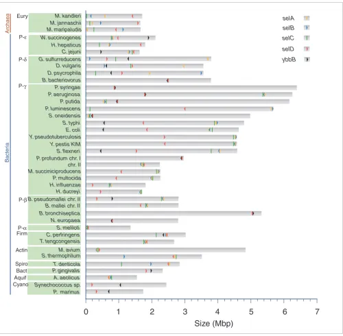
A search across genomes for ybbB (indicated in gray in Figure 1) revealed that six out of seven selD-containing and non-Sec-decoding species also contained ybbB. In addition, all ybbB-containing organisms also possess selD, including cyanobac-teria. Furthermore, in most of these species, except P. gingi-valis and cyanobacteria, both genes are located contiguously and arranged in an operon (Figure 3), as has been previously suggested [13]. This gene organization is also seen in some species that incorporate Sec and possess ybbB: selD is contig-uous to ybbB in some genomes, but is rarely contigcontig-uous to the selA-selB operon (Figure 3).
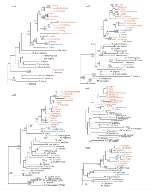
Phylograms of selA, selB, selC, selD and ybbB
Figure 2 (see following page)
comm en t re v ie w s re ports refer e e d re sear ch de p o si te d r e se a rch interacti o ns inf o rmation
Figure 2 (see legend on previous page)
Y. pestis Y. pseudotuberculosis 100 P. luminicum 100 E. coli S. flexeneri 100 S. typhi 100 98 M. succiniciproducens P. multocida 75 H. influenzae 100 H. ducreyi 100 S. oneidensis 100 P. putida P. aeruginosa 100 B. mallei B. pseudomallei 100 60 S. melioti 83 100 M. avium S. thermophilum T. tengcongensis C. perfringens D. vulgaris D. psycrophila G. sulfurreducens P. profundum T. denticola 100 W. succinogenes H. hepaticus 100 C. jejuni 95 A. aeolicus selA 1.00 1.00 1.00 1.00 1.00 1.00 1.00 1.00 1.00 1.00 0.60 1.00 1.00 1.00 1.00 1.00 E. coli S.flexeneri 100/1.00 S. typhi 99 Y. pseudotuberculosis Y. pestis 100 P. luminicum 73 98 S. oneidensis P. multocida H. influenzae M. succiniciproducens 93 H. ducreyi 100 81 B. pseudomallei B. mallei 100 S. meliloti 71 P. putida P. aeruginosa 100 88 M. avium G. sulfurreducens D. psycrophila D. vulgaris 89 T. tengcongensis C. perfringens 81 S. thermophilum W. succinogenes C. jejuni H. hepaticus 100 P. profundum T. denticola 100 A. aelicus 100 H. sapiens D. melanogaster 97 C. elegans 100 M. maripaludis M. jannaschii 100 88 selB 1.00 1.00 1.00 1.00 1.00 1.00 1.00 1.00 1.00 0.99 1.00 0.95 1.00 1.00 1.00 1.00 M. kandleri 1.00 1.00 1.00 1.00 0.99 S. flexneri S. typhi E. coli Y. pestis Y. pseudotuberculosis P. luminicum S. oneidensis M. succiniciproducens H. ducreyi H. influenzae P. multocida P. aeruginosa P. putida B. pseudomallei B. mallei S. meliloti D. vulgaris G. sulfurreducens C. perfringens T. tengcongensis S. termophilus D. psycrophila M. avium T. denticola P. profundum C. jejuni W. succinogens H. hepaticus A. aeolicus M. jannaschii M. kandleri M. maripaludis C. elegans D. melanogaster H. sapiens M. succiniciproducens H. influenzae 73 P. multocida H. ducreyi 96 Y. pseudotuberculosis Y. pestis 100 P. luminicum E. coli S. flexneri S. typhi 97 P. profundum1 72 P. aeruginosa P. putida P. syringae B. bronchiseptica 92 S. oneidensis 96 B. mallei B. pseudomallei 92 S. meliloti 100 M. avium 85 W. succinogenes H. hepaticus 100 C. jejuni A. aeolicus D. psycrophila T. denticola E. faecalis T. tengcongensis C. perfringens P. profundum G. sulfurreducens N. europeae B. bacteriovorus 100 D. vulgaris P. gingivalis S. termophilum 100 M. maripaludis M. jannaschii 100 M. kandleri 79
H. sapiens (SPS1)
D. melanogaster (SPS1) 78
C. elegans 100
100 100
H. sapiens (SPS2)
[image:5.612.54.555.84.718.2]The analysis of selA, selB, selC, selD and ybbB genes also revealed that, within the set of species that incorporate Sec, many, but not all, organisms, possess ybbB and vice versa. In other words, the set of species that incorporate Sec into pro-tein overlaps with, but is different from, the set of species that possess ybbB (Figure 1). It is important to note that a low-identity homolog to bacterial ybbB is present in
Methanococ-cus jannaschii and Methanopyrus kandleri, and absent in
other archaea, suggesting that this base modification might not be unique to bacteria.
Finally, we investigated the presence of additional genes linked to the selenouridine synthesis trait by searching genomes for genes that occur in organisms possessing ybbB and are absent in organisms lacking ybbB. This search did not identify any additional gene associated with this trait. Thus, the overall analysis allows us to corroborate that the two products of these genes form a pathway with 2-selenouridine in the tRNA as the final product. However, only ybbB is the gene signature of this trait. On the other hand, the dual use of selenophosphate (for Sec decoding and 2-selenouridine
bio-synthesis) makes selD a signature of a broader trait of
sele-nium utilization, and our data suggest that both Sec decoding and selenouridine traits are independently maintained, but both require selD.
Phylogeny of selA, selB, selC, selD and ybbB: evidence of horizontal gene transfer (HGT) of Sec-decoding and selenouridine synthesis traits
The phylogenies of selA, selB, selC, selD and ybbB shown in Figure 2 are neither mutually coherent nor match the 'species tree' (Figure 1). This does not necessarily imply an error in the phylogenetic reconstruction since the evolutionary history of each gene could be different. Many nodes are mutually con-sistent across different methods and have high statistical sup-port. Certain anomalous situations occur with distantly related organisms (deep nodes), which could be due to the limitations of these analyses. However, some of the inconsist-encies may be considered as 'genuine' and raise HGT as the most likely alternative explanation.
A striking observation is the clustering of P. profundum (a γ
-proteobacterium) with T. denticola (a spirochete) at a basal position of the selA, selB and selC trees. This topology is con-sistent in various phylogenetic reconstruction methods and has high statistical support in all cases. The congruence of the trees sustains the idea that these genes were horizontally transferred to P. profundum. Several facts provide further support for this proposition. The P. profundum genome encodes four selenoproteins: two glycine reductases A, one glycine reductase B and selenophosphate synthetase. This
selenoproteome is entirely distinct from that of γ
-proteobac-teria and very similar to that of T. denticola, which consists of glycine reductase A, two glycine reductases B, selenophos-phate synthetase, glutathione peroxidase and thioredoxin. Furthermore, glycine reductase is absent in every other
pro-teobacterial genome. In addition, P. profundum is the single prokaryotic genome that has two selDs: one encodes a Sec-containing isoform that is located next to the selAB operon, on chromosome II; the second encodes a Cys-containing enzyme that is adjacent to ybbB on chromosome I. The
phyl-ogeny of selD places the Cys isoform within the γ
-proteobac-terial clade as expected according to the organismal phylogeny, whereas the Sec isoform does not cluster with T.
denticola or with γ-proteobacteria. Altogether, these results
indicate that it is highly unlikely that P. profundum has acquired the Sec-decoding trait by vertical descent, raising HGT as the obvious alternative. In addition, we analyzed the codon usage of selA and selB, looking for an anomalous pat-tern, using the method described by García-Vallvé [22]. These genes do not display biased values of codon usage with respect to the rest of the genes. This result could indicate that P. profundum has already adapted the codon usage of these genes to its internal values. A recent paper suggested that the compatible codon usage between foreign genes and recipient genomes increases the probability of HGT [23]. Since T. den-ticola and P. profundum do not share the same environment, it is likely that the HGT took place from a species of spiro-chete, a bacterial phylum exhibiting great variability in habi-tat and physiology [24].
An additional incongruence is observed when the selA, selB, selC and the species trees are compared among
Pseudomo-mas spp. (γ-proteobacteria), Sinorhizobium meliloti (α
-pro-teobacterium) and Burkholderia spp. (β-proteobacteria)
(Figure 2). The evolutionary history of these genes is, how-ever, difficult to solve.
Conflicts relating to the selenouridine synthesis trait were also observed. The consistent and statistically supported
clus-ter between Bordetella bronchiseptica (a β-proteobacterium)
and Pseudomonas spp., within the γ-proteobacteria clade in
both selD and ybbB gene trees, strongly suggests an event of HGT from Pseudomonas spp. to B. bronchiseptica. A situa-tion that cannot be explained by vertical descent is also the
cluster of Nitrosomonas europae (a β-proteobacterium) and
Bdellovibrio bacteriovorus (δ-proteobacteria) in selD and
ybbB phylogenies (Figure 2). The location of ybbB and selD genes also supports this possibility: while arranged in an operon in N. europeae and B. bacteriovorus; they are distant
in the genomes of the other δ-proteobacteria (Figure 3).
Fur-thermore, B. bacteriovorus is a predatory bacterium with a multiplication phase within many Gram-negative bacteria [25]. Thus, the ready access to the prey's genetic information and vice versa might be a possible explanation for this HGT event.
Discussion
comm
en
t
re
v
ie
w
s
re
ports
refer
e
e
d
re
sear
ch
de
p
o
si
te
d r
e
se
a
rch
interacti
o
ns
inf
o
rmation
selC and selD in the domain Bacteria. This is based on the
absence of close paralogs for Sec-decoding genes in bacteria, the high bootstrap value for the bacterial node in all phyloge-nies, and the presence of bacterial sequence signatures in selA, selB, selC and selD sequences (see Additional data files). The phylogenies of selB, selC and selD also indicate that the archaeal and eukaryal Sec-decoding genes cluster together. This is further supported by the similar overall organization of the Sec-decoding machinery in Archaea and Eukarya
[image:7.612.55.555.84.574.2][26-28]. The emergence of the Sec-decoding trait before the divi-sion of the three domains has been previously postulated [18,29]. The evolution of the Sec insertion system only once is certainly the most parsimonious evolutionary scenario. How-ever, this does not necessarily imply that every gene involved in Sec-decoding has a common origin. This is exemplified by selA: no clear ortholog has been found in Archaea and Eukarya. This suggests that the mechanism of Sec biosynthe-sis and insertion could have been adjusted during evolution. Genome location of selA, selB, selC, selD and ybbB
Figure 3
Genome location of selA, selB, selC, selD and ybbB. Each bar represents one replicon of a species. On the vertical axis the species name, phylum, and domain are specified. The horizontal axis corresponds to the replicon size. Location of selA (yellow), selB (blue), selC (green), selD (red) and ybbB (black) is indicated; arrows denote direction of transcription.
Synechococcus sp.
T. tengcongensis C. perfringens B. bronchiseptica P. luminescens B. bacteriovorus G. sulfurreducens W. succinogenes M. maripaludis
0
1
2
3
4
5
6
7
Size (Mbp)
P. marinusA. aeolicus P. gingivalis T. denticola M. avium S. meliloti N. europaea H. ducreyi H. influenzae P. multocida S. flexneri Y. pestis KIM E. coli S. typhi S. oneidensis P. putida P. aeruginosa P. syringae D. vulgaris C. jejuni H. hepaticus M. jannaschii M. kandleri
Archaea
P-ε
P-δ
P-γ
P-β
P-α Firm
Actin
Spiro Bact
Cyano Aquif
selA
selB
selC
selD
ybbB
Bacter
ia
Eury
D. psycrophila
Y. pseudotuberculosis
P. profundum chr. I chr. II
M. succiniciproducens
B. pseudomallei chr. II
B. mallei chr. II
Assuming the common origin of the Sec-decoding trait, it is possible to sketch a scenario compatible with our results in order to explain the pattern of presence/absence of the Sec-decoding trait. We propose that this pattern is the result of two mechanisms, primarily speciation and differential gene loss, with some contribution from HGT. Regarding the sele-nouridine synthesis trait, the results also suggest a common origin in the bacterial domain, as well as the possibility that 2-selenouridine pathway can be acquired by HGT.
An important issue in the evolution of Se utilization traits relates to the selective forces operating to maintain, loose or acquire the traits. Although it is not possible to draw conclu-sions, the search for a common biochemical, physiological or ecological trait in organisms possessing/lacking either or both traits provides interesting clues. The analysis of the prokaryotic selenoproteome revealed that formate dehydro-genase is present in most organisms capable of Sec decoding,
exceptions being T. denticola, P. profundum, Clostridium
perfringens and Thermoanerobacter tengcongensis [6]. For-mate dehydrogenase plays a key role in anaerobic respiration. Indeed, most of these species are obligatory anaerobes or fac-ultative aerobes; the sole exception was S. meliloti, a symbi-otic nitrogen-fixing obligatory aerobe that lives in the oxygen-limited environment of the nodule [30]. Formate dehydroge-nase is the single Sec-containing polypeptide encoded in the Sinorhizobium meliloti genome [6,30], suggesting that the presence of the trait may be important for respiration under conditions of restricted oxygen supply. On the other hand,
glycine reductase is present in T. denticola, P. profundum
and T. tengcongensis and several species of the genera
Clostridium except C. perfringens. Glycine reductase is an
enzymatic complex that allows certain anaerobic bacteria to conserve energy via a soluble substrate level phosphorylation system [31]. Sec is more reactive than Cys by virtue of the lower pKa and higher nucleophilicity of selenol group com-pared to that of the thiol group [12], and can increase the pH range at which certain enzymes are active [32]. This might have conferred a selective advantage improving catalytic effi-ciency of proteins.
Regarding selective forces operating on the evolution of the selenouridine synthesis trait, we begin from the fact that syn-thesis of 2-selenouridine is carried out exclusively at the wob-ble position (first of the anticodon) of the tRNAs for lysine, glutamate and glutamine (the only amino acids encoded by twofold purine-ending codons). Several modifications of this base have been reported to be essential for correct decoding; thiouridine, in particular, would convert the base into an ion-ized form that would favor pairing with A and G, and avoid pairing with U or C, contributing to the discrimination of two-fold codons ending in purine from those ending in pyrimidine [33]. The low pKa value of 2-selenouridine of these tRNAs would be consistent with this argument and it has been sug-gested that this would also favor base-pairing with G [34]. Thus, we postulate that selenium modification of tRNAs
matching twofold codons might be a refinement in the base discrimination at the wobble position. The interaction of the first base of the anticodon with the third base of the codon plays an important role in the efficiency and accuracy of the translation process, suggesting that this base modification could be linked to certain aspects of codon usage. In any case, it should be stressed that ybbB null E. coli has no apparent phenotypic differences to wild type-E. coli and does not alter nonsense suppression phenotype [13].
One of the driving forces for the loss of the traits probably relates to the variability of selenium abundance in the envi-ronment. The absolute dependence of organisms on Se can compromise their existence if dietary Se becomes limiting. In these situations, enzymes containing Sec as catalytic residues could have evolved into Cys-containing proteins or, alterna-tively, both Sec-containing and Cys-containing forms could be maintained. This latter case is exemplified by the genome
of M. maripaludis, which encodes several Sec-containing
proteins and also homologs that contain cysteine in place of Sec. In a medium that contains adequate amounts of sele-nium, this organism represses the synthesis of the cysteine homologs, but this repression is not observed in a mutant
with disrupted selB [35], suggesting that the cysteine
homologs are a backup system in case of selenium scarcity. Nevertheless, the existence of organisms carrying only one of the selenium-utilization traits suggests that selenium availa-bility might not be the sole factor involved in the loss of either trait. It is also possible that the higher reactivity of selenium over sulfur in biological molecules might have had a role in counterselecting the pervasive use of Sec and/or selenourid-ine in living systems.
Conclusion
This paper provides an organismal map for Sec-decoding and 2-selenouridine synthesis traits within the tree of life, and defines selB and selC as the gene signature of the
Sec-decod-ing trait, ybbB as the gene signature of selenouridine
synthesis, with selD defining overall selenium utilization. We show that the set of species that incorporate Sec overlaps with, yet is distinct from, the set of species that synthesize selenouridine, and our data suggest that Sec decoding and 2-selenouridine traits can be independently maintained, and both require selD.
comm
en
t
re
v
ie
w
s
re
ports
refer
e
e
d
re
sear
ch
de
p
o
si
te
d r
e
se
a
rch
interacti
o
ns
inf
o
rmation
organism and the amino-acid repertoire can be 'laterally' expanded.
The study of selenium-utilization traits, which directly associ-ate protein synthesis with a discrete set of genes, can contrib-ute to the understanding of basic questions regarding the evolution of the genetic code and the translation machinery.
Materials and methods
Sequences of selA, selB, selC, selD and ybbB
Complete genome sequences of 194 prokaryotes were retrieved from GenBank [36] as of 20 October 2004, repre-senting 151 species.
Annotated sequences corresponding to selA, selB, selD and
ybbB prokaryotic genes were retrieved from GenBank, and used as queries to perform local BLAST searches across a database generated with the 194 genomes. For selA, selD and
ybbB, hits with an e-value below e-15 were recovered; for selB,
the cutoff e-value was e-30 to decrease the number of hits
cor-responding to other translation factors. A total of 242 selB, 48
selA, 47 selD and 25 ybbB sequences were recovered. The
sequences were aligned using ClustalW [37], a raw phyloge-netic analysis was conducted and clear nonorthologous sequences were discarded. This dataset was manually curated, and the number of sequences was reduced to 29 selA, 32 selB, 41 selD, and 21 ybbB sequences. These datasets were used as queries for BLAST searches but no new sequences were identified. Finally, only one sequence by strain was included and the set was supplemented with sequences of three representative eukaryotes (Caenorhabditis elegans, Drosophila melanogaster and Homo sapiens).
Most sequences of selC were retrieved from GenBank or iden-tified using tRNAscan software with default parameters
[38,39]. The sequences of Wolinella succinogenes,
Helico-bacter hepaticus, Burkholderia mallei and
Thermoanaero-bacter tencongensis, were found changing the parameters to 'Cove-only search mode' and lowering the tRNA Cove cutoff score to 6.
All sequences are provided aligned in the Additional data files.
Alignments and phylogenetic reconstruction of gene trees
Several phylogenetic gene trees were built using different inference methods performed on different sequence align-ments. Sequences were aligned with T-coffee version 1.37 [40], ClustalW 1.8 and Dialign-2 [41] using different parame-ters. The 'score' option of the T-coffee software was enabled to assess alignment quality. The alignments were then visually inspected, compared and uncertain sites were removed. In another approach, we applied the g-blocks software [42] to remove unstable blocks with 2 different sets of parameters.
The final alignment sets were the following: i) raw alignments using each software with two different sets of parameters ii) 'sub-alignments' obtained removing the unstable blocks from the raw alignments using g-blocks software, and iii) 'sub-alignment' obtained using the '-score' option of T-coffee for evaluation of the alignment, then the low scoring regions were removed manually.
For each of these alignments, we applied several phylogenetic reconstruction methods including Neighbor Joining using MEGA software [43], Maximum Likelihood (ML) using phyML 2.4 software [44] and Bayesian approaches using MrBayes 3.0b4 [45]. For each of these methods, different transition matrices (WAG and JTT) and evolutionary models were tested. In total, more than 80 trees were analyzed for each gene. The gene trees presented in Figure 2 were built using the T-coffee alignment evaluated with the '-score' option and manually refined. The ML and Bayesian trees were built using WAG matrix and gamma+invar model of evolutionary change. In the ML method, the assessment of node reliability was done using 100 bootstrap replicates. In the case of Bayesian analyses, four heated Markov chains were started from random trees and run for 1,000,000 gener-ations each. Chains were sampled every 500 genergener-ations to assure independence. Sample points prior to reach stationary (200) were discarded as 'burn-in'.
Almost all trees yielded similar topologies and, more impor-tant, all of them supported the conclusions. In particular, the HGT results were reproduced with any of the alignments and phylogenetic trees.
Species tree
Different species trees were initially constructed, based on
small-subunit (SSU) rRNA, EF-Tu/EF-1α (a highly conserved
translation elongation factor present in all organisms), and a concatenated set of 9 ortholog sequences present in all prokaryotes. A set of aligned SSU-rRNA sequences was retrieved from the Ribosomal Database Project (RDP) [46], release 2.1, missing sequences were retrieved from Genbank and aligned against the set from the RDP using the profile option of ClustalW.
EF-Tu and EF-1α were recovered using a similar approach to
that described for selA, selB, selD and ybbB, and aligned
The different 'species trees' display some discrepancies. In any case, the conclusions drawn are maintained with any of the above mentioned 'species tree'. The SSU-rRNA was finally adopted because it is by large the commonest used, and trees inferred for this gene are sound descriptors of the general evolutionary history of prokaryotes. This tree also recovers the major groups described in Bergey's Manual [47].
Codon bias analyses
Codon bias was evaluated according to the method described in [22]. This method uses the Mahalanobis distance measure for detecting outliers in a multivariate distribution.
Search for additional genes linked to Se-U trait
To study the possible association of a certain gene with ybbB we run an all-against-all BLAST search with an e-value
threshold of e-10 among organisms carrying ybbB, to pick up
homologs present in all these genomes. Then we used this set of genes to run a new BLAST search against a control set of closely related species lacking a ybbB homolog. This search detected no gene. When we excluded Sec-decoding species from the control set, we were able to recover a single gene: selD.
Additional data files
Additional data are available with the online version of this paper. Additional data file 1 is a table containing the gene
locations of selA, selB, selC selD and ybbB in the genomes
analyzed in this work. Additional data file 2 contains the sequence alignments of selA, selB, selC selD and ybbB of the genomes analyzed in this work.
Additional data file 1
A table containing the gene location of selA, selB, selC selD and
ybbB in the genomes analyzed in this work
A table containing the gene location of selA, selB, selC selD and
ybbB in the genomes analyzed in this work
Click here for file Additional data file 2
Sequence alignments of selA, selB, selC selD and ybbB of the genomes analyzed in this work.
Sequence alignments of selA, selB, selC selD and ybbB of the genomes analyzed in this work.
Click here for file
Acknowledgements
This work was supported by Fogarty International Research Collaboration Award TW006959 and NIH GM061603 grant to V.N.G. We thank Dr Alexey Lobanov for help in identifying tRNASec sequences and Héctor Musto (Universidad de la República, Uruguay) for critical reading of the manuscript. We also thank the faculty, teaching assistants and students of the 'Workshop on Molecular Evolution' 2004 at the Marine Biological Lab-oratory, Woodshole, attended by HR, for valuable general discussion.
References
1. Low SC, Berry MJ: Knowing when not to stop: selenocysteine incorporation in eukaryotes. Trends Biochem Sci 1996, 21:203-208.
2. Hatfield DL, Gladyshev VN: How selenium has altered our understanding of the genetic code. Mol Cell Biol 2002, 22:3565-3576.
3. Driscoll DM, Copeland PR: Mechanism and regulation of seleno-protein synthesis. Annu Rev Nutr 2003, 23:17-40.
4. Lee SR, Bar-Noy S, Kwon J, Levine RL, Stadtman TC, Rhee SG: Mam-malian thioredoxin reductase: oxidation of the C-terminal cysteine/selenocysteine active site forms a thioselenide, and replacement of selenium with sulfur markedly reduces cata-lytic activity. Proc Natl Acad Sci USA 2000, 97:2521-2526. 5. Kryukov GV, Castellano S, Novoselov SV, Lobanov AV, Zehtab O,
Guigo R, Gladyshev VN: Characterization of mammalian selenoproteomes. Science 2003, 300:1439-1443.
6. Kryukov GV, Gladyshev VN: The prokaryotic selenoproteome. EMBO Rep 2004, 5:538-543.
7. Leinfelder W, Zehelein E, Mandrand-Berthelot MA, Bock A: Gene for a novel tRNA species that accepts L-serine and cotrans-lationally inserts selenocysteine. Nature 1988, 331:723-725. 8. Forchhammer K, Rucknagel KP, Bock A: Purification and
bio-chemical characterization of SELB, a translation factor involved in selenoprotein synthesis. J Biol Chem 1990, 265:9346-9350.
9. Bock A: Biosynthesis of selenoproteins - an overview. Biofac-tors 2000, 11:77-78.
10. Copeland PR, Driscoll DM: Purification, redox sensitivity, and RNA binding properties of SECIS-binding protein 2, a pro-tein involved in selenopropro-tein biosynthesis. J Biol Chem 1999, 274:25447-25454.
11. Carlson BA, Xu XM, Kryukov GV, Rao M, Berry MJ, Gladyshev VN, Hatfield DL: Identification and characterization of phos-phoseryl-tRNA[Ser]Sec kinase. Proc Natl Acad Sci U S A 2004, 101:12848-12853.
12. Stadtman TC: Selenocysteine. Annu Rev Biochem 1996, 65:83-100. 13. Wolfe MD, Ahmed F, Lacourciere GM, Lauhon CT, Stadtman TC, Larson TJ: Functional diversity of the rhodanese homology domain: the Escherichia coli ybbB gene encodes a seleno-phosphate-dependent tRNA 2-selenouridine synthase. J Biol Chem 2004, 279:1801-1809.
14. Castellano S, Morozova N, Morey M, Berry MJ, Serras F, Corominas M, Guigo R: In silico identification of novel selenoproteins in the Drosophila melanogaster genome. EMBO Rep 2001, 2:697-702.
15. Lescure A, Gautheret D, Carbon P, Krol A: Novel selenoproteins identified in silico and in vivo by using a conserved RNA struc-tural motif. J Biol Chem 1999, 274:38147-38154.
16. Martin-Romero FJ, Kryukov GV, Lobanov AV, Carlson BA, Lee BJ, Gladyshev VN, Hatfield DL: Selenium metabolism in Drosophila: selenoproteins, selenoprotein mRNA expression, fertility, and mortality. J Biol Chem 2001, 276:29798-29804.
17. Jukes TH: Genetic code 1990. Outlook. Experientia 1990, 46:1149-1157.
18. Bock A, Forchhammer K, Heider J, Baron C: Selenoprotein syn-thesis: an expansion of the genetic code. Trends Biochem Sci 1991, 16:463-467.
19. Gladyshev VN, Kryukov GV: Evolution of selenocysteine-con-taining proteins: significance of identification and functional characterization of selenoproteins. Biofactors 2001, 14:87-92. 20. Venter JC, Remington K, Heidelberg JF, Halpern AL, Rusch D, Eisen
JA, Wu D, Paulsen I, Nelson KE, Nelson W, et al.: Environmental genome shotgun sequencing of the Sargasso Sea. Science 2004, 304:66-74.
21. Deng W, Burland V, Plunkett G 3rd, Boutin A, Mayhew GF, Liss P, Perna NT, Rose DJ, Mau B, Zhou S, et al.: Genome sequence of Yersinia pestis KIM. J Bacteriol 2002, 184:4601-4611.
22. Garcia-Vallve S, Romeu A, Palau J: Horizontal gene transfer in bacterial and archaeal complete genomes. Genome Res 2000, 10:1719-1725.
23. Medrano-Soto A, Moreno-Hagelsieb G, Vinuesa P, Christen JA, Col-lado-Vides J: Successful lateral transfer requires codon usage compatibility between foreign genes and recipient genomes. Mol Biol Evol 2004, 21:1884-1894.
24. Seshadri R, Myers GS, Tettelin H, Eisen JA, Heidelberg JF, Dodson RJ, Davidsen TM, DeBoy RT, Fouts DE, Haft DH, et al.: Comparison of the genome of the oral pathogen Treponema denticola with other spirochete genomes. Proc Natl Acad Sci USA 2004, 101:5646-5651.
25. Rendulic S, Jagtap P, Rosinus A, Eppinger M, Baar C, Lanz C, Keller H, Lambert C, Evans KJ, Goesmann A, et al.: A predator unmasked: life cycle of Bdellovibrio bacteriovorus from a genomic perspective. Science 2004, 303:689-692.
26. Fagegaltier D, Hubert N, Yamada K, Mizutani T, Carbon P, Krol A: Characterization of mSelB, a novel mammalian elongation factor for selenoprotein translation. EMBO J 2000, 19:4796-4805.
27. Hubert N, Sturchler C, Westhof E, Carbon P, Krol A: The 9/4 sec-ondary structure of eukaryotic selenocysteine tRNA: more pieces of evidence. RNA 1998, 4:1029-1033.
28. Foster CB: Selenoproteins and the metabolic features of the archaeal ancestor of eukaryotes. Mol Biol Evol 2005, 22:383-386. 29. Rao M, Carlson BA, Novoselov SV, Weeks DP, Gladyshev VN, Hat-field DL: Chlamydomonas reinhardtii selenocysteine tRNA[Ser]Sec. RNA 2003, 9:923-930.
comm
en
t
re
v
ie
w
s
re
ports
refer
e
e
d
re
sear
ch
de
p
o
si
te
d r
e
se
a
rch
interacti
o
ns
inf
o
rmation
Bowser L, Capela D, Galibert F, Gouzy J, et al.: Nucleotide sequence and predicted functions of the entire Sinorhizobium meliloti pSymA megaplasmid. Proc Natl Acad Sci USA 2001, 98:9883-9888.
31. Andreesen JR: Glycine reductase mechanism. Curr Opin Chem Biol 2004, 8:454-461.
32. Gromer S, Johansson L, Bauer H, Arscott LD, Rauch S, Ballou DP, Williams CH Jr, Schirmer RH, Arner ES: Active sites of thiore-doxin reductases: why selenoproteins? Proc Natl Acad Sci USA 2003, 100:12618-12623.
33. Takai K, Yokoyama S: Roles of 5-substituents of tRNA wobble uridines in the recognition of purine-ending codons. Nucleic Acids Res 2003, 31:6383-6391.
34. Ching WM: Characterization of selenium-containing tRNA-Glu from Clostridium sticklandii. Arch Biochem Biophys 1986, 244:137-146.
35. Rother M, Mathes I, Lottspeich F, Bock A: Inactivation of the selB gene in Methanococcus maripaludis: effect on synthesis of selenoproteins and their sulfur-containing homologs. J Bacteriol 2003, 185:107-114.
36. GenBank [http://ftp.ncbi.nlm.nih.gov/genomes/Bacteria]
37. Thompson JD, Higgins DG, Gibson TJ: CLUSTAL W: improving the sensitivity of progressive multiple sequence alignment through sequence weighting, position-specific gap penalties and weight matrix choice. Nucleic Acids Res 1994, 22:4673-4680. 38. Lowe TM, Eddy SR: tRNAscan-SE: A program for improved detection of transfer RNA genes in genomic sequence. Nucl Acids Res 1997, 25:955-964.
39. tRNAscan [ftp://ftp.genetics.wustl.edu/pub/eddy/software/tRNAs can-SE.tar.Z]
40. Notredame C, Higgins DG, Heringa J: T-Coffee: A novel method for fast and accurate multiple sequence alignment. J Mol Biol 2000, 302:205-217.
41. Morgenstern B: DIALIGN 2: improvement of the segment-to-segment approach to multiple sequence alignment. Bioinfor-matics 1999, 15:211-218.
42. Castresana J: Selection of conserved blocks from multiple alignments for their use in phylogenetic analysis. Mol Biol Evol 2000, 17:540-552.
43. Kumar S, Tamura K, Jakobsen IB, Nei M: MEGA2: molecular evo-lutionary genetics analysis software. Bioinformatics 2001, 17:1244-1245.
44. Guindon S, Gascuel O: A simple, fast, and accurate algorithm to estimate large phylogenies by maximum likelihood. Syst Biol 2003, 52:696-704.
45. Ronquist F, Huelsenbeck JP: MrBayes 3: Bayesian phylogenetic inference under mixed models. Bioinformatics 2003, 19:1572-1574.
46. Cole JR, Chai B, Farris RJ, Wang Q, Kulam SA, McGarrell DM, Garrity GM, Tiedje JM: The Ribosomal Database Project (RDP-II): sequences and tools for high-throughput rRNA analysis. Nucleic Acids Res 2005, 33:D294-D296.