ORIGINAL RESEARCH ARTICLE
Open Access
Tissue Engineering Whole Bones Through
Endochondral Ossification:
Regenerating the Distal Phalanx
Eamon J. Sheehy,1,2Tariq Mesallati,1,2Lara Kelly,1,2Tatiana Vinardell,3Conor T. Buckley,1,2and Daniel J. Kelly1,2,4,5,*
Abstract
Novel strategies are urgently required to facilitate regeneration of entire bones lost due to trauma or disease. In this study, we present a novel framework for the regeneration of whole bones by tissue engineering anatomically shaped hypertrophic cartilaginous graftsin vitrothat subsequently drive endochondral bone formationin vivo. To realize this, we first fabricated molds from digitized images to generate mesenchymal stem cell-laden alginate hydrogels in the shape of different bones (the temporomandibular joint [TMJ] condyle and the distal phalanx). These constructs could be stimulatedin vitroto generate anatomically shaped hypertrophic cartilaginous tissues that had begun to calcify around their periphery. Constructs were then formed into the shape of the distal pha-lanx to create the hypertrophic precursor of the osseous component of an engineered long bone. A layer of car-tilage engineered through self-assembly of chondrocytes served as the articular surface of these constructs. Following chondrogenic priming and subcutaneous implantation, the hypertrophic phase of the engineered phalanx underwent endochondral ossification, leading to the generation of a vascularized bone integrated with a covering layer of stable articular cartilage. Furthermore, spatial bone deposition within the construct could be modulated by altering the architecture of the osseous component before implantation. These findings open up new horizons to whole limb regeneration by recapitulating key aspects of normal bone development.
Key words:anatomical; biomaterials; stem cells; tissue engineering; endochondral; alginate
Introduction
A number of clinical situations exist where bone regen-eration is required in large quantities, such as for the reconstruction of large bone defects caused by trauma, infection, and skeletal abnormalities, or in circum-stances where the regenerative process is compromised, such as in avascular necrosis and atrophic nonunions.1 Tissue engineering involves using a combination of cells, three-dimensional (3D) scaffolds, and signaling molecules to repair or regenerate such damaged or dis-eased tissues.2,3 Since no biological therapies exist for whole bone regeneration, tissue-engineered
anatomi-cally shaped bone grafts have been proposed as func-tional replacements for bones lost due to trauma or disease.4–12 The approaches adopted in these studies have varied from the selective placement of perios-teum, chondrocytes, and tenocytes into a biodegradable synthetic polymer scaffold4 to the use of anatomi-cally shaped scaffolds generated from decellularized trabecular bone that were seeded with mesenchymal stem cells (MSCs) and maintained in a flow perfusion bioreactor.11 To date, cell-based bone tissue engineering strategies have generally focused on the direct osteogenic priming of MSC-seeded scaffolds in a process resembling
1
Trinity Centre for Bioengineering, Trinity Biomedical Sciences Institute, and2
Department of Mechanical and Manufacturing Engineering, School of Engineering, Trinity College Dublin, Dublin, Ireland.
3
School of Agriculture and Food Science, University College Dublin, Belfield, Dublin, Ireland.
4
Department of Anatomy, Royal College of Surgeons in Ireland, Dublin, Ireland.
5
Advanced Materials and Bioengineering Research Centre (AMBER), Royal College of Surgeons in Ireland and Trinity College Dublin, Dublin, Ireland.
*Address correspondence to: Daniel J. Kelly, PhD, Department of Mechanical and Manufacturing Engineering, School of Engineering, Trinity College Dublin, Dublin 2, Ireland, E-mail: [email protected]
ªEamon J. Sheehyet al. 2015; Published by Mary Ann Liebert, Inc. This Open Access article is distributed under the terms of the Creative Commons License (http://creativecommons.org/licenses/by/4.0), which permits unrestricted use, distribution, and reproduction in any medium, provided the original work is properly credited.
intramembranous ossification.13This approach, however, has been hampered by insufficient vascularization of the graft followingin vivoimplantation, thus preventing the necessary delivery of oxygen and nutrients required to en-sure cell survival.14 The development of a necrotic core within such grafts is a significant challenge in the field of bone tissue engineering,15and one which will be exa-cerbated by the scaling up of grafts to regenerate whole bones and joints.
The long bones of the body form not by intramem-branous, but by endochondral, ossification, whereby chondrocytes in the developing cartilaginous template undergo hypertrophy and direct remodeling of the car-tilage into bone.16 Cells progressing down the endo-chondral route are programmed to survive low-oxygen conditions,17such as those experienced by tissue-engineered grafts upon implantation. Furthermore, cells undergo-ing hypertrophy release proangiogenic factors such as vascular endothelial growth factor for the conversion of avascular tissue to vascularized tissue.18 Chondro-genically primed MSCs have been shown to possess an inherent hypertrophic capacity,19leading to an in-creased interest in the engineering of hypertrophic car-tilaginous grafts for bone regeneration.17,20–27 This endochondral approach has also been leveraged to engineer osteochondral tissues by spatially regulating endochondral ossification within chondrogenically primed constructs.28 These advances pave the way for the engineering of scaled-up anatomically shaped grafts, equipped with functional articular surfaces, for the po-tential regeneration of whole joints and bones.
The objective of this study was to tissue engineer hypertrophic cartilaginous constructs in the shape of specific bones in vitro, which, we hypothesized, would provide a template for the development of an entire bone in vivo by recapitulating the process of endochondral ossification. To realize this goal, we fabricated molds from digitized images to generate MSC-laden alginate hydrogels in the shape of various bones. To test our hypothesis, MSC-seeded hydrogels were cast into the shape of the distal phalanx to form the hypertrophic precursor of the osseous compo-nent of an engineered long bone. A layer of hyaline cartilage engineered through self-assembly of chon-drocytes served as the articular surface of these con-structs. The capacity of these anatomically shaped constructs to generate a functional bone was then eval-uated by subcutaneous implantation of the engineered phalanx into nude mice following chondrogenic prim-ingin vitro.
Materials and Methods Cell isolation and expansion
Bone marrow-derived MSCs were isolated from the femoral shafts of 4-month-old pigs and expanded according to a modified method for human MSCs29 in high-glucose Dulbecco’s modified Eagle’s medium GlutaMAX (hgDMEM) supplemented with 10% v/v fetal bovine serum (FBS), 100 U/mL penicillin–100lg/ mL streptomycin (all Gibco; Biosciences), and 2.5lg/ mL amphotericin B (Sigma-Aldrich) at 20% pO2.
Fol-lowing colony formation, MSCs were trypsinized, counted, seeded at density of 5·103 cells/cm2 in 500-cm2triple flasks (Thermo Fisher Scientific), supplemented with hgDMEM, 10% v/v FBS, 100 U/mL penicillin– 100lg/mL streptomycin, 2.5lg/mL amphotericin B, and 5 ng/mL human fibroblastic growth factor-2 (FGF-2; Prospec-Tany TechnoGene Ltd.), and expanded to passage 2. At the end of passage 2, MSCs were frozen in 90% v/v FBS and 10% dimethyl sulfoxide (Sigma-Aldrich) and stored in liquid nitrogen. Porcine chondrocytes were also isolated from the articular cartilage of the femoropatellar joints. Cartilage slices were rinsed with Dulbecco’s phosphate-buffered saline (PBS; Sigma-Aldrich) supplemented with 100lg/mL streptomycin and 2.5lg/ mL amphotericin B and digested with hgDMEM con-taining collagenase type II (350 U/mL) (Worthington; Langanbach Services) for 12–14 h under constant rota-tion at 37C. The resulting cell suspension was filtered through a 40-lm-pore-size cell sieve (Fisher Scientific), centrifuged, rinsed with PBS, and counted using a he-macytometer. Chondrocytes were then frozen in 90% v/v FBS and 10% dimethyl sulfoxide (Sigma-Aldrich) and stored in liquid nitrogen. Before fabrication of anatomically shaped constructs (details below), MSCs and chondrocytes were thawed and expanded for one additional passage (i.e., MSCs to passage 3, chondro-cytes to passage 1).
Chondrocyte self-assembly
L-proline, 50lg/mLL-ascorbic acid-2-phosphate, 4.7lg/ mL linoleic acid, 1.5 mg/mL bovine serum albumin, 1·
insulin–transferrin–selenium, 100 nM dexamethasone (all from Sigma-Aldrich), 2.5lg/mL amphotericin B, and 10 ng/mL of human transforming growth
factor-b3 (TGF-b3) (Prospec-Tany TechnoGene Ltd.) at 20% pO2for a period of 4 weeks to form the chondral
layer of the tissue-engineered phalanx.
Fabrication and culture of tissue-engineered alginate phalanx constructs
The distal phalanx of a human skeleton model was scanned using a PICZA 3D Laser Scanner model LPX-250. 3D computer-aided design software was used to render the scans, and the designs were rapid prototyped using the Stratasys Dimension Fused Dep-osition Modeler to produce a two-part acrylonitrile– butadiene–styrene (ABS) reverse mold. The two-part ABS mold was infiltrated with a 4% agarose/50 mM CaCl2 solution and allowed to set. The resulting
two-part 4% agarose/50 mM CaCl2 mold was assembled
and injected with MSC-laden 2% alginate (Pronova; FMC Biopolymer) at a cell density of 20·106 MSCs/ mL and allowed to gel for 30 min at 37C. Constructs were cultured in a CM at 5% pO2 for 4 weeks to
form the osseous component of the engineered pha-lanx. The osseous and chondral components were at-tached using a fibrin glue (same formulation as fibrin hydrogel described below) to form the tissue-engineered phalanx construct. Channeled tissue-engineered phalanx constructs were fabricated by generating a single axially aligned channel (Ø1.6 mm) within the MSC-seeded hydrogel immediately before the attachment of the osse-ous and chondral components. This axial channel was created by inserting a hypodermic needle into the engi-neered phalanx construct. The tissue-engiengi-neered alginate phalanx constructs (regular and channeled) were cul-tured for an additional week in a CM at 20% pO2
be-fore subcutaneous implantation in nude mice.
Fabrication and culture of tissue-engineered fibrin phalanx constructs
This study also explored if such engineered constructs could be generated using fibrin hydrogels, as opposed to alginate hydrogels. To this end, a two-part ABS pha-lanx mold was infiltrated with a 4% agarose solution and allowed to set. The resulting two-part 4% agarose mold was assembled and injected with an MSC-laden 50 mg/mL fibrinogen, 2.5 U/mL thrombin, 5,000 KIU/
mL aprotinin, 17 mg/mL sodium chloride, and 20 mM CaCl2 solution at a cell density of 20·106 MSCs/mL
and allowed to gel for 30 min at 37C. Constructs were cultured in a CM at 5% pO2for 4 weeks to form a fibrin
osseous component. The osseous and chondral compo-nents were attached using a fibrin sealant, maintained in a CM at 20% pO2 for an additional week, and
implanted subcutaneously into nude mice.
Fabrication and culture of tissue-engineered alginate TMJ condyle constructs
A two-part ABS mold was recapitulated from a 3D dig-itized image of a temporomandibular joint (TMJ) con-dyle, as described above. The two-part ABS mold was infiltrated with a 4% agarose/50 mM CaCl2 solution
and allowed to set. The resulting two-part 4% agarose/ 50 mM CaCl2 mold was assembled and injected with
MSC-laden 2% alginate at a cell density of 20·106 MSCs/mL. TMJ condyle constructs were cultured in a CM at 5% pO2for a period of 5 weeks, followed by
culture in a hypertrophic medium consisting of hgDMEM GlutaMAX supplemented with 100 U/mL penicillin/ streptomycin, 100lg/mL sodium pyruvate, 40lg/mL L-proline, 50lg/mLL-ascorbic acid-2-phosphate, 4.7lg/ mL linoleic acid, 1.5 mg/mL bovine serum albumin, 1·
insulin–transferrin–selenium, 1 nM dexamethasone, 2.5lg/mL amphotericin B, 1 nM L-thyroxine, and 20lg/mL b-glycerophosphate (both Sigma-Aldrich) at 20% pO2for an additional 3 weeks.
In vivosubcutaneous implantation
Tissue-engineered phalanx constructs were implanted subcutaneously into the back of nude mice (Balb/c; Harlan). Two subcutaneous pockets were made on either side of the spine, and a single construct was inserted into each pocket. Four constructs were implanted per group and constructs were harvested 8 weeks postim-plantation. Mice were sacrificed by CO2 inhalation,
and the animal protocol was reviewed and approved by the ethics committee of Trinity College Dublin.
formation, picrosirius red to assess collagen distribu-tion, 1% alizarin red to assess calcium accumuladistribu-tion, and aldehyde fuchsin/alcian blue to assess sGAG con-tent. Collagen types I, II, and X were evaluated using a standard immunohistochemical technique; briefly, collagen I and II sections were treated with peroxi-dase, followed by treatment with chondroitinase ABC (Sigma-Aldrich) in a humidified environment at 37C to enhance permeability of the extracellular matrix. Sections were incubated with goat serum to block non-specific sites, and collagen type I (ab6308, 1:400; 1 mg/ mL) or collagen type II (ab3092, 1:100; 1 mg/mL) pri-mary antibodies (mouse monoclonal; Abcam) were ap-plied overnight at 4C, followed by incubation with the secondary antibody (anti-mouse IgG biotin conjugate, 1:200; 2.1 mg/mL) (Sigma-Aldrich) at room tempera-ture for 1 h. Collagen type X sections were treated with peroxidase, pronase, and goat serum before incu-bation with the collagen type X primary antibody (ab49945, 1:100; 1.4 mg/mL) overnight at 4C and ap-plication of the secondary antibody (ab49760, 1:100) at room temperature for 1 h. Thereafter, all sections were incubated with ABC reagent (Vectastain PK-400; Vec-tor Labs) for 45 min. Finally, sections were developed with DAB peroxidase (Vector Labs) for 5 min. Positive and negative controls were included in the immunohis-tochemical staining protocols for each batch.
Microcomputed tomography
Microcomputed tomography (lCT) scans were per-formed on constructs using a Scanco Medical 40lCT system (Scanco Medical). Constructs were scanned in PBS, at a voxel resolution of 30lm, a voltage of 70 kVp, and a current of 114lA. A Gaussian filter (sigma=0.8, support=1) was used to suppress noise, and a global threshold corresponding to a density of 399.5 mg hy-droxyapatite/cm3was applied. 3D evaluation was carried out on the segmented images to reconstruct a 3D image. Evaluation was also carried out on center sections of the constructs corresponding to a thickness of 300lm. Four constructs were analyzed per experimental group.
Results
Development andin vitroculture of anatomically
shaped cartilaginous grafts
The engineering of scaled-up, anatomically shaped, hy-pertrophic cartilaginous grafts to act as soft tissue tem-plates for the regeneration of whole bones first required the fabrication of anatomical molds, which could be used to create MSC-laden hydrogels in the shape of
dif-ferent bones. To that end, the distal phalanx of a skel-eton model was scanned using a 3D laser scanner and the resultant scans were rendered and meshed to re-construct a 3D solid model (Fig. 1a). This model was sectioned in half and used to create a two-part reverse mold. These molds were filled with a 4% agarose/ 50 mM CaCl2solution and, when set, were assembled
and injected with MSC-laden 2% alginate to produce a template for the osseous or endochondral component of a tissue-engineered phalanx (Fig. 1c).
A further requirement of a functional engineered long bone is the development of a stable layer of artic-ular cartilage at the articulating ends. To this end, pri-mary chondrocytes were used to tissue engineer a chondral layer using a self-assembly approach (Fig. 1b), which was maintained in chondrocyte culture con-ditions for a period of 4 weeks. The osseous component and chondral layer were then attached (Fig. 1d) using a fibrin sealant and maintained in chondrocyte culture con-ditions for an additional week, resulting in a totalin vitro culture period of 5 weeks. At the end of the 5-week in vitroculture period, the two components remained intact and generated a matrix rich in sGAG and colla-gen, as demonstrated by positive staining for aldehyde fuchsin/alcian blue and picrosirius red (Fig. 1e).
To demonstrate the broad utility of this approach, the same methodology was also applied to engineer an MSC-laden alginate TMJ condyle. These constructs were cultured in a CM for 5 weeks, followed by culture in a hypertrophic medium for an additional 3 weeks (Fig. 2a). This led to the development a calcified car-tilaginous construct in vitro, consisting of an inner cartilaginous matrix positively stained for aldehyde fuchsin/alcian blue and picrosirius red (Fig. 2b, d) and an outer calcified matrix positively stained for alizarin red (Fig. 2c).
In vivodevelopment of tissue-engineered phalanx constructs
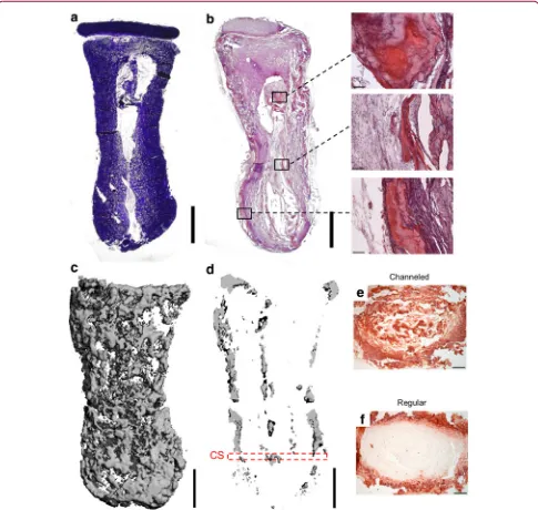
layer stained positive for cartilage-specific extracellular matrix components—sGAGs and collagen type II (Fig. 3c; top inset)—and negative for the hypertrophic marker, collagen type X (data not shown). The calcified tissue around the periphery of the osseous component appeared to be bone forming through endochondral ossification, as evident by collagen type X immuno-staining (Fig. 3c; middle inset), collagen type I immu-nostaining (Fig. 3c; bottom inset), and H&E staining, indicating the presence of bone-like tissue (Fig. 3d; bottom inset). Furthermore, a reduction in aldehyde fuchsin/alcian blue staining was observed in this region, indicating the transition from cartilage into
bone, which was followed by infiltration of blood vessel structures (Fig. 3d; middle inset). Central regions in the osseous component appeared to retain the morpholog-ical characteristics of cartilage (Fig. 3d; top inset).lCT imaging confirmed the development of a heavily calci-fied outer matrix in the osseous component (Fig. 3e), with the central regions remaining uncalcified (Fig. 3f). No evidence of calcification in the chondral layer was apparent in lCT scans.
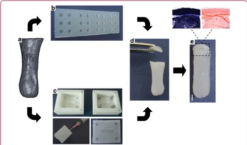
[image:5.612.59.549.86.374.2]Anatomically shaped phalanx constructs consisting of an MSC-laden fibrin osseous component and a self-assembled chondrocyte chondral layer were also evaluated in vivo.Eight weeks postimplantation, such FIG. 1. Anatomically shaped MSC-seeded alginate phalanx constructs were fabricated and cultured
chondrogenically for 5 weeks.(a)3D solid model of anatomically shaped phalanx construct.(b)Chondral layer molding system. Chondrocytes were self-assembled in cylindrical agarose wells.(c)Osseous component molding system. Clockwise from bottom; The two-part negative ABS mold, one half of the resultant positive 4% agarose/50 mM CaCl2mold, assembly of the two-part agarose/CaCl2, and injection with MSC-laden alginate
hydrogel.(d)Anatomically shaped constructs were formed by attaching a chondral layer (self-assembled chondrocytes, top) to an osseous component (MSC-laden alginate, bottom) using fibrin glue.(e)Macroscopic image of the anatomically shaped construct at the end of the in vitro culture period. Scale bar is 2 mm. Insets show the interface of the osseous and chondral components stained with aldehyde fuchsin/alcian blue (left) and picrosirius red (right). Inset scale bars are 500lm. ABS, acrylonitrile–butadiene–styrene; MSC,
constructs also showed evidence of a vascular network surrounding a heavily calcified osseous component, with the chondral layer remaining intact (Fig. 4), con-firming that the molding system developed in this study can be leveraged to tissue engineer anatomically shaped constructs using different hydrogels.
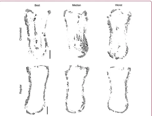
Modifying the architecture of tissue-engineered phalanx constructs to accelerate bone formation After 8 weeks in vivo, bone formation was limited to the periphery of the engineered phalanx (Fig. 3). In an attempt to accelerate bone formation throughout the tissue-engineered phalanx construct, we next mod-ulated the architecture of the graft in an attempt to facilitate infiltration of host cells and vasculature. A single axially aligned cylindrical channel (Ø1.6 mm) was inserted into the longitudinal axis of the alginate osseous component of the construct before the attach-ment of the chondral layer (Fig. 5a). The channel remained partially patent for the duration of the in vivo study. H&E staining of the construct
postim-plantation demonstrated the formation of bone around the periphery and also in central regions adjacent to the channel (Fig. 5b).lCT imaging also demonstrated bone formation in peripheral and central regions of channeled constructs (Fig. 5c, d). Alizarin red staining of cross sections confirmed the enhancement of calcifi-cation in the center of channeled constructs (Fig. 5e) compared with regular nonchanneled constructs (Fig. 5f).lCT analysis (Fig. 6), however, revealed no signifi-cant difference in total bone density between channeled and regular nonchanneled constructs (channeled, 158.7–
30.2 mg hydroxyapatite/cm3; regular, 162.3–13.3 mg hydroxyapatite/cm3).
Discussion
[image:6.612.66.550.87.340.2]the shape of the human distal phalanx and TMJ con-dyle. The MSC-seeded alginate constructs could be cultured in vitro to generate a cartilaginous matrix, which stained homogenously for sGAG and collagen, surrounded by a peripheral layer of calcified tissue.
[image:7.612.62.550.85.530.2]Chondrogenically primed tissue-engineered phalanx constructs, consisting of an MSC-laden alginate hydro-gel with an overlapping layer of articular cartilage gen-erated by chondrocyte self-assembly, were found to undergo spatially regulated endochondral ossification FIG. 3. Anatomically shaped alginate phalanx constructs, culturedin vitrofor 5 weeks, were implanted
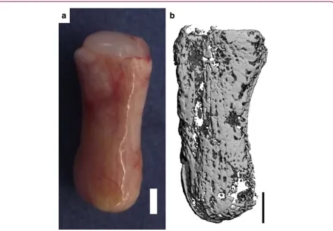
subcutaneously into nude mice and harvested 8 weeks postimplantation.(a)Macroscopic image of construct.
in vivo with the chondral layer retaining its stable chondrogenic phenotype and the osseous component proceeding along the endochondral pathway and form-ing bone around its periphery. Modifyform-ing the architec-ture of the tissue-engineered phalanx construct, by inserting a single axially aligned channel into the algi-nate hydrogel before implantation, augmented the spa-tial distribution of bone formation within the hydrogel, leading to the development of a more homogenous osseous component. While this proof-of-principal study is limited to distal phalanx tissue engineering, we believe this endochondral strategy could be scaled to treat other conditions beyond phalanx regeneration.
A well-documented challenge with the scaling up of engineered tissues is the associated issue of nutri-ent limitation and waste removal. Recnutri-ent work utilizing MSC-seeded collagen scaffolds for endochondral bone
[image:8.612.73.550.85.417.2]FIG. 5. Before the attachment of the chondral layer, a single channel (Ø1.6 mm) was cored into the osseous component of the alginate phalanx construct. After 5 weeks ofin vitroculture, constructs were subcutaneously implanted into nude mice for a period of 8 weeks.(a)Aldehyde fuchsin/alcian blue staining of channeled phalanx construct preimplantation.(b)H&E staining of channeled phalanx construct postimplantation.(c)lCT image of whole channeled construct.(d)lCT image of the center section of the channeled construct postimplantation corresponding to a thickness of 300lm.(e)Alizarin red staining of the cross section of a channeled construct postimplantation.(f)Alizarin red staining of the cross section of a regular nonchanneled construct postimplantation. Cross sections were taken from the region CS, as illustrated in(d). Scale bars in
the outer regions of the construct, thus preventing pe-ripheral cell and tissue growth and their associated ef-fects on nutrient transport. In spite of this, it should be noted that inhomogeneous tissues do form in MSC-laden hydrogels of a significant scale,34 and perhaps, at such very large dimensions, additional strategies such as bioreactor culture may be beneficial.
As the osseous component of the tissue-engineered phalanx construct, the current study employed an alginate hydrogel, which degrades over time,35 encapsu-lated with chondrogenically primed bone marrow-de-rived MSCs capable of surviving the initial hypoxic conditions experienced by engineered tissues upon in vivoimplantation.17This hypertrophic precursor of the engineered phalanx underwent endochondral
[image:10.612.65.553.84.456.2]alginate hydrogels through gamma irradiation has been shown to enhance bone regeneration.36 Inflammatory cytokines may also be harnessed to efficiently remodel engineered cartilaginous grafts into bone.37
An alternative approach for the engineering of a more homogenous osseous tissue is modifying the archi-tecture of the scaffold. We have recently demonstrated that the incorporation of channeled arrays into cylindri-cal hydrogels facilitates vascularization and enhances calcification of engineered hypertrophic cartilaginous constructsin vivo.38In the current study, a single axially aligned channel inserted into the MSC-seeded alginate hydrogel promoted bone formation in the central region of the engineered phalanx. It would appear therefore that optimization of scaffold or hydrogel architecture is a key design criterion in the scaling up of large ana-tomically shaped grafts for whole bone regeneration.
As previously noted, a further requirement of a func-tional engineered long bone is the development of a stable layer of articular cartilage at the articulating ends. We have previously demonstrated that it is pos-sible to engineer osteochondral tissues by implanting chondrogenically primed bilayered constructs contain-ing both chondrocytes and MSCs and spatially regulat-ing endochondral ossification.28 Motivated by this finding, primary chondrocytes were used to tissue engineer a chondral layer using a self-assembly or scaf-fold-free approach,39–44which retained a stable chon-drogenic phenotype following implantation, forming a matrix consisting of sGAG and collagen type II and void of collagen type X. There are, however, difficulties associated with obtaining sufficient numbers of chon-drocytes for the engineering of large cartilaginous lay-ers.45,46Previous studies have demonstrated,in vitro, a beneficial effect of coculturing a small number of chon-drocytes with a larger number of MSCs.47–50 Future work in our laboratory will investigate if cocultures of chondrocytes and MSCs can be utilized to generate scaled-up stable cartilaginous graftsin vivo.
This study employed a subcutaneous environment to facilitate the development of an engineered cartilagi-nous construct into an endochondral bone tissue. From a translational perspective, this approach may also be adopted in the clinic, that is, using an ectopic environment as anin vivobioreactor51,52to allow mat-uration of an engineered tissue, with functional vascu-lature and marrow components,24which can then be implanted into an orthotopic defect site. The alterna-tive would be to forsake the ectopic transplantation and implant the engineered cartilaginous graft directly
into the defect site, allowing endochondral ossification to occur orthotopically. This approach has been dem-onstrated using a coral scaffold, which would have an inherent advantage over a hydrogel in performing an immediate mechanical function, for the replacement of an avulsed phalanx.5 The relatively lower load-bearing environment of the upper limbs may allow for direct implantation of an engineered cartilaginous construct into a bone defect, although it may be more challenging if applied in a mechanically loaded de-fect site in the lower limb, which would require the engineered tissue to perform a more demanding bio-mechanical function. (Previous studies from our labo-ratory have shown that the equilibrium compressive modulus of cartilaginous tissues engineered using bone marrow-derived MSCs is typically less than 100 kPa,33,53–55 potentially limiting their use in mechanically challeng-ing environments). Further studies uschalleng-ing larger animal models are required to compare the efficacy of ectopic and orthotopic strategies in endochondral bone tissue engineering applications.
In conclusion, this work demonstrates the potential of utilizing anatomically shaped cartilaginous grafts for the tissue engineering of whole bones through en-dochondral ossification. An MSC-laden alginate hydro-gel served as the osseous or endochondral component of an engineered phalanx construct, and a self-assembly strategy was used to engineer the overlapping articular cartilage layer using primary chondrocytes, as opposed to MSCs. The chondrogenically primed phalanx constructs were found to undergo spatially regulated endochondral ossificationin vivo, with the osseous component engi-neered using bone marrow-derived MSCs proceeding along the endochondral pathway, and no evidence of calcification being observed in the integrated layer of self-assembled chondrocytes. Modifying the architec-ture of phalanx constructs, by inserting a single chan-nel into the alginate hydrogel before implantation, accelerated bone formation in the center of the engi-neered construct and facilitated the development of a more homogenous osseous tissue.
Acknowledgments
This work was supported by the Science Founda-tion Ireland (SFI/08/Y15/B1336) and the European Research Council (StemRepair-Project No: 258463).
Author Disclosure Statement
References
1. Dimitriou R, Jones E, McGonagle D, et al. Bone regeneration: current concepts and future directions. BMC Med. 2011;9:66.
2. Koh CJ, Atala A. Tissue engineering, stem cells, and cloning: opportunities for regenerative medicine. J Am Soc Nephrol. 2004;15:1113–1125. 3. Langer R. Tissue engineering. Mol Ther. 2000;1:12–15.
4. Isogai N, Landis W, Kim TH, et al. Formation of phalanges and small joints by tissue-engineering. J Bone Joint Surg. 1999;81:306–316.
5. Vacanti CA, Bonassar LJ, Vacanti MP, et al. Replacement of an avulsed phalanx with tissue-engineered bone. N Engl J Med. 2001;344:1511–1514. 6. Sedrakyan S, Zhou ZY, Perin L, et al. Tissue engineering of a small hand phalanx with a porously casted polylactic acid-polyglycolic acid copoly-mer. Tissue Eng. 2006;12:2675–2683.
7. Alhadlaq A, Elisseeff JH, Hong L, et al. Adult stem cell driven genesis of human-shaped articular condyle. Ann Biomed Eng. 2004;32:911–923. 8. Alhadlaq A, Mao JJ. Tissue-engineered osteochondral constructs in the
shape of an articular condyle. J Bone Joint Surg. 2005;87:936–944. 9. Hung CT, Lima EG, Mauck RL, et al. Anatomically shaped osteochondral
constructs for articular cartilage repair. J Biomech. 2003;36:1853–1864. 10. Lee CH, Marion NW, Hollister S, et al. Tissue formation and vascularization
in anatomically shaped human joint condyle ectopicallyin vivo. Tissue Eng Part A. 2009;15:3923–3930.
11. Grayson WL, Frohlich M, Yeager K, et al. Engineering anatomically shaped human bone grafts. Proc Natl Acad Sci U S A. 2010;107:3299–3304. 12. Grayson WL, Chao PH, Marolt D, et al. Engineering custom-designed
osteochondral tissue grafts. Trends Biotechnol. 2008;26:181–189. 13. Meijer GJ, De Bruijn JD, Koole R, et al. Cell-based bone tissue engineering.
PLoS Med. 2007;4:0260–0264.
14. Santos MI, Reis RL. Vascularization in bone tissue engineering: physiology, current strategies, major hurdles and future challenges. Macromol Biosci. 2010;10:12–27.
15. Lyons FG, Al-Munajjed AA, Kieran SM, et al. The healing of bony defects by cell-free collagen-based scaffolds compared to stem cell-seeded tissue engineered constructs. Biomaterials. 2010;31:9232–9243.
16. Kronenberg HM. Developmental regulation of the growth plate. Nature. 2003;423:332–336.
17. Farrell E, Both SK, Odo¨rfer KI, et al.In-vivogeneration of bone via endo-chondral ossification byin-vitrochondrogenic priming of adult human and rat mesenchymal stem cells. BMC Musculoskelet Disord. 2011;12. 18. Gerber HP, Vu TH, Ryan AM,. VEGF couples hypertrophic cartilage
remodeling, ossification and angiogenesis during endochondral bone formation. Nat Med. 1999;5:623–628.
19. Pelttari K, Winter A, Steck E, et al. Premature induction of hypertrophy duringin vitrochondrogenesis of human mesenchymal stem cells cor-relates with calcification and vascular invasion after ectopic transplanta-tion in SCID mice. Arthritis Rheum. 2006;54:3254–3266.
20. Farrell E, Van Der Jagt OP, Koevoet W, et al. Chondrogenic priming of human bone marrow stromal cells: a better route to bone repair? Tissue Eng Part C Methods. 2009;15:285–295.
21. Huang JI, Durbhakula MM, Angele P, et al. Lunate arthroplasty with au-tologous mesenchymal stem cells in a rabbit model. J Bone Joint Surg. 2006;88:744–752.
22. Scotti C, Tonnarelli B, Papadimitropoulos A, et al. Recapitulation of en-dochondral bone formation using human adult mesenchymal stem cells as a paradigm for developmental engineering. Proc Natl Acad Sci U S A. 2010;107:7251–7256.
23. Janicki P, Kasten P, Kleinschmidt K, et al. Chondrogenic pre-induction of human mesenchymal stem cells onb-TCP: enhanced bone quality by endochondral heterotopic bone formation. Acta Biomater. 2010;6: 3292–3301.
24. Scotti C, Piccinini E, Takizawa H, et al. Engineering of a functional bone organ through endochondral ossification. Proc Natl Acad Sci U S A. 2013;110:3997–4002.
25. Yang W, Yang F, Wang Y, et al.In vivobone generation via the endo-chondral pathway on three-dimensional electrospun fibers. Acta Bio-mater. 2013;9:4505–4512.
26. Lau TT, Lee LQP, Vo BN, et al. Inducing ossification in an engineered 3D scaffold-free living cartilage template. Biomaterials. 2012;33:8406–8417. 27. Sheehy EJ, Mesallati T, Vinardell T, et al. Engineering cartilage or endo-chondral bone: a comparison of different naturally derived hydrogels. Acta Biomater. 2015;13:245–253.
28. Sheehy EJ, Vinardell T, Buckley CT, et al. Engineering osteochondral constructs through spatial regulation of endochondral ossification. Acta Biomater. 2013;9:5484–5492.
29. Sheehy EJ, Buckley CT, Kelly DJ. Oxygen tension regulates the osteogenic, chondrogenic and endochondral phenotype of bone marrow derived mesenchymal stem cells. Biochem Biophys Res Commun. 2012;417: 305–310.
30. Martin I, Wendt D, Heberer M. The role of bioreactors in tissue engi-neering. Trends Biotechnol. 2004;22:80–86.
31. Mauck RL, Soltz MA, Wang CCB, et al. Functional tissue engineering of articular cartilage through dynamic loading of chondrocyte-seeded agarose gels. J Biomech Eng. 2000;122:252–260.
32. Mauck RL, Nicoll SB, Seyhan SL, et al. Synergistic action of growth factors and dynamic loading for articular cartilage tissue engineering. Tissue Eng. 2003;9:597–611.
33. Thorpe SD, Buckley CT, Vinardell T, et al. The response of bone marrow-derived mesenchymal stem cells to dynamic compression following
tgf-b3 induced chondrogenic differentiation. Ann Biomed Eng. 2010;38:2896–2909.
34. Buckley CT, Meyer EG, Kelly DJ. The influence of construct scale on the composition and functional properties of cartilaginous tissues engi-neered using bone marrow-derived mesenchymal stem cells. Tissue Eng Part A. 2012;18:382–396.
35. Drury JL, Mooney DJ. Hydrogels for tissue engineering: scaffold design variables and applications. Biomaterials. 2003;24:4337–4351.
36. Simmons CA, Alsberg E, Hsiong S, et al. Dual growth factor delivery and controlled scaffold degradation enhancein vivobone formation by transplanted bone marrow stromal cells. Bone. 2004;35:562–569. 37. Mumme M, Scotti C, Papadimitropoulos A, et al. Interleukin-1bmodulates
endochondral ossification by human adult bone marrow stromal cells. Eur Cell Mater. 2012;24:224–236.
38. Sheehy EJ, Vinardell T, Toner ME, et al. Altering the architecture of tissue engineered hypertrophic cartilaginous grafts facilitates vascularisation and accelerates mineralisation. PLoS One. 2014;9.
39. Mesallati T, Buckley CT, Kelly DJ. Engineering articular cartilage-like grafts by self-assembly of infrapatellar fat pad-derived stem cells. Biotechnol Bioeng. 2014;111:1686–1698.
40. Hoben GM, Hu JC, James RA, et al. Self-assembly of fibrochondrocytes and chondrocytes for tissue engineering of the knee meniscus. Tissue Eng. 2007;13:939–946.
41. Revell CM, Reynolds CE, Athanasiou KA. Effects of initial cell seeding in self assembly of articular cartilage. Ann Biomed Eng. 2008;36:1441–1448. 42. Hu JC, Athanasiou KA. A self-assembling process in articular cartilage
tissue engineering. Tissue Eng. 2006;12:969–979.
43. Mesallati T, Buckley CT, Kelly DJ. A comparison of self-assembly and hydrogel encapsulation as a means to engineer functional cartilaginous grafts using culture expanded chondrocytes. Tissue Eng Part C Methods. 2014;20:52–63.
44. Naumann A, Dennis JE, Aigner J, et al. Tissue engineering of autologous cartilage grafts in three-dimensionalin vitromacroaggregate culture system. Tissue Eng. 2004;10:1695–1706.
45. Barbero A, Grogan S, Scha¨fer D, et al. Age related changes in human ar-ticular chondrocyte yield, proliferation and post-expansion chondrogenic capacity. Osteoarthritis Cartilage. 2004;12:476–484.
46. Diaz-Romero J, Gaillard JP, Grogan SP, et al. Immunophenotypic analysis of human articular chondrocytes: changes in surface markers associated with cell expansion in monolayer culture. J Cell Physiol. 2005;202:731– 742.
47. Cooke ME, Allon AA, Cheng T, et al. Structured three-dimensional co-culture of mesenchymal stem cells with chondrocytes promotes chon-drogenic differentiation without hypertrophy. Osteoarthritis Cartilage. 2011;19:1210–1218.
48. Bian L, Zhai DY, Mauck RL, et al. Coculture of human mesenchymal stem cells and articular chondrocytes reduces hypertrophy and enhances functional properties of engineered cartilage. Tissue Eng Part A. 2011;17:1137–1145.
49. Wu L, Leijten JCH, Georgi N, et al. Trophic effects of mesenchymal stem cells increase chondrocyte proliferation and matrix formation. Tissue Eng Part A. 2011;17:1425–1436.
51. Stevens MM, Marini RP, Schaefer D, et al.In vivoengineering of organs: the bone bioreactor. Proc Natl Acad Sci U S A. 2005;102: 11450–11455.
52. Emans PJ, van Rhijn LW, Welting TJ, et al. Autologous engineering of cartilage. Proc Natl Acad Sci U S A. 2010;107:3418–3423.
53. Thorpe SD, Nagel T, Carroll SF, et al. Modulating Gradients in Regulatory Signals within Mesenchymal Stem Cell Seeded Hydrogels: a Novel Strat-egy to Engineer Zonal Articular Cartilage. PLoS One. 2013;8.
54. Vinardell T, Buckley CT, Thorpe SD, et al. Composition-function relations of cartilaginous tissues engineered from chondrocytes and mesenchymal stem cells isolated from bone marrow and infrapatellar fat pad. J Tissue Eng Regen Med. 2011;5:673–683.
55. Carroll SF, Buckley CT, Kelly DJ. Cyclic hydrostatic pressure promotes a stable cartilage phenotype and enhances the functional development of cartilaginous grafts engineered using multipotent stromal cells iso-lated from bone marrow and infrapatellar fat pad. J Biomech. 2014; 47:2115–2121.
Cite this article as:Sheehy EJ, Mesallati T, Kelly L, Vinardell T, Buckley CT, Kelly DJ (2015) Tissue engineering whole bones through endo-chondral ossification: regenerating the distal phalanx,BioResearch Open Access4:1, 229–241, DOI: 10.1089/biores.2015.0014.
Abbreviations Used
ABS¼acrylonitrile–butadiene–styrene CM¼chondrogenic medium FBS¼fetal bovine serum H&E¼hematoxylin and eosin
hgDMEM¼high-glucose Dulbecco’s modified Eagle’s medium MSC¼mesenchymal stem cell
lCT¼microcomputed tomography PBS¼phosphate-buffered saline
3D¼three-dimensional TMJ¼temporomandibular joint
Publish in BioResearch Open Access
-Broad coverage of biomedical research
-Immediate, unrestricted online access
-Rigorous peer review
-Compliance with open access mandates
-Authors retain copyright
-Highly indexed
-Targeted email marketing