Knowledge of Undergraduate and Graduate Dentists and
Dental Therapists concerning Panoramic Radiographs:
Knowledge of Panoramic Radiographs
Scott McNab
1, Paul Monsour
1,*, Daniel Madden
2, Deanne Gannaway
21School of Dentistry, University of Queensland, Australia
2Teaching and Education Development Institute, University of Queensland, Australia
Copyright © 2015 Horizon Research Publishing All rights reserved.
Abstract Background:
Increasing numbers of panoramic radiographs (PRs) are being taken every year worldwide. This study was designed to assess the ability of dental students, bachelor of oral health (BOH) students, graduate dentists and graduate oral health therapists (OHT) in Queensland, Australia, in the interpretation of PRs in order to assess future teaching needs. Methods: This study was conducted as a web-based survey. Final year dental students, final year BOH students, graduate dentists and graduate OHTs in Queensland were invited to participate in this study. The study examined three topics; 1) radiographic anatomy, 2) positioning errors, and 3) pathology/anomalies relating to PRs. Results: No significant difference was found between any of the four groups regarding identification of radiographic anatomy on PRs. Undergraduate dental students correctly identified significantly more positioning errors than graduated dentists. Undergraduate OHTs identified significantly more positioning errors than graduated OHTs. Graduate dentists scored significantly higher than final year dental students in the identification of pathology/anomalies in PRs. Graduated dentists who had access to a PR machine or had completed a refresher course in extra-oral radiography displayed significantly higher performance in identifying positioning errors. Generally the scores were low in all areas. Conclusions: Additional teaching of the three areas assessed would be beneficial in both the undergraduate curriculum and in the form of continuing education courses. Findings from this study support the findings from similar international studies [1-7] and have the potential to be extrapolated to teaching and learning initiatives in other Australian States.Keywords Panoramic Radiography, Positioning Errors,
Anatomy, Pathology, Education1. Introduction
Panoramic radiography (PR) is the most common extra
oral radiographic examination used in dental practice. Figures available for the National Health Service in England and Wales show that in 1991-92 the number of panoramic examinations performed within general dental practices was estimated to be over 1.5 million.[8] By 1997, this number had risen to over 1.7 million and for 2000-01, 2.2 million panoramic radiographs were taken in these practices alone. [6, 9] Surveys conducted in other countries have found the number of practices operating PR machines to vary between 7% and 99%.[1]
One of the strengths of this radiograph is that it displays both jaws and the dentition on a single film.[8] PRs display many anatomical structures apart from the dentition and the supporting bone, including the temporomandibular joints (TMJ’s), the maxillary sinuses, the nasal cavity, parts of the cervical spine and cranial base, the pharyngeal airway and neck. Knowledge of the anatomy of these regions is necessary to be able to detect an abnormality and to then determine whether it represents a variant of normal or a disease process.
The aim of this study was to compare the ability of undergraduates and practitioners in Queensland, Australia, to interpret PRs with a view to identifying needs for future undergraduate curriculum changes and the provision of continued professional learning. The information obtained could be used to support any changes required in the current curriculum regarding the teaching of extra-oral radiography at undergraduate level.
2. Materials and Methods
This study was conducted as an anonymous web-based survey, designed to take approximately fifteen minutes to complete. The research protocol for this survey was approved by a suitably constituted Ethics Committee of the institution within which the research was undertaken conforming to the provisions of the Declaration of Helsinki (as revised in Tokyo 2004).
The study sample targeted four groups: 1) final year dental students; 2) final year Bachelor of Oral Health (BOH) students; 3) graduate dentists, and 4) graduate Oral Health Therapists (OHT).
Three methods were used to obtain the sample: a paper-based mail-out to all dentists and therapists in Queensland whose address could be obtained through public-access documents; two advertisements in the newsletter of the Queensland branch of the Australian Dental Association placed inviting registered graduated dentists (N = 2,780) and OHT (hygienists and therapists) to participate (N= 337); and an emailed invitation to all final year dental (N= 106) and BOH students (N= 37) at the University of
Queensland. These administration processes resulted in 219 responses across the following groups: 1) final year dental students (N=55); 2) final year Bachelor of Oral Health (BOH) students (N=27); 3) graduate dentists (N=123), and 4) graduate Oral Health Therapists (OHT) (N=14).
Before completing the survey, respondents consented to participate in the study and provided demographic information on their year of graduation, institution, degree, whether they had a PR machine at their place of work and whether they had completed any additional courses on panoramic radiography since graduation.
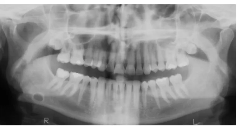
Figure 1. Example of anatomy question showing the region of interest circled (a) and then an unaltered copy of the same OPG (b)
Figure 2. Example of OPG for technique error
[image:3.595.115.496.519.721.2]Statistical analyses included between groups ANOVA and standard multiple regression. ANOVA was used to investigate whether undergraduates have superior PR interpretation abilities when compared to graduate practitioners overall. It was hypothesised that there would be a significant difference in performance across groups on identifying 1) radiographic anatomy, 2) positioning errors, and 3) pathology/anomalies relating to PRs. Specifically, it was predicted that undergraduate dental students would outperform graduated dentists in all three areas and undergraduate OHT students would outperform graduated OHTs on anatomy and positioning error questions. Assumptions were tested prior to using ANOVA, showing mixed results. Whilst inspection of Q-Q plots revealed that data was approximately normally distributed for all three dependent measures, the homogeneity of variance assumption was only met for the anatomy and pathology measures according to Levene’s test for equality of variances. Levene’s test revealed that the homogeneity of variance assumption was not met for the positioning errors measure,
F(3,193) = 9.71, p < .001. In accordance with Kesleman [10], the Welch’s F test[11] was therefore used for the analysis of positioning errors. Tukey HSD multiple comparisons were used to follow up significant omnibus tests, except in the case of heterogeneous variances for the positioning error variable. Games-Howell pairwise multiple comparisons [12] were used to follow up the Welch’s F test for positioning errors, as this approach is appropriate even in the case of heterogeneous variances in an unbalanced design [13].
Inspection of scatterplots revealed that linear regression was an appropriate approach for the given data. Inspection of residuals also indicated that homoscedasticity and normality of errors assumptions were met. A standard multiple regression analysis was therefore used to investigate whether various forms of post-graduate experience affected the three areas of performance in graduated dentists. It was hypothesised that performance in anatomy, positioning and pathology would increase with experience, including having an extra-oral radiography licence, having a PR machine at their place of work and completing a refresher course in extra-oral radiography.
3. Results
A between groups ANOVA on anatomy item scores showed that there was no significant difference between the different groups of respondents, F(3,202) = 1.70, p = .168, ω2
= .01. This indicated that contrary to predictions, undergraduate dentists (M=3.80, SD=1.64) and undergraduate OHTs (M=3.50, SD=1.36) did not significantly outperform graduated dentists (M=3.47,
SD=1.95) or graduated OHTs (M=2.86, SD=1.89) on the 10 radiographic anatomy questions.
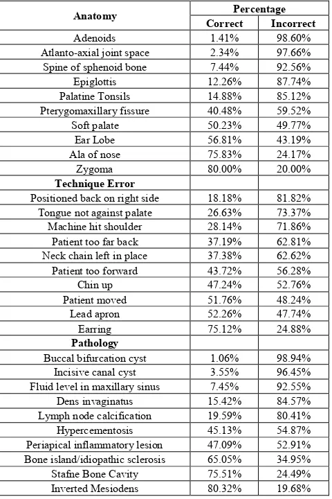
Descriptive statistics (Table 1) showed the radiographic anatomy items varied greatly in the number of correct responses, ranging between 1.41% (identifying adenoids) and 80.00% (identifying the zygoma) (3 items < 10.00%
correct; 3 items < 50.00% correct; and 4 items ≤ 80.00% correct). Out of a maximum score of 10, undergraduate dental students had the highest average anatomy score of 3.80 (SD = 1.64), while graduate OHTs had the lowest average score of 2.86 (SD = 1.88) (Table 2). However, when comparing the four groups (see aim of study) on identification of radiographic anatomy no significant difference was observed.
Table 1. Combined scores from all respondents for each question
Anatomy Correct Percentage Incorrect
Adenoids 1.41% 98.60%
Atlanto-axial joint space 2.34% 97.66% Spine of sphenoid bone 7.44% 92.56%
Epiglottis 12.26% 87.74%
Palatine Tonsils 14.88% 85.12%
Pterygomaxillary fissure 40.48% 59.52%
Soft palate 50.23% 49.77%
Ear Lobe 56.81% 43.19%
Ala of nose 75.83% 24.17%
Zygoma 80.00% 20.00%
Technique Error
Positioned back on right side 18.18% 81.82% Tongue not against palate 26.63% 73.37% Machine hit shoulder 28.14% 71.86% Patient too far back 37.19% 62.81% Neck chain left in place 37.38% 62.62% Patient too forward 43.72% 56.28%
Chin up 47.24% 52.76%
Patient moved 51.76% 48.24%
Lead apron 52.26% 47.74%
Earring 75.12% 24.88%
Pathology
Buccal bifurcation cyst 1.06% 98.94% Incisive canal cyst 3.55% 96.45% Fluid level in maxillary sinus 7.45% 92.55%
Dens invaginatus 15.42% 84.57%
Lymph node calcification 19.59% 80.41%
Hypercementosis 45.13% 54.87%
Periapical inflammatory lesion 47.09% 52.91% Bone island/idiopathic sclerosis 65.05% 34.95%
Stafne Bone Cavity 75.51% 24.49%
[image:4.595.313.553.198.558.2]Inverted Mesiodens 80.32% 19.68%
Table 2. Mean scores for each of the 4 groups for the three areas of knowledge examined.
N Mean Std. Dev.
Anatomy
Graduated dentist 97 3.474 1.948
Graduated therapist 29 2.862 1.885 Undergraduate dentist 54 3.796 1.641 Undergraduate therapists 26 3.500 1.364
Positioning Errors
Graduated dentist 89 3.708 2.681
Graduated therapist 30 2.433 1.994 Undergraduate dentist 51 6.098 1.640 Undergraduate therapists 27 4.296 1.636
Pathology
Graduated dentist 79 4.684 1.472
[image:4.595.312.551.587.755.2]A separate between groups ANOVA conducted on positioning error scores revealed a significant difference in performance among respondent groups, FW(3,79.64) = 29.06,
p < .001, ω2 = .30. This was followed up with Games-Howell
multiple comparisons to address the individual hypotheses. In accordance with predictions, follow up comparisons showed that undergraduate dentists (M=6.10, SD=1.64) significantly outperformed graduated dentists (M=3.71,
SD=2.68, p < .001). It was also found that undergraduate OHTs (M=4.30, SD=1.64) significantly outperformed graduated OHTs (M=2.43, SD=2.00, p = .002).
The third between groups ANOVA on pathology scores indicated that there was a significant difference in performance among respondent groups, F(3,180) = 27.97, p
< .001, ω2 = .31. This was followed up using Tukey HSD
multiple comparisons. Follow up tests revealed that contrary to predictions, graduated dentists (M=4.68, SD=1.47) significantly outperformed undergraduate dental students (M=3.45, SD=1.45) on pathology items, p < .001.
Pathology is not part of the undergraduate Bachelor of Oral Health curriculum at the University of Queensland Dental School (UQDS). Subsequently, items identifying pathology or anomalies on PRs were compared between final year dental students and graduate dentists only. Buccal bifurcation cysts were correctly identified in only 1.06% of responses and the presence of a mesiodens was correctly identified in 80.32% of answers (Table 1).
Table 3. Effect of possession of extra-oral licence on mean scores of graduated dentists
Have
License Number Mean Score Std Dev. Anatomy Yes No 42 55 4.024 3.055 2.078 1.747 Positioning
Error Yes No 39 50 4.897 2.780 2.827 2.169
Pathology Yes No 32 47 5.094 4.404 1.329 1.513
Table 4. Effect of having a PR machine at the place of work on mean score of graduated dentists
Have PR
machine Number Mean Score Std Dev.
Anatomy Yes 31 3.968 2.089
No 66 3.242 1.849
Positioning
Error Yes No 29 60 5.414 2.883 2.557 2.344
Pathology Yes 26 5.269 1.185
[image:5.595.58.298.655.756.2]No 53 4.396 1.523
Table 5. Effect of completion of continuing education course on mean scores of graduated dentists
Continuing Education
Course Number
Mean
Score Deviation Std
Anatomy Yes 35 3.971 2.079
No 62 3.194 1.827
Positioning
Error Yes No 31 58 5.194 2.914 2.833 2.242
Pathology Yes 28 5.036 1.427
No 51 4.490 1.475
Further analysis was conducted on the results obtained for graduated dentists only. The three predictors included whether the graduated dentist had 1) an extra-oral radiology licence (Table 3); 2) a PR machine at their place of work or study (Table 4); and 3) completed any continuing education courses in extra-oral radiography (Table 5). As answers of no
were coded as 0 and answers of yes were coded as 1, a mean value of .44 can be taken to indicate that 44% of graduated dentists who responded to the positioning questions answered yes to having an extra-oral radiology licence. Similarly, 33% have a PR machine at their place of work or study and 35% have completed a refresher course in extra-oral radiography.
Regression analysis showed that the three predictors together explained 28% of the variance in positioning scores,
F(3,85) = 11.24, p < .001. Whether the respondent had a PR machine significantly accounted for 6% of the variance in positioning scores, such that having an PR machine predicted higher scores (M=5.41, SD=2.56) than not having a machine (M=2.88, SD=2.34), β = .29, p = .008. Having an PR machine was therefore the most important predictor of positioning scores, followed by the 5% of variance uniquely accounted for by whether the respondent had completed courses in extra-oral radiography. Answering yes predicted higher scores (M=5.19, SD=2.83) than answering no
(M=2.91, SD=2.24), β = .24, p = .021. Having an extra-oral radiology licence did not significantly predict positioning scores, β = .14, p = .214, sr2 = .01. A further 16% of variance
in positioning scores accounted for by the model was not uniquely contributed by any one predictor.
Conversely, standard multiple regression analyses showed that together, the same three predictors did not significantly predict anatomy scores, R2 = .07, F(3,93) = 2.43, p = .071, or
pathology scores, R2 = .09, F(3,75) = 2.43, p = .072.
Anatomy scores were not significantly predicted by having an extra-oral radiology licence (β = .18, p = .151, sr2 = .02),
having an PR machine at work (β = .05, p = .667, sr2 < .01),
or by having completed courses in extra-oral radiography (β = .10, p = .368, sr2 = .01). Similarly, pathology scores were
not significantly predicted by having an extra-oral radiology licence (β = .08, p = .562, sr2 < .01), having an PR machine at
work (β = .21, p = .128, sr2 = .03, or by having completed
courses in extra-oral radiography (β = .06, p = .639, sr2
< .01).
4. Discussion
The scores of all groups in this study were lower than that reported by Razmus et al (93).[5] This difference may be in part due to the fact that the current study was not in a multi-choice format and so the correct answer was not present as a prompt. In addition, the anatomical structures selected in the current study were a combination of both hard and soft tissues and may have been more difficult to identify. The anatomical structures that proved to be the hardest to identify were the adenoids (1.4%) , the atlanto-axial joint spaces (2.3%) and the spine of the sphenoid bone (7.4% correct). It is conceivable that these structures were more difficult to identify than structures such as the head of the condyle, maxillary sinus or mental foramen which were included in the Razmus et al (93) study. The anatomical structures included in this study were selected because of their relevance to interpretation of PRs.
The current study dealt only with the identification of positioning errors and did not test the knowledge of respondents in correcting them. While this is the logical next step, it was not included as the study was designed to test the respondents’ ability to correctly identify anatomy and pathology/anomalies in PRs. The inclusion of additional questions examining the correction of positioning errors would have risked making the survey too time consuming with a potential drop in the completion rate. Furthermore, it was possible that respondents would be unable to consistently identify the faults present, in which case further questions that required them to detail how to correct the faults would be meaningless.
In accordance with predictions, undergraduate dental students correctly identified significantly more positioning errors than graduated dentists, and undergraduate BOH students identified more errors than graduated OHTs.
The mean score of 6.1/10 for dental students is surprisingly similar to the mean percentage of correct responses of 61.7% reported by Razmus et al,[5] however, that study also included questions on error correction. Razmus et al found that undergraduates at schools that were taught this subject for three hours or more performed better than those at which it was taught for one or two hours. Students who had clinical experience with PRs also performed better than those that did not.
Rushton et al [7] found a large difference between the abilities of undergraduates at two different UK dental schools in their ability to identify PR faults (albeit under different testing conditions). For example, “incorrect antero-posterior position” was present in two PRs in their study. The undergraduates from University A identified this error correctly in 8.3% and 14.6% of cases. The students at University B identified them in 30.4% and 54.3% of cases. In our study, questions 13 (patient positioned too far forward) and 17 (patient positioned too far back) were identified in 43.7% and 37.2% of responses, falling between the results of the two UK dental schools. Rushton et al [7] concluded that limitations existed at that time in the undergraduate teaching of the identification (and correction) of film faults. Our findings also indicate that additional teaching of this topic in
the undergraduate curriculum would be of benefit.
Contrary to predictions, graduate dentists scored significantly higher than undergraduate dentists in the identification of pathology/anomalies on PRs (mean score 4.7/10 v 3.5/10). The result may indicate an area of the undergraduate curriculum that requires additional attention. While significant time is allocated to the teaching of oral pathology, it seems possible that additional focus on the radiographic appearances of these entities may be advantageous.
There are no comparative studies that have directly examined the ability of students or graduate dentists to diagnose pathology on PRs. Rushton et al [14] reported that of the 67 cases of TMJ abnormalities identified by dental radiologists in their study, none were identified by the general dentists who took the radiograph. Furthermore, pathology of the maxillary antra was identified in 255 cases by dental radiologists and in 11 cases by graduated dentists. In our study, pathology in the maxillary sinus was only correctly identified in 7.4% of cases. Rushton et al [14] concluded that on the whole, abnormalities were often not detected by general practitioners and that more emphasis needs to be placed on radiological diagnosis in undergraduate dental education and for qualified dentists. Our results are in agreement with their finding.
It was hypothesised that graduate dentists’ performance in anatomy, positioning and pathology would increase with post-graduate experience, including having an extra-oral radiography licence, having an PR machine at their place of work and completing a course in extra-oral radiography. The standard multiple regression analysis revealed that graduated dentists having an PR machine at their place of work or completing a course in extra-oral radiography predicted significantly higher positioning error performance, but having an extra-oral radiography licence did not. Contrary to predictions, none of these types of post-graduate experience were found to predict anatomy or pathology scores.
participate.
5. Conclusions
The trends identified by the survey align with the aim of the study to focus on future teaching needs. Results indicate that additional teaching and or changes to the current methods of teaching employed for the three areas tested may be beneficial both in the undergraduate curriculum and in the form of continuing education courses for graduates who take and/or interpret PRs. Perhaps for a similar reason, analysis within the graduated dentists found that the presence of an PR machine at their place of work and attending courses were a significant contributing factor to the higher scores obtained by these dentists in the identification of positioning errors. The results of this study offer a suggested approach to curriculum changes in the undergraduate programs and potential topics for future continued professional learning activities in Queensland. Findings from this study support the findings from similar international studies 1,2,4,5,6,7,8 and
have the potential to be extrapolated to teaching and learning initiatives in other Australian states.
Abbreviations and Acronyms
BOH = bachelor of oral health; OHT = oral health therapists; PR = panoramic radiograph; University of Queensland Dental School = UQDS
Acknowledgements
This research was funded by a research grant from the Dental Board of Queensland
REFERENCES
Aps, J.K., Flemish general dental practitioners' knowledge of [1]
dental radiology. Dentomaxillofac Radiol, 2010. 39(2): p. 113-8.
Brezden, N.A. and S.L. Brooks, Evaluation of panoramic [2]
dental radiographs taken in private practice. Oral Surg Oral Med Oral Pathol, 1987. 63(5): p. 617-21.
Granlund, C.M., et al., Frequency of errors and pathology in [3]
panoramic images of young orthodontic patients. Eur J Orthod, 2012.
Hellen-Halme, K., M. Nilsson, and A. Petersson, Digital [4]
radiography in general dental practice: a field study. Dentomaxillofac Radiol, 2007. 36(5): p. 249-55.
Razmus, T.F., G.F. Williamson, and M.L. Van Dis, [5]
Assessment of the knowledge of graduating American dental students about the panoramic image. Oral Surg Oral Med Oral Pathol, 1993. 76(3): p. 397-402.
Tugnait, A., V. Clerehugh, and P.N. Hirschmann, [6]
Radiographic equipment and techniques used in general dental practice: a survey of general dental practitioners in England and Wales. J Dent, 2003. 31(3): p. 197-203.
Rushton, V.E., P.N. Hirschmann, and D.R. Bearn, The [7]
effectiveness of undergraduate teaching of the identification of radiographic film faults. Dentomaxillofac Radiol, 2005.
34(6): p. 337-42.
Rushton, V.E. and K. Horner, The use of panoramic [8]
radiology in dental practice. J Dent, 1996. 24(3): p. 185-201. Rushton, V.E., K. Horner, and H.V. Worthington, The [9]
quality of panoramic radiographs in a sample of general dental practices. Br Dent J, 1999. 186(12): p. 630-3.
Keselman, J.C., et al., Statistical Practices of Educational [10]
Researchers: An Analysis of Their ANOVA, MANOVA, and ANCOVA Analyses. REVIEW OF EDUCATIONAL RESEARCH, 1998 68(3): p. 350-386.
Welch, B.L., On the Comparison of Several Mean Values: [11]
An Alternative Approach. Biometrika, 1951. 38(3/4): p. 330-336.
Games, P.A. and J.F. Howell, Pairwise Multiple Comparison [12]
Procedures with Unequal N's and/or Variances: A Monte Carlo Study. Journal of Educational Statistics, 1976. 1(2): p. 113-125.
Raftery, J.A., M.L. Abelly, and J.P. Braseltony, Multiple [13]
Comparison Methods for Means. SIAM REVIEW 2002.
44(2): p. 259 - 278.
Rushton, V.E., K. Horner, and H.V. Worthington, Screening [14]