HIF1
is a central regulator of collagen hydroxylation and secretion under hypoxia during
bone development
Lital Bentovim, Roy Amarilio and Elazar Zelzer
There was an error published in the supplementary material accompanying Development 139, 4473-4483.
The Greek symbols were missing in Table S2. The correct version of the table is shown below and has been corrected online.
We apologise to the authors and readers for this mistake.
Development 140, 248 (2013) doi:10.1242/dev.092023 © 2013. Published by The Company of Biologists Ltd
qRT-PCR primers
Primer name Forward Reverse
Hif-1 AGATCTCGGCGAAGCAAAGAGT CGGCATCCAGAAGTTTTCTCACAC
Tbp GCAGCCTCAGTACAGCAATCAACA GGTGCAGTGGTCAGAGTTTGAGAA
18S rRNA GTAACCCGTTGAACCCCATT CCATCCAATCGGTAGTAGCG
Chop TGTTGAAGATGAGCGGGTGGCA GGACCAGGTTCTGCTTTCAGGTGT
BiP GGGGACCACCTATTCCTGCGTC ATACGACGGCGTGATGCGGT
Pdk1 GACTGTGAAGATGAGTGACCGGGG CGTTTCAACACGAGGCCGGG
P4h1 AGGACATGTCGGATGGCTTCATCT TCTTGCAGCCGAAACAGAGCTT
P4h2 AGGTGTTGGTGTTGGTGTTGCT TGTACCAGGTCCTTCTCTGCGTAA
P4h CAGATGAGCTGACGGCTGAGAAAA CTTCAAAGTTCGCCCCAACCAGTA
ChIP primers
Primer name Forward Reverse
Pdk1 site A-C AACTTCACACGTGGCAGGATAGT ACCCCACGAAAATCACGTCTGTCT
Pdk1 site D-G CTGGAAGGCCGGGCACGTAA AGACACCAGGTCCCCAAGCG
P4h1 site A GTGTCCCACCACGAGATGCCA GCCAGGTGTAGCAGGCTCACAAT
P4h1 site B ACAGAGCGCACGTAGCGAGG TGCGACTGGGCAGTAGAGGGA
P4h2 site A TGGTGCCGGTCCCACGC CGAGCCACTGGAGCCTTCGG
P4h2 site B ATCACCCTGAGTGGCCGCAA GTGGGGCCCTTGGACAGCTA
P4h site A-C TCCCACGCCTTCCACACGTC CCACTGCCACGTTCGACGGA
P4h site D TCGGGGTCGGTGTTCTGTGC TGGTGGACAGGAGCCTCGGA
Development 139, 4473-4483 (2012) doi:10.1242/dev.083881 © 2012. Published by The Company of Biologists Ltd
INTRODUCTION
Hypoxic conditions occur normally in various tissues during embryonic development, as well as postnatally (Dunwoodie, 2009; Maltepe and Simon, 1998; Mitchell and Yochim, 1968; Rodesch et al., 1992; Simon and Keith, 2008). To maintain homeostasis in this oxygen-poor environment, mechanisms that allow cells to adjust their metabolism and function had to evolve. Extensive studies performed over the past two decades have identified the complex mechanisms that cells use to adapt their metabolism to changing oxygen levels (Semenza, 2009; Semenza, 2012; Weidemann and Johnson, 2008). However, we know little about the mechanisms that underlie the ability of cells to perform their specific roles, such as synthesis and secretion of extracellular matrix (ECM) proteins, under hypoxia.
Low oxygen levels are found in the growth plates, located at the two extremities of developing bones (Schipani et al., 2001). The growth plate regulates bone ossification and elongation by maintaining balance between chondrocyte proliferation and differentiation (Mackie et al., 2008; Provot and Schipani, 2005). Chondrocytes are professional secretory cells that produce large amounts of ECM, which includes collagen type II (COL2A1), the most prevalent collagen in cartilage, as well as non-collagenous components, such as the proteoglycans aggrecan and perlecan (Kiani et al., 2002; Kvist et al., 2008). The ECM that surrounds the
cells is essential for the mechanical properties and integrity of cartilage, as manifested by various pathological conditions associated with abnormal biosynthesis, secretion or turnover of ECM components (Bateman et al., 2009; Kiani et al., 2002; Roughley, 2006; Schwartz and Domowicz, 2002).
One of the crucial steps in the biosynthesis of ECM proteins is their folding in the endoplasmic reticulum (ER). The integrity of this process usually depends on post-translational modifications (PTMs), which are alterations to the composition or structure of proteins. For example, a key step in collagen production is the hydroxylation of proline residues (Berg and Prockop, 1973; Cooper and Prockop, 1968; Juva et al., 1966; Lamandé and Bateman, 1999). This reaction is catalyzed by a group of enzymes known as collagen prolyl 4-hydroxylases (cP4Hs) (Myllyharju, 2003; Myllyharju and Kivirikko, 2004). Aberrant hydroxylation of collagen results in reduced secretion and its accumulation in the ER (Uitto et al., 1975). Interestingly, and perhaps paradoxically, although collagen secretion requires oxygen-dependent proline hydroxylation, the growth plate that hosts secreting chondrocytes is avascular – hence, hypoxic (Myllyharju, 2003; Schipani et al., 2001). The growth plate can therefore serve as an excellent model system with which to study mechanisms that ensure collagen synthesis and secretion under hypoxia.
The basic helix-loop-helix (bHLH)-PAS transcription factor hypoxia-inducible factor 1 (HIF1) is a key regulator of cellular and physiological adaptation to hypoxia. It is a heterodimer consisting of HIF1, the oxygen-sensitive subunit, and the constitutively expressed HIF1, also known as ARNT. Under hypoxic conditions, HIF1 activates, for example, the transcription of glucose transporters and glycolytic enzymes, which are important for transition to anaerobic metabolism. HIF1 can also increase oxygen delivery and thereby facilitate Department of Molecular Genetics, Weizmann Institute of Science, Rehovot 76100,
Israel.
*Author for correspondence (eli.zelzer@weizmann.ac.il) Accepted 26 August 2012
SUMMARY
Collagen production is fundamental for the ontogeny and the phylogeny of all multicellular organisms. It depends on hydroxylation of proline residues, a reaction that uses molecular oxygen as a substrate. This dependency is expected to limit collagen production to oxygenated cells. However, during embryogenesis, cells in different tissues that develop under low oxygen levels must produce this essential protein. In this study, using the growth plate of developing bones as a model system, we identify the transcription factor hypoxia-inducible factor 1 (HIF1) as a central component in a mechanism that underlies collagen hydroxylation and secretion by hypoxic cells. We show that Hif1aloss of function in growth plate chondrocytes arrests the secretion of extracellular matrix proteins, including collagen type II. Reduced collagen hydroxylation and endoplasmic reticulum stress induction in Hif1a -depleted cells suggests that HIF1regulates collagen secretion by mediating its hydroxylation and consequently its folding. We demonstrate in vivo the ability of Hif1to drive the transcription of collagen prolyl 4-hydroxylase, which catalyzes collagen hydroxylation. We also show that, concurrently, HIF1maintains cellular levels of oxygen, most likely by controlling the expression of pyruvate dehydrogenase kinase 1, an inhibitor of the tricarboxylic acid cycle. Through this two-armed mechanism, HIF1acts as a central regulator of collagen production that allows chondrocytes to maintain their function as professional secretory cells in the hypoxic growth plate. As hypoxic conditions occur also during pathological conditions such as cancer, our findings may promote the understanding not only of embryogenesis, but also of pathological processes.
KEY WORDS: HIF1, Collagen, Hydroxylation, Secretion, Chondrocytes, Growth plate, Bone development, cP4H, Hypoxia, Pdk1, ER stress, Extracellular matrix, Mouse
HIF1
is a central regulator of collagen hydroxylation and
secretion under hypoxia during bone development
Lital Bentovim, Roy Amarilio and Elazar Zelzer*D
E
V
E
LO
P
M
E
N
adaptation to hypoxia by inducing the expression of genes such as erythropoietin, which regulates erythrocyte production, and vascular endothelial growth factor (Vegf), which regulates angiogenesis (Semenza, 2009; Semenza, 2012; Weidemann and Johnson, 2008).
The importance of HIF1 in chondrocyte biology was demonstrated at the onset of chondrogenesis, as well as after the growth plate is established (Amarilio et al., 2007; Provot et al., 2007; Schipani et al., 2001). In all these studies, loss of Hif1a resulted in massive cell death in the hypoxic regions of cartilaginous elements. These studies firmly establish that HIF1 is necessary for chondrocyte survival under hypoxia. However, the cell death phenotype has hampered further investigation into the involvement of HIF1in the function of growth plate chondrocytes during development.
Although the deciphering of mechanisms that regulate ECM composition and secretion is fundamental for the understanding of embryogenesis, as well as of human pathologies such as cancer, little is known about these mechanisms. In this study, we identify a novel role for HIF1in the regulation of cartilage ECM secretion during bone development. We show that Hif1aloss of function in growth plate chondrocytes results in ER stress and arrested secretion of ECM proteins, including COL2A1. We then demonstrate that HIF1 regulates collagen hydroxylation and subsequent secretion by maintaining the fine balance between the hydroxylation enzyme and its substrate oxygen. This molecular mechanism explains how chondrocytes maintain their functionality as professional secretory cells under the challenging conditions of the hypoxic growth plate.
MATERIALS AND METHODS
Animals
The generation of floxed-Hif1a (Ryan et al., 2000), Col2-CreERT (Nakamura et al., 2006) and Rosa26R-lacZ reporter mice (Soriano, 1999) has been described previously. In all timed pregnancies, plug date was defined as E0.5. For harvesting of embryos, timed-pregnant female mice were sacrificed by CO2intoxication. The gravid uterus was dissected out and suspended in a bath of ice-cold PBS and the embryos were harvested after amnionectomy and removal of the placenta. Tail genomic DNA was used for genotyping. Tamoxifen (Sigma) was dissolved in corn oil (Sigma) at a concentration of 20 mg/ml. The solution was administered to time-pregnant female mice by intraperitoneal injection of 300 l at embryonic day (E) 15.5.
Histology, immunofluorescence, in situ hybridization and TUNEL assay
For in situ hybridization, aggrecan immunofluorescence, TUNEL assay and Hematoxylin and Eosin staining, embryos were fixed overnight in 4% PFA-PBS, decalcified in a solution containing equal parts of 0.5 M EDTA (pH 7.4) and 4% PFA in PBS overnight, dehydrated to 100% ethanol, embedded in paraffin and sectioned at 7 m. Hematoxylin and Eosin staining was performed following standard protocols. TUNEL assay was performed using In Situ Cell Death Detection Kit (Roche) according to the manufacturer’s protocol. Section in situ hybridization was performed as described previously (Murtaugh et al., 1999; Riddle et al., 1993). All probes are available by request.
For immunofluorescence of aggrecan, sections were rehydrated to PBS and then digested with chondroitin ABC lyase [0.1 U/ml in 0.01% BSA, 50 mM Tris/HCl (pH 8.0), 60 mM NaAc; Sigma] for 40 minutes at 37°C. Sections were then washed twice for 5 minutes with PBS, permeabilized with 0.5% Triton X-100, 1% BSA in PBS for 15 minutes and blocked with 5% goat serum, 1% BSA in PBT for 1 hour. Following blockage, sections were incubated overnight at 4°C with primary antibody rabbit anti-aggrecan (Millipore AB1031, 1:100). Then, sections were washed three times for 5 minutes with PBS and incubated with biotin donkey anti rabbit
secondary antibody (Jackson ImmunoResearch, 1:100) for 1 hour at room temperature. After washing with PBS (three times for 5 minutes), sections were incubated with streptavidin Cy3 (Jackson ImmunoResearch, 1:100) for 1 hour at room temperature and washed again with PBS (three times for 5 minutes). The sections were subsequently stained with DAPI, washed with PBS three times for 5 minutes and mounted with Immu-Mount (Thermo Scientific).
For immunofluorescence of COL2A1 and perlecan, embryos were embedded in OCT (Tissue-Tek) and 10 m cryostat sections were made. Alternatively, primary chondrocytes were fixed in 1% PFA-PBS for 5 minutes. The rest of the procedure was performed as described previously (Smits et al., 2010), with some adjustments. Digestion with chondroitin ABC lyase was limited to 40 minutes and biotinylated secondary antibodies (1:100) and streptavidin Cy3 (1:100) were used. Primary antibodies used were mouse anti-COL2A1 (Developmental Studies Hybridoma Bank, the University of Iowa, II-II6B3, 1:100) and rat anti-perlecan/HSPG2 (Lifespan Biosciences LS-C15756, 1:100). Secondary antibodies used were biotin donkey anti-rat (Jackson ImmunoResearch) and biotin goat anti-mouse (Jackson ImmunoResearch).
Chondrocyte primary culture, viral transfer and ER stress induction
For chondrocyte primary culture, hindlimbs of E17.5 floxed-Hif1aembryos were dissected and soft tissue, skin and particularly muscles were removed. The growth plates were dissected and placed in DMEM 4500 mg/l glucose (Invitrogen) with 1% pen-strep solution (Biological Industries). Growth plates were digested in trypsin containing 0.25% EDTA (Biological Industries) for 30 minutes at 37°C and in 1 mg/ml collagenase type V (Sigma) in DMEM 4500 mg/l glucose with 1% pen-strep solution for 2 hours. Chondrocytes were plated at a density of 125⫻103cells/ml and grown in monolayer cultures in high glucose DMEM supplemented with 10% fetal bovine serum (FBS, Biological Industries) and 1% pen-strep solution. Cells were infected with 300 viral particles/cell of Ad5CMVeGFP or Ad5CMVCre-eGFP virus (Gene Transfer Vector Core, University of Iowa). After viral infection, cells were cultured either in 20% oxygen (normoxia) or in 1% oxygen (hypoxia) balanced with N2 in a 3-Gas incubator (Heraeus) in a humidified atmosphere for 48 hours. During all experiments, except for exposure to hypoxia or normoxia, medium was changed daily. As a positive control, chondrocytes (primary cells) were treated with the ER stress inducer Brefeldin-A (Sigma) at a final concentration of 0.4 g/ml for 24 hours.
Electron microscopy
Tibiae growth plates were prepared for electron microscopy analysis as described previously (Hunziker et al., 1983), with some adjustments. The growth plates were shaken in a primary fixative solution containing 2% glutaraldehyde, 0.05 M sodium cacodylate buffer (pH 7.4) with 0.7% ruthenium hexammine trichloride (RHT) (Polysciences) for 3 hours at room temperature, then washed three times for 10 minutes with 0.1 M sodium cacodylate buffer (pH 7.4). Post fixation was carried out with 1% osmium tetroxide in 0.1 M sodium cacodylate buffer (pH 7.4) with 0.7% RHT for 2 hours at room temperature, and samples were washed three times for 5 minutes with the same buffer. For the control, primary fixation and post fixation were carried out without RHT. Following fixation, samples were dehydrated in ethanol (70% twice for 15 minutes, 96% once for 15 minutes, 100% twice for 25 minutes) and washed with propylene oxide twice for 15 minutes at room temperature. The tissue was then subjected to propylene oxide with increasing concentration of epon (30%, 50% and 75%, each overnight at room temperature) and then 100% epon for 5 hours before polymerization. Ultrathin sections (80 nm) were examined with a transmission electron microscope operated at 120 kV.
Quantitative real-time (qRT-) PCR and Xbp1reverse transcription Total RNA was purified from long bone growth plates of E17.5 embryos or from tissue culture cells using the RNeasy Kit (Qiagen). Reverse transcription was performed with high capacity reverse transcription kit (Applied Biosystems) according to the manufacturer’s protocol. Analysis of Xbp1splicing was performed as described previously (Ho et al., 2007).
D
E
V
E
LO
P
M
E
N
qRT-PCR was performed using Fast SYBR Green master mix (Applied Biosystems) on the StepOnePlus machine (Applied Biosystems). Values were calculated using the StepOne software version 2.2, according to the relative standard curve method. Data was normalized to TATA-box binding protein (Tbp) in all cases, except in chondrocyte primary culture experiments, where it was also normalized to 18SrRNA. Primer sequences can be found in supplementary material Table S2. Statistical analysis was carried out using Prism 5 software and statistical significance was determined by Student’s t-test as P<0.05.
Western blot analysis
For western blot analysis, protein was extracted from long bone growth plates of E17.5 embryos or from primary chondrocytes. Protein concentration was determined by Bio-Rad protein assay (Bio-Rad). GADD 153 (R-20; Santa Cruz Biotechnology, 1:500) and -tubulin (Sigma, 1:10,000) antibodies were used, followed by the appropriate HRP-conjugated secondary antibodies (1:10,000; Jackson ImmunoResearch) and HRP detection using the EZ-ECL kit (Biological Industries).
Amino acid analysis
E17.5 embryos were sacrificed and growth plates from the long bones were isolated and subjected to collagenase type V digestion (Sigma), diluted 1 mg/ml in PBS for 20 minutes at 37°C. Samples were then washed with PBS three times and hydrolyzed, using constant-boiling vapor HCl under vacuum at 110°C for 30 hours. Alternatively, primary chondrocytes were subjected to trypsin containing 0.25% EDTA digestion for 6 minutes at 37°C and were separated from medium by centrifugation. Cell samples were also hydrolyzed for 30 hours. Amino acid analysis was performed on Waters Alliance 2695 instrument equipped with Waters 474 fluorescence detector (Waters, Milford, MA, USA), using Waters AccqTag amino acids analysis kit (Waters). The ratio between areas of hydroxyproline and proline was calculated for each tissue sample. Statistical analysis was carried out using Prism 5 software and statistical significance was determined by Student’s t-test as P<0.05.
X-gal staining
X-gal staining was performed as described previously (Eshkar-Oren et al., 2009).
Hypoxia detection and BrdU assay
Hypoxia detection and BrdU assay were performed as described previously (Amarilio et al., 2007).
Bioinformatics analysis
Transcription factor binding site prediction was performed with MatInspector (Cartharius et al., 2005), in the Genomatix Genome Analyzer package (Genomatix Software GmbH, Germany) with the search set specifically to find HIF binding sites (V$HIFF). Conservation of binding sites was analyzed using the UCSC genome browser (genome version mm9) 30-way Conservation track (Fujita et al., 2011).
In vivo ChIP
Growth plates from E17.5 wild-type embryos were digested in trypsin containing 0.25% EDTA for 15 minutes at 37°C and in 1 mg/ml collagenase type V in DMEM 4500 mg/l glucose with 1% Pen-Strep solution for approximately 1 hour. During collagenase digestion, cells were collected every 10 minutes for crosslinking, suspended with high glucose DMEM supplemented with 10% FBS and 1% Pen-Strep solution containing 1% formaldehyde and kept shaking on ice. After all cells were collected (3⫻106cells), they were left for additional 10 minutes shaking at room temperature. ChIP was the performed as described previously (Ainbinder et al., 2002), with some adjustments. Chromatin collected from the cells was sonicated (10 cycles of 30 seconds each) by Bioruptor UCD-200 sonicator (Diagenode). For immunoprecipitation, rabbit polyclonal anti-sera to HIF1(PM14, kindly provided by Prof. Christopher W. Pugh, University of Oxford, UK) and whole rabbit serum (IgG control, kindly provided by Prof. Groner Yoram, Weizmann Institute, Israel) were used. HIF1binding was analyzed using PCR; primer sequences can be found in supplementary material Table S2.
RESULTS
Temporally controlled Hif1adeletion in the growth plate provides a time window of chondrocyte viability
The centrality of HIF1in cell adaptation to hypoxia makes it a strong candidate to partake in mechanisms that allow professional secretory cells, such as chondrocytes, to maintain functionality under these conditions. However, previous attempts to explore the role of HIF1in the growth plate of developing bones by a loss-of-function approach resulted in massive chondrocyte cell death, which prevented further analysis (Schipani et al., 2001). To overcome this obstacle, we used Cre recombinase fused to an estrogen receptor under regulation of collagen type II promoter, allowing for temporal control of recombinase activity in chondrocytes (Hif1af/fCol2-CreERT) (Nakamura et al., 2006).
Using TUNEL assay, we assessed chondrocyte cell death in the Hif1af/fCol2-CreERT growth plate. At E18.5, 72 hours after tamoxifen (TM) administration, cell death was prominent in the center of the growth plate (Fig. 1A-B⬘). This result was similar to the cell death phenotype previously reported (Schipani et al., 2001). Importantly, at E17.5, 48 hours after TM administration, although Cre activity and Hif1a ablation were verified (Fig. 1C,D), chondrocytes were viable, as was evident by the expression of growth plate markers and TUNEL analysis (supplementary material Fig. S1A-D,F-I; and data not shown). The viability of the cells 48 hours after TM administration provided us with the opportunity to study the role of HIF1in the hypoxic growth plate. All the studies described hereafter were performed on E17.5 embryos, 48 hours after TM administration.
To study the effect of HIF1on growth plate chondrocytes, we first analyzed histological sections of the hypoxic central region of Hif1-deficient growth plate. Analysis revealed that chondrocytes in this region lost their columnar organization and their number was reduced (Fig. 1E,F). BrdU assay confirmed this observation by demonstrating reduced cell proliferation in that region (supplementary material Fig. S1E,J). Growth plate hypocellularization was accompanied by reduced Hematoxylin and Eosin staining of the matrix around the chondrocytes (Fig. 1E,F), indicating abnormal matrix composition.
Extracellular reduction and intracellular
accumulation of ECM components in Hif1
-deficient growth plate
In order to examine more closely matrix distribution and secretion in Hif1-deficient growth plate, we performed electron microscopy (EM) analysis. Chondrocytes in Hif1-deficient growth plates exhibited dilated and fragmented ER and excessive intracellular vacuoles containing ECM-like material (Fig. 2A-D), implying impaired ECM secretion.
To verify the suspected accumulation of ECM components inside Hif1-deficient chondrocytes, we analyzed the material retained within these cells. First, we examined by EM sections of Hif1-deficient growth plates that were fixed in the presence of ruthenium hexamine trichloride (RHT). RHT binds to proteoglycans, retains them within the cartilaginous tissue and prevents cell shrinkage and detachment from the pericellular matrix upon fixation (Hunziker et al., 1983; Nuehring et al., 1991). Examination of RHT-fixed chondrocytes showed that, in control growth plates, the pericellular matrix was intact, forming a ring-like shape that surrounded the cells. By contrast, chondrocytes in the hypoxic region of Hif1-deficient growth plate exhibited a dramatic reduction in ECM content, particularly in the pericellular
D
E
V
E
LO
P
M
E
N
zone (Fig. 2E,F). Comparison between RHT-treated and untreated tissues revealed a significant increase in proteoglycans inside vacuoles at the Golgi area in Hif1a-deficient chondrocytes, but not in control cells (Fig. 2G-J).
To support the EM observation, we immunostained Hif1a -deficient growth plates using antibodies for two major cartilage proteoglycans: aggrecan and perlecan. In agreement with the EM results, aggrecan and perlecan accumulated inside chondrocytes in the hypoxic central region of Hif1a-deficient growth plates, unlike in control growth plates (Fig. 2K-N⬙). Moreover, immunostaining for collagen type II revealed that it also accumulated in these cells (Fig. 2O-P⬙). Intracellular accumulation of ECM components concomitantly with a reduction in ECM extracellularly and loss of
cell-matrix attachment imply that in the hypoxic growth plate, HIF1is necessary for proper ECM secretion by chondrocytes.
Hif1aloss of function in chondrocytes results in ER stress and UPR activation
It is well established that abnormal folding may interfere with protein secretion and lead to the accumulation of unfolded proteins, a condition termed ER stress (Lin et al., 2008). We therefore hypothesized that the impaired ECM secretion by Hif1a-deficient chondrocytes was a consequence of defective protein folding. Consistent with our hypothesis, the ER in those cells appeared distended and fragmented (Fig. 2B), a typical structure under ER stress.
Another well-known feature of ER stress is the activation of the unfolded protein response (UPR) (Ron and Walter, 2007). To determine whether Hif1a-deficient chondrocytes were under ER stress, we analyzed the expression of two key factors activated during UPR: BiP (HSPA5 – Mouse Genome Informatics) and CHOP (DDIT3 – Mouse Genome Informatics). In situ hybridization demonstrated increased Bipand Chopexpression in Hif1a-deficient growth plate, compared with the control (Fig. 3A-D); qRT-PCR and western blot analysis verified this elevation (Fig. 3E,F). To further validate these observations, we examined the expression of Bipand Chopupon Hif1adepletion in chondrocyte primary culture. Under hypoxic conditions, Hif1adepletion led to an increase in Bip and Chop expression, when compared with control cells (Fig. 3G). Western blot analysis showed a compatible increase in CHOP protein level in Hif1a-depleted cultures incubated under hypoxia (Fig. 3H). Finally, we analyzed mRNA splicing of Xbp1, another typical marker for ER stress (Calfon et al., 2002; Yoshida et al., 2001). Although in control cells there was a mild activation of Xbp1splicing under hypoxia, Hif1adepletion led to increased transcription of Xbp1and elevated levels of Xbp1 splicing (Fig. 3I).
Taken together, these results strongly suggest that Hif1aloss of function in chondrocytes induces ER stress, implying that protein folding in these cells is defective.
HIF1regulates collagen hydroxylation
Our finding that Hif1a-deficient chondrocytes are under ER stress and accumulate collagen type II suggested that HIF1plays a role in collagen folding. Hydroxylation of proline residues is absolutely necessary for proper folding and secretion of collagen (Berg and Prockop, 1973; Cooper and Prockop, 1968; Juva et al., 1966; Lamandé and Bateman, 1999; Uitto et al., 1975). To test the hypothesis that HIF1 is required for collagen proline hydroxylation in chondrocytes, we examined the level of hydroxylation in Hif1a-deficient growth plates. Amino acid analysis on whole growth plate extracts demonstrated a 9% reduction in hydroxyproline/proline ratio in Hif1a-deficient growth plates, when compared with the control (Fig. 4A). Considering that Hif1a-deficient growth plates contained hydroxylated collagen that had been secreted prior to the depletion of Hif1ain chondrocytes, it is most likely that this result represents reduction in the hydroxylation of collagen that was formed post-Hif1adepletion and was retained within the cells (Fig. 2P-P⬙).
[image:5.612.52.296.59.435.2]To support this possibility, we studied collagen hydroxylation and secretion in control and Hif1a-depleted chondrocyte primary cultures. Under hypoxic conditions, immunostaining for collagen type II in control cells demonstrated its presence both intracellularly and extracellularly. By contrast, in Hif1a-depleted chondrocytes it was detected only intracellularly (Fig. 4B,C). This Fig. 1. Temporally controlled Hif1adeletion in the growth plate.
(A-B⬘) TUNEL analysis of control (A,A⬘) and mutant (B,B⬘) growth plates 72 hours post tamoxifen injection demonstrates beginning of cell death in the center of Hif1a-deficient growth plates (marked by an arrow). (C)lacZstaining of radius (r) and ulna (u) sections from Col2-CreERT,
Rosa26Rembryo demonstrates efficient recombination through the growth plate. (D)qRT-PCR analysis demonstrates efficient deletion of
Hif1a[n4, P<0.05, data normalized to TATA box binding protein (Tbp) and represented as mean±s.e.m.]. (E,F)Histological analysis of control (E) and mutant (F) growth plates 48 hours post tamoxifen injection, demonstrating reduced cell number and reduced Hematoxylin and Eosin staining of the matrix in the hypoxic central region of Hif1a -deficient growth plate.
D
E
V
E
LO
P
M
E
N
result supported our in vivo finding that Hif1adepletion arrests collagen secretion. Next, we compared the amount of hydroxyproline in control and Hif1a-depleted cells under hypoxia. Although collagen type II was clearly produced in Hif1a-deficient cells, hydroxyproline level in those cells was undetectable, unlike in control cells. Importantly, the levels of proline were comparable between Hif1a-depleted and control cells (Fig. 4D).
Together, these results indicate that HIF1 is required for collagen hydroxylation and secretion under hypoxia, and therefore supports the possibility that collagen folding is impaired in its absence, resulting in its intracellular accumulation and in ER stress.
HIF1controls oxygen consumption in the growth
plate by regulating Pdk1expression
Our finding that collagen hydroxylation is impaired in Hif1 -deficient chondrocytes prompted us to investigate the mechanism by which HIF1regulates this reaction. First, we examined the effect of HIF1on the bioavailability of oxygen, which is the substrate of the reaction. To this end, we analyzed chondrocyte oxygenation in Hif1a-deficient and control growth plates by using hypoxyprobe, a marker of hypoxic cells (Arteel et al., 1998). Staining revealed a
significant increase in the hypoxic signal in Hif1a-deficient growth plates, compared with the control (Fig. 5A-B⬙).
[image:6.612.51.411.63.490.2]HIF1 can affect oxygen level in the growth plate either by increasing the number of flanking blood vessel, or by reducing oxygen consumption in chondrocytes through inhibition of the mitochondrial tricarboxylic acid (TCA) cycle (Fukuda et al., 2007; Kim et al., 2006; Papandreou et al., 2006; Semenza, 2007). As it was recently demonstrated that elevated Vegfexpression in Hif1a -deficient growth plates had little effect on oxygen bioavailability (Maes et al., 2012), we focused on the effect of HIF1on oxygen consumption. HIF1can directly regulate oxygen consumption through induction of pyruvate dehydrogenase kinase 1 (PDK1), which , in turn, inhibits the activity of pyruvate dehydrogenase and thereby blocks the TCA cycle. qRT-PCR analysis revealed that the expression of Pdk1 in Hif1a-deficient growth plates was dramatically reduced, relative to the control (Fig. 5C). Previously, it has been shown that HIF1 directly regulates human PDK1gene expression by binding to several HIF1 consensus binding sites, also referred to as hypoxia response elements (HRE) (Kim et al., 2006; Wenger et al., 2005). In order to determine whether HIF1directly regulates mouse Pdk1 expression in vivo in chondrocytes, a
Fig. 2. Extracellular reduction and intracellular accumulation of ECM components in Hif1a-deficient growth plate. (A-D)EM analysis of control (A,C) and mutant (B,D) chondrocytes demonstrating
accumulation of fibrous macromolecules, resembling ECM components, in large vacuoles inside mutant cells. Red arrows in B indicate distended and fragmented ER in mutant cells. (E,F)RHT fixation reveals lack of ECM and cell-matrix detachment in Hif1a-deficient growth plate (F), compared with the control (E). (G-J)Increased electron-dense material, indicating areas enriched with
proteoglycans, was detected in vacuoles (yellow arrows) at the Golgi area of
Hif1a-deficient RHT-treated chondrocytes (H), compared with the control (G) and with untreated Hif1a-deficient (J) or control (I) tissues. Dashed yellow line marks cell border. Boxed area in the bottom right-hand corner in I is a magnification of the boxed area indicated by a yellow arrow. (K-P⬙) Immunostaining for aggrecan (K-L⬙), perlecan (M-N⬙) and collagen type II (O-P⬙) demonstrates increased staining for all three
components inside columnar proliferating chondrocytes in the hypoxic central region of Hif1a-deficient growth plate (L-L⬙, N-N⬙and P-P⬙), in comparison with the control (K-K⬙, M-M⬙and O-O⬙). The boxed areas in the upper right-hand corners in K⬙,L⬙,M⬙,N⬙,O⬙,P⬙are magnifications of the areas demarcated by dashed line. White arrowheads in N⬙
indicate increased intracellular staining of perlecan. Scale bars: 2m in A,B; 1m in C; 0.5m in D; 5m in E,F; 500 nm in G-J; 200nm in the magnified boxed area in I; 20m in K-P⬙.
D
E
V
E
LO
P
M
E
N
chromatin immunoprecipitation (ChIP) assay was performed on chondrocyte lysate extracted from wild-type growth plates. We found two active HIF1binding areas on the mouse Pdk1gene, each containing several HRE sequences (Fig. 5D; supplementary material Table S1). This result provides a mechanistic explanation for the reduction in oxygen levels and for the consequent reduction in collagen hydroxylation we observed in Hif1a-deficient growth plates.
HIF1regulates cP4H expression in the growth
plate
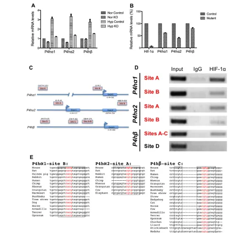
Having found an effect of HIF1on the hydroxylation substrate oxygen, we proceeded to explore its involvement in regulation of the enzyme cP4H. As mentioned, cP4Hs catalyze collagen
[image:7.612.56.477.57.384.2]hydroxylation in the ER. These enzymes are tetramers and consist of two subunits and two subunits. While cP4HI is the main subunit in most cell types, in chondrocytes it is cP4HII (Myllyharju, 2003; Myllyharju and Kivirikko, 2004). Previous in vitro studies have raised the possibility that HIF1may regulate cP4Hs in chondrocytes and other cell types (Copple et al., 2011; Grimmer et al., 2006; Hofbauer et al., 2003; Takahashi et al., 2000). Therefore, the expression of cP4H subunits was first analyzed in chondrocyte primary culture. Under hypoxia, mRNA levels of P4ha1, P4ha2and P4hbwere elevated relative to cells under normoxic conditions (Fig. 6A). This indicated that cP4H subunit genes are hypoxia responsive, consistent with the hypothesis that under hypoxic conditions, an elevation in their expression is necessary to maintain collagen hydroxylation. Fig. 3. ER stress and UPR activation in Hif1a-deficient growth plate. (A,B)In situ hybridization for Bipdemonstrates an elevation in mRNA levels in Hif1a-deficient growth plate (B) relative to the control (A). (C,D)In situ hybridization for Chopdemonstrates increased expression in Hif1a -deficient growth plate (D), compared with the control (C). (E)qRT-PCR shows a 3.7-fold increase in Chop mRNA expression in Hif1a-deficient growth plate (n4, P<0.05 for both genes, data normalized to Tbpand represented as mean±s.e.m.). (F)Western blot analysis indicating an elevation of CHOP protein levels in Hif1a-deficient growth plate (‘Mutant’ lane) compared with the control. An increase in CHOP protein levels is also detected in primary chondrocytes that were treated, as a positive control, with the ER stress inducer brefeldin A (‘+Br-A’ lane), in comparison with untreated control cells (‘no Br-A’ lane). (G)qRT-PCR analysis indicates a 3-fold and a 4.7-fold increase in Bipand ChopmRNA, respectively, in
Hif1a-depleted primary chondrocyte culture under hypoxia (Hyp KO) relative to control cells (Hyp control; P<0.05 in both cases). This elevation is not detected under normoxia (Nor Control/KO; n3, data normalized to Tbpand 18S rRNA and represented as mean±s.e.m.). For mRNA levels of Hif1a, see supplementary material Fig. S2. (H)Western blot analysis demonstrates an increase in CHOP protein levels in Hif1a-depleted primary
chondrocyte culture under hypoxia (Hyp KO), relative to control cells (Hyp control) and relative to control and Hif1a-depleted cells under normoxia (Nor Control/KO). An elevation in CHOP protein levels is also detected in primary chondrocytes that were treated with brefeldin A (+Br-A) as a positive control, in comparison with untreated control cells (no Br-A). (I)Reverse transcription (RT) PCR of Xbp1mRNA products reveals stronger bands of Xbp1unspliced variant (Xbp1U, 220 bp) and Xbp1spliced variant (Xbp1S, 194 bp) in Hif1a-depleted primary chondrocyte culture under hypoxia (Hyp KO), relative to control cells (Hyp Control), indicating an induction of ER stress signaling upon Hif1ainactivation. Splicing of Xbp1is not detected under normoxic conditions (Nor Control/KO). Xbp1spliced variant is also detected in primary chondrocytes that were treated with brefeldin A (+Br-A) as a positive control, but not in untreated control cells (no Br-A). Scale bars: 200m in A-D.
D
E
V
E
LO
P
M
E
N
Next, we analyzed the expression of cP4H subunits in cells where Hif1aexpression was blocked. As can be seen in Fig. 6A, the elevation in the expression of cP4H subunits was lost in Hif1a -depleted cells, suggesting an involvement of HIF1 in their expression by chondrocytes under hypoxia. To validate this hypothesis, we examined in vivo the expression levels of these subunits in Hif1a-deficient growth plates. A significant reduction in P4ha1and P4hbmRNA levels was detected in Hif1a-deficient growth plates, when compared with the control (Fig. 6B). Interestingly, the expression level of P4ha2, the main subunit in chondrocytes, was dramatically reduced by 60% in the absence of Hif1a, when compared with control growth plates. These results imply that in chondrocytes, HIF1regulates the expression of cP4H subunits under hypoxic conditions.
In order to examine whether HIF1directly regulates cP4H expression, we searched the mouse cP4H subunits promoters for HRE sequences. We identified several potential binding sites on P4ha1, P4ha2and P4hbgenes (Fig. 6C; supplementary material Table S1). To determine whether HIF1binds to these putative sequences, a ChIP assay was performed on chondrocyte lysate extracted from wild-type growth plates. We found two active
HIF1-binding sites on P4ha1promoter (sites A and B) and two active binding sites on P4ha2gene (sites A and B). Moreover, one area on P4hbpromoter that contained three potential binding sites (sites A-C) also demonstrated active binding to HIF1(Fig. 6D). Analysis of those sequences, using the conservation track of the UCSC genome browser (Fujita et al., 2011), revealed conservation among species for P4ha1site B, P4ha2site A and P4hbsite C (Fig. 6E). This suggests that the direct regulation of HIF1on the expression of cP4H subunits is evolutionarily conserved.
[image:8.612.53.301.57.338.2]Collectively, these results provide a molecular mechanism for the regulation of collagen hydroxylation by HIF1by showing that this factor controls both the bioavailability of the substrate oxygen and the expression levels of cP4H enzymes.
[image:8.612.316.559.58.420.2]Fig. 4. HIF1regulates collagen hydroxylation. (A)Amino acid analysis on whole growth plate extracts demonstrates a 9% reduction in hydroxyproline/proline ratio (Hyd-Pro/Pro) in Hif1a-deficient growth plates (Mutant) (n3, P<0.05, data represented as mean±s.e.m.). (B,C)Immunostaining for collagen type II (red) on control (B) and Hif1a -depleted (C) primary chondrocytes incubated under hypoxia. Collagen type II staining is detected both intracellularly and extracellularly in control cells, whereas in Hif1a-depleted cells it is detected only intracellularly. Nuclei are stained with DAPI (blue). (D)Amino acid analysis on control (Hyp Control) and Hif1a-depleted (Hyp KO) primary chondrocytes incubated under hypoxia. Hydroxyproline (Hyd-Pro) was undetectable (UD) in Hif1a-depleted cells but not in control cells, whereas the amount of proline (Pro) was comparable (n3, data represented as mean±s.e.m.).
Fig. 5. HIF1controls oxygen consumption in the growth plate by directly regulating Pdk1expression. (A-B”) Hypoxyprobe staining of control (A-A”) and Hif1a-deficient (B-B”) growth plates indicates a substantially increased signal in the mutant. (C)qRT-PCR demonstrates 53% reduction in Pdk1expression levels in Hif1a -deficient growth plate, compared with the control (n5, P<0.05 for both genes, data were normalized to Tbpand are represented as mean±s.e.m.). (D)In vivo ChIP analysis on chondrocyte lysate extracted from wild-type growth plates demonstrates active HIF1binding to two areas on the mouse Pdk1gene (sites A-C and D-G; +1 is the
transcription start site, also marked by the blue arrow). For the predicted sequences of HIF1-binding sites, see supplementary material Table S1. As a positive control, the known binding site of HIF1to the promoter of Vegf(Hu et al., 2006) was analyzed.
D
E
V
E
LO
P
M
E
N
DISCUSSION
Post-translational modifications (PTM) are essential for protein secretion and function both during development and through the life of the organism. In humans, collagens are the most abundant proteins and hydroxylation of proline residues is the most common PTM during collagen production. As collagen proline
[image:9.612.56.505.57.517.2]hydroxylation is dependent on bioavailability of molecular oxygen, variation in tissue oxygen levels dictates the need for a mechanism that would secure the robustness of this pivotal process. In this study, we identify HIF1as a central component in a mechanism that allows collagen hydroxylation in hypoxic cells by controlling the level of both the catalyst and the substrate of the reaction. Fig. 6. HIF1directly regulates cP4H expression. (A)qRT-PCR demonstrates an approximate threefold increase of collagen prolyl 4-hydroxylase subunits P4ha1, P4ha2and P4hbmRNA levels in chondrocyte primary culture under hypoxia (Hyp Control), when compared with normoxic conditions (Nor Control). Upon Hif1ainactivation (Hyp KO), this elevation is lost (n3, P<0.05 for all genes in both cases, data normalized to Tbp
and 18S rRNA and represented as mean±s.e.m.). For mRNA levels of Hif1a, see supplementary material Fig. S2. (B)qRT-PCR reveals an ~39%, 60% and 20% reduction in P4ha1, P4ha2and P4hbmRNA levels, respectively, in Hif1a-deficient growth plate compared with the control (n3, P<0.05 for all genes, data normalized to Tbpand represented as mean±s.e.m.). (C)The positions of several HIF1potential binding sites identified on mouse P4ha1, P4ha2and P4hbgenes (+1 is the transcription start site, also marked by blue arrows, for the sites sequences see supplementary material Table S1). (D)In vivo ChIP analysis on chondrocyte lysate extracted from wild-type growth plates demonstrates active HIF1binding to sites A and B on P4ha1promoter, sites A and B on P4ha2gene and to the P4hbpromoter sequence that includes sites A-C (active sites are marked in red). As a positive control, known binding site of HIF1on the promoter of Vegf(Hu et al., 2006) is demonstrated in Fig. 5D. (E)Conservation among species of the following mouse sequences containing the HIF1binding site: P4ha1site B (chr10:58786018-58786042), P4ha2site A (chr11:53914340-53914360), P4hbsite C (chr11:120434609-120434631). The HIF1-binding site is written in bold letters on the mouse sequence and marked with a box in all sequences. Highlighted in red is the core consensus sequence (four nucleotides).
D
E
V
E
LO
P
M
E
N
The evolutionary origin of HIF1coincides with the divergence of metazoans (Loenarz et al., 2011; Rytkönen et al., 2011). During the evolution of multicellular organisms, oxygen supply to all cells has become a major challenge owing to environmental constraints and an increase in organism complexity and size. Metazoan cells had to overcome two fundamental problems. The first was to adapt their metabolism in order to ensure survival under varying oxygen levels. The second necessity was to produce under these conditions collagen, which serves as a ‘glue’ that holds the cells together and guarantees the integrity of connective tissues (Towe, 1970). The need to overcome these problems fits well with the emergence of HIF1as a major regulator of oxygen homeostasis. The role of HIF1in metabolic adaptation to hypoxia was extensively studied and described. By contrast, its involvement in collagen production has thus far been neglected.
Here, we demonstrate in vivo the ability of HIF1to regulate directly the transcription of the cP4H subunits cP4HI, cP4HII and cP4Hin hypoxic growth plate chondrocytes. We also show that, concurrently, HIF1maintains cellular levels of the substrate
oxygen, most likely by controlling the expression of Pdk1, a key component in the mechanism that regulates cellular oxygen level (Fukuda et al., 2007; Kim et al., 2006; Papandreou et al., 2006; Semenza, 2007). Hence, HIF1acts both to increase the level of the enzyme and to maintain the level of the substrate. In the absence of HIF1, intracellular oxygen levels and cP4H subunits expression are reduced, leading to reduction in collagen type II hydroxylation and, consequently, to an arrest in its secretion and its intracellular accumulation (Fig. 7).
Our finding that HIF1regulates cP4H subunits corresponds with previous in vitro studies in different cell types, including fibroblasts and hepatoma cells, which suggest that HIF1regulates cP4HI and cP4HII expression (Copple et al., 2011; Grimmer et al., 2006; Hofbauer et al., 2003; Takahashi et al., 2000). This raises the possibility that HIF1-mediated regulation of cP4Hs is a broad phenomenon that is not restricted to chondrocytes.
Although our results explain collagen hydroxylation under hypoxic conditions, it may be just the tip of the iceberg regarding the involvement of HIF1 in ECM secretion. Several pieces of evidence support this supposition. First, in vitro studies demonstrate the involvement of HIF1in regulating the expression of procollagen lysyl-hydroxylases (Plod) that catalyze hydroxylation of lysine residues, which is also required for collagen maturation (Hofbauer et al., 2003). Another indication is our finding that HIF1regulates the expression of cP4H, which is identical to protein disulfide isomerase (PDI). In addition to its role in collagen hydroxylation, cP4Hcatalyzes the formation and rearrangement of disulfide bonds, which are required for proper folding of collagen and other proteins (Feige and Hendershot, 2011; Forster and Freedman, 1984; Hatahet and Ruddock, 2009; Koivu and Myllylä, 1987). Moreover, it was previously reported that HIF1regulates the expression of ERO1L, which is also involved in disulfide bond formation (May et al., 2005). As the formation of disulfide bonds is also oxygen-dependent, HIF1 may also regulate this process indirectly by maintaining oxygen homeostasis.
Further support for the central role of HIF1in ECM secretion is our finding that blockage of Hif1aexpression also leads to accumulation of proteoglycans, such as aggrecan and perlecan, within chondrocytes. It was previously reported that interference with proteoglycan glycosylation in the Golgi inhibits their secretion (Hameetman et al., 2007; Ratcliffe et al., 1985). HIF1is known to regulate uptake and consumption of glucose, which is necessary for proteoglycan glycosylation (Mobasheri et al., 2002; Peansukmanee et al., 2009; Ren et al., 2008). This suggests that HIF1 may regulate not only collagen hydroxylation, but also proteoglycan glycosylation. It is also possible that HIF1regulates a battery of genes that are necessary for the secretion of a variety of ECM components under hypoxia. Moreover, previous in vitro studies suggested that HIF1 is involved in regulating proteoglycans and collagen expression (Pfander et al., 2003). Collectively, these reports and the findings we present here may indicate a major involvement of HIF1in ECM production and secretion under hypoxic conditions.
[image:10.612.52.298.64.280.2]ER stress is known to have severe deleterious effects that significantly contribute to the pathology of a variety of diseases (Bateman et al., 2009; Boot-Handford and Briggs, 2010; Lisse et al., 2008; Rajpar et al., 2009; Tsang et al., 2010). However, several recent studies indicate that during bone development, ER stress in hypoxic chondrocytes plays a role in the normal sequence of their differentiation (Patra et al., 2007; Saito et al., 2009). Our findings that link among hypoxic conditions, PTMs, HIF1and ER stress may implicate HIF1as an essential molecule in this mechanism. Fig. 7. HIF1regulates collagen hydroxylation and secretion in
the hypoxic growth plate.A model that summarizes our findings on the role of HIF1in regulating collagen hydroxylation and secretion. Left panel, wild-type chondrocyte; right panel, Hif1a-deficient chondrocyte. (I, left) In the hypoxic growth plate, HIF1directly activates the transcription of P4ha1, P4ha2and P4hb, the subunits of collagen prolyl 4-hydroxylase enzyme (P4H, marked in orange). In addition, HIF1directly activates the transcription of pyruvate dehydrogenase kinase 1 (Pdk1). (II, left) PDK1 inhibits oxygen consumption through inhibition of the TCA cycle to maintain oxygen homeostasis. (III, left) Oxygen is used by P4H to hydroxylate collagen. Collagen hydroxylation is required for the formation of the triple helix (procollagen). (IV, left) Normal collagen secretion. (I, right) The expression of P4ha1, P4ha2, P4hband Pdk1is reduced in
Hif1a-deficient chondrocyte. (II, right) Oxygen levels are reduced as a result of reduced Pdk1expression and increased oxygen consumption by the TCA cycle. (III, right) Collagen hydroxylation and folding is impaired due to reduced P4H levels and reduced oxygen levels. Unfolded collagen accumulates in the ER. (IV, right) The accumulation of unfolded collagen induces ER stress and results in impaired collagen secretion in
Hif1a-deficient chondrocyte.
D
E
V
E
LO
P
M
E
N
Further support for this notion comes from a recent finding that HIF1can induce ER stress in chondrocytes (Yang et al., 2008). In light of this, changes in HIF1levels may determine the level of ER stress in those cells.
Finally, the connection we found between HIF1and ER stress may provide a new explanation for the massive chondrocyte cell death observed upon Hif1ainactivation, which was previously attributed to impaired glycolysis and to the role of HIF1as a regulator of cell metabolism (Maes et al., 2012; Schipani et al., 2001). Another possible explanation is our finding that impaired matrix secretion in Hif1a-deficient chondrocytes results in loss of the vital cell-matrix attachment (Aszodi et al., 2003; Hirsch et al., 1997; Pulai et al., 2002; Raducanu et al., 2009; Svoboda, 1998; Woods et al., 2007).
This work describes a novel role for HIF1as a central regulator of ECM secretion, which ensures the function of chondrocytes as professional secretory cells in the hypoxic growth plate. Focusing on the secretion of collagen type II, a major ECM component in cartilage, we demonstrate that HIF1 controls a two-armed mechanism to maintain collagen PTM in chondrocytes under hypoxia. It operates by regulating both the expression of cP4H subunits, which catalyze collagen hydroxylation, and the availability of oxygen – the substrate of the reaction. The identification of HIF1 as a pivotal regulator of ECM secretion under hypoxia significantly increases the extent of its known involvement in cellular homeostasis and may shed new light on metazoan evolution.
Acknowledgements
We thank N. Konstantin for expert editorial assistance, S. Krief for expert technical support, and all members of the Zelzer laboratory for advice and suggestions. We thank Prof. R. S. Johnson for kindly providing us with
floxed-Hif1amice. We appreciate the collaboration and fruitful discussions with Prof. D. R. Eyre, M. Weis and Prof. E. B. Hunziker. We thank Prof. C. W. Pugh for kindly providing us with the anti-sera to HIF1. We are grateful to R. Kramer, Dr A. Tishbee and Dr V. Shinder from the Department of Chemical Research Support, and to Dr S. Ben-Dor from the Biological Services Unit at the Weizmann Institute of Science. We also acknowledge the help and advice of S. Viukov, K. B. Umansky and Dr O. Meir. The electron microscopy studies were conducted at the Irving and Cherna Moskowitz Center for Nano and Bio-Nano Imaging at the Weizmann Institute of Science.
Funding
This work was supported by grants from the United States-Israel Binational Science Foundation (BSF) [2007307], Israel Science Foundation (ISF) [1206/09], Minerva Foundation [M1138], The Y. Leon Benoziyo Institute for Molecular Medicine, Helen and Martin Kimmel Institute for Stem Cell Research, J and R Center for Scientific Research, Estate of Raymond Lapon, Estate of David Levinson, The Leo and Julia Forchheimer Center for Molecular Genetics, and Marla L. Schaefer (New York, NY, USA). E.Z. is the incumbent of the Martha S. Sagon Career Development Chair.
Competing interests statement
The authors declare no competing financial interests.
Supplementary material
Supplementary material available online at
http://dev.biologists.org/lookup/suppl/doi:10.1242/dev.083881/-/DC1
References
Ainbinder, E., Revach, M., Wolstein, O., Moshonov, S., Diamant, N. and Dikstein, R.(2002). Mechanism of rapid transcriptional induction of tumor necrosis factor alpha-responsive genes by NF-kappaB. Mol. Cell. Biol.22, 6354-6362.
Amarilio, R., Viukov, S. V., Sharir, A., Eshkar-Oren, I., Johnson, R. S. and Zelzer, E.(2007). HIF1alpha regulation of Sox9 is necessary to maintain differentiation of hypoxic prechondrogenic cells during early skeletogenesis.
Development134, 3917-3928.
Arteel, G. E., Thurman, R. G. and Raleigh, J. A.(1998). Reductive metabolism of the hypoxia marker pimonidazole is regulated by oxygen tension independent of the pyridine nucleotide redox state. Eur. J. Biochem.253, 743-750.
Aszodi, A., Hunziker, E. B., Brakebusch, C. and Fässler, R.(2003). Beta1 integrins regulate chondrocyte rotation, G1 progression, and cytokinesis. Genes Dev.17, 2465-2479.
Bateman, J. F., Boot-Handford, R. P. and Lamandé, S. R.(2009). Genetic diseases of connective tissues: cellular and extracellular effects of ECM mutations. Nat. Rev. Genet.10, 173-183.
Berg, R. A. and Prockop, D. J.(1973). The thermal transition of a non-hydroxylated form of collagen. Evidence for a role for hydroxyproline in stabilizing the triple-helix of collagen. Biochem. Biophys. Res. Commun.52, 115-120.
Boot-Handford, R. P. and Briggs, M. D.(2010). The unfolded protein response and its relevance to connective tissue diseases. Cell Tissue Res.339, 197-211.
Calfon, M., Zeng, H., Urano, F., Till, J. H., Hubbard, S. R., Harding, H. P., Clark, S. G. and Ron, D.(2002). IRE1 couples endoplasmic reticulum load to secretory capacity by processing the XBP-1 mRNA. Nature415, 92-96.
Cartharius, K., Frech, K., Grote, K., Klocke, B., Haltmeier, M., Klingenhoff, A., Frisch, M., Bayerlein, M. and Werner, T.(2005). MatInspector and beyond: promoter analysis based on transcription factor binding sites.
Bioinformatics21, 2933-2942.
Cooper, G. W. and Prockop, D. J.(1968). Intracellular accumulation of protocollagen and extrusion of collagen by embryonic cartilage cells. J. Cell Biol.
38, 523-537.
Copple, B. L., Bai, S., Burgoon, L. D. and Moon, J. O.(2011). Hypoxia-inducible factor-1regulates the expression of genes in hypoxic hepatic stellate cells important for collagen deposition and angiogenesis. Liver Int.31, 230-244.
Dunwoodie, S. L.(2009). The role of hypoxia in development of the Mammalian embryo. Dev. Cell17, 755-773.
Eshkar-Oren, I., Viukov, S. V., Salameh, S., Krief, S., Oh, C. D., Akiyama, H., Gerber, H. P., Ferrara, N. and Zelzer, E.(2009). The forming limb skeleton serves as a signaling center for limb vasculature patterning via regulation of Vegf. Development136, 1263-1272.
Feige, M. J. and Hendershot, L. M.(2011). Disulfide bonds in ER protein folding and homeostasis. Curr. Opin. Cell Biol.23, 167-175.
Forster, S. J. and Freedman, R. B.(1984). Catalysis by protein disulphide-isomerase of the assembly of trimeric procollagen from procollagen polypeptide chains. Biosci. Rep.4, 223-229.
Fujita, P. A., Rhead, B., Zweig, A. S., Hinrichs, A. S., Karolchik, D., Cline, M. S., Goldman, M., Barber, G. P., Clawson, H., Coelho, A. et al.(2011). The UCSC Genome Browser database: update 2011. Nucleic Acids Res.39, D876-D882.
Fukuda, R., Zhang, H., Kim, J. W., Shimoda, L., Dang, C. V. and Semenza, G. L.(2007). HIF-1 regulates cytochrome oxidase subunits to optimize efficiency of respiration in hypoxic cells. Cell129, 111-122.
Grimmer, C., Balbus, N., Lang, U., Aigner, T., Cramer, T., Müller, L., Swoboda, B. and Pfander, D.(2006). Regulation of type II collagen synthesis during osteoarthritis by prolyl-4-hydroxylases: possible influence of low oxygen levels.
Am. J. Pathol.169, 491-502.
Hameetman, L., David, G., Yavas, A., White, S. J., Taminiau, A. H., Cleton-Jansen, A. M., Hogendoorn, P. C. and Bovée, J. V.(2007). Decreased EXT expression and intracellular accumulation of heparan sulphate proteoglycan in osteochondromas and peripheral chondrosarcomas. J. Pathol.211, 399-409.
Hatahet, F. and Ruddock, L. W.(2009). Protein disulfide isomerase: a critical evaluation of its function in disulfide bond formation. Antioxid. Redox Signal.
11, 2807-2850.
Hirsch, M. S., Lunsford, L. E., Trinkaus-Randall, V. and Svoboda, K. K.(1997). Chondrocyte survival and differentiation in situ are integrin mediated. Dev. Dyn.
210, 249-263.
Ho, M. S., Tsang, K. Y., Lo, R. L., Susic, M., Mäkitie, O., Chan, T. W., Ng, V. C., Sillence, D. O., Boot-Handford, R. P., Gibson, G. et al.(2007). COL10A1 nonsense and frame-shift mutations have a gain-of-function effect on the growth plate in human and mouse metaphyseal chondrodysplasia type Schmid.
Hum. Mol. Genet.16, 1201-1215.
Hofbauer, K. H., Gess, B., Lohaus, C., Meyer, H. E., Katschinski, D. and Kurtz, A.(2003). Oxygen tension regulates the expression of a group of procollagen hydroxylases. Eur. J. Biochem.270, 4515-4522.
Hu, C. J., Iyer, S., Sataur, A., Covello, K. L., Chodosh, L. A. and Simon, M. C.
(2006). Differential regulation of the transcriptional activities of hypoxia-inducible factor 1 alpha (HIF-1alpha) and HIF-2alpha in stem cells. Mol. Cell. Biol.
26, 3514-3526.
Hunziker, E. B., Herrmann, W. and Schenk, R. K.(1983). Ruthenium hexammine trichloride (RHT)-mediated interaction between plasmalemmal components and pericellular matrix proteoglycans is responsible for the preservation of chondrocytic plasma membranes in situ during cartilage fixation.
J. Histochem. Cytochem.31, 717-727.
Juva, K., Prockop, D. J., Cooper, G. W. and Lash, J. W.(1966). Hydroxylation of proline and the intracellular accumulation of a polypeptide precursor of collagen. Science152, 92-94.
Kiani, C., Chen, L., Wu, Y. J., Yee, A. J. and Yang, B. B.(2002). Structure and function of aggrecan. Cell Res.12, 19-32.
Kim, J. W., Tchernyshyov, I., Semenza, G. L. and Dang, C. V.(2006). HIF-1-mediated expression of pyruvate dehydrogenase kinase: a metabolic switch
required for cellular adaptation to hypoxia. Cell Metab.3, 177-185.
D
Koivu, J. and Myllylä, R.(1987). Interchain disulfide bond formation in types I and II procollagen. Evidence for a protein disulfide isomerase catalyzing bond formation. J. Biol. Chem.262, 6159-6164.
Kvist, A. J., Nyström, A., Hultenby, K., Sasaki, T., Talts, J. F. and Aspberg, A.
(2008). The major basement membrane components localize to the chondrocyte pericellular matrix – a cartilage basement membrane equivalent? Matrix Biol.27, 22-33.
Lamandé, S. R. and Bateman, J. F.(1999). Procollagen folding and assembly: the role of endoplasmic reticulum enzymes and molecular chaperones. Semin. Cell Dev. Biol.10, 455-464.
Lin, J. H., Walter, P. and Yen, T. S.(2008). Endoplasmic reticulum stress in disease pathogenesis. Annu. Rev. Pathol.3, 399-425.
Lisse, T. S., Thiele, F., Fuchs, H., Hans, W., Przemeck, G. K., Abe, K., Rathkolb, B., Quintanilla-Martinez, L., Hoelzlwimmer, G., Helfrich, M. et al.(2008). ER stress-mediated apoptosis in a new mouse model of osteogenesis imperfecta.
PLoS Genet.4, e7.
Loenarz, C., Coleman, M. L., Boleininger, A., Schierwater, B., Holland, P. W., Ratcliffe, P. J. and Schofield, C. J.(2011). The hypoxia-inducible transcription factor pathway regulates oxygen sensing in the simplest animal, Trichoplax adhaerens. EMBO Rep.12, 63-70.
Mackie, E. J., Ahmed, Y. A., Tatarczuch, L., Chen, K. S. and Mirams, M.
(2008). Endochondral ossification: how cartilage is converted into bone in the developing skeleton. Int. J. Biochem. Cell Biol.40, 46-62.
Maes, C., Araldi, E., Haigh, K., Khatri, R., Van Looveren, R., Giaccia, A. J., Haigh, J. J., Carmeliet, G. and Schipani, E.(2012). VEGF-independent cell-autonomous functions of HIF-1alpha regulating oxygen consumption in fetal cartilage are critical for chondrocyte survival. J. Bone Miner. Res.27, 596-609.
Maltepe, E. and Simon, M. C.(1998). Oxygen, genes, and development: an analysis of the role of hypoxic gene regulation during murine vascular development. J. Mol. Med. (Berl.)76, 391-401.
May, D., Itin, A., Gal, O., Kalinski, H., Feinstein, E. and Keshet, E.(2005). Ero1-L alpha plays a key role in a HIF-1-mediated pathway to improve disulfide bond formation and VEGF secretion under hypoxia: implication for cancer.
Oncogene24, 1011-1020.
Mitchell, J. A. and Yochim, J. M.(1968). Measurement of intrauterine oxygen tension in the rat and its regulation by ovarian steroid hormones. Endocrinology
83, 691-700.
Mobasheri, A., Vannucci, S. J., Bondy, C. A., Carter, S. D., Innes, J. F., Arteaga, M. F., Trujillo, E., Ferraz, I., Shakibaei, M. and Martín-Vasallo, P.
(2002). Glucose transport and metabolism in chondrocytes: a key to
understanding chondrogenesis, skeletal development and cartilage degradation in osteoarthritis. Histol. Histopathol.17, 1239-1267.
Murtaugh, L. C., Chyung, J. H. and Lassar, A. B.(1999). Sonic hedgehog promotes somitic chondrogenesis by altering the cellular response to BMP signaling. Genes Dev.13, 225-237.
Myllyharju, J.(2003). Prolyl 4-hydroxylases, the key enzymes of collagen biosynthesis. Matrix Biol.22, 15-24.
Myllyharju, J. and Kivirikko, K. I.(2004). Collagens, modifying enzymes and their mutations in humans, flies and worms. Trends Genet.20, 33-43.
Nakamura, E., Nguyen, M. T. and Mackem, S.(2006). Kinetics of tamoxifen-regulated Cre activity in mice using a cartilage-specific CreER(T) to assay temporal activity windows along the proximodistal limb skeleton. Dev. Dyn.235, 2603-2612.
Nuehring, L. P., Steffens, W. L. and Rowland, G. N.(1991). Comparison of the Ruthenium hexammine trichloride method to other methods of chemical fixation for preservation of avian physeal cartilage. Histochem. J.23, 201-214.
Papandreou, I., Cairns, R. A., Fontana, L., Lim, A. L. and Denko, N. C.(2006). HIF-1 mediates adaptation to hypoxia by actively downregulating mitochondrial oxygen consumption. Cell Metab.3, 187-197.
Patra, D., Xing, X., Davies, S., Bryan, J., Franz, C., Hunziker, E. B. and Sandell, L. J.(2007). Site-1 protease is essential for endochondral bone formation in mice. J. Cell Biol.179, 687-700.
Peansukmanee, S., Vaughan-Thomas, A., Carter, S. D., Clegg, P. D., Taylor, S., Redmond, C. and Mobasheri, A.(2009). Effects of hypoxia on glucose transport in primary equine chondrocytes in vitro and evidence of reduced GLUT1 gene expression in pathologic cartilage in vivo. J. Orthop. Res.27, 529-535.
Pfander, D., Cramer, T., Schipani, E. and Johnson, R. S.(2003). HIF-1alpha controls extracellular matrix synthesis by epiphyseal chondrocytes. J. Cell Sci.
116, 1819-1826.
Provot, S. and Schipani, E.(2005). Molecular mechanisms of endochondral bone development. Biochem. Biophys. Res. Commun.328, 658-665.
Provot, S., Zinyk, D., Gunes, Y., Kathri, R., Le, Q., Kronenberg, H. M., Johnson, R. S., Longaker, M. T., Giaccia, A. J. and Schipani, E.(2007). Hif-1alpha regulates differentiation of limb bud mesenchyme and joint
development. J. Cell Biol.177, 451-464.
Pulai, J. I., Del Carlo, M., Jr and Loeser, R. F.(2002). The alpha5beta1 integrin provides matrix survival signals for normal and osteoarthritic human articular chondrocytes in vitro. Arthritis Rheum.46, 1528-1535.
Raducanu, A., Hunziker, E. B., Drosse, I. and Aszódi, A.(2009). Beta1 integrin deficiency results in multiple abnormalities of the knee joint. J. Biol. Chem.284, 23780-23792.
Rajpar, M. H., McDermott, B., Kung, L., Eardley, R., Knowles, L., Heeran, M., Thornton, D. J., Wilson, R., Bateman, J. F., Poulsom, R. et al.(2009). Targeted induction of endoplasmic reticulum stress induces cartilage pathology.
PLoS Genet.5, e1000691.
Ratcliffe, A., Fryer, P. R. and Hardingham, T. E.(1985). Proteoglycan
biosynthesis in chondrocytes: protein A-gold localization of proteoglycan protein core and chondroitin sulfate within Golgi subcompartments. J. Cell Biol.101, 2355-2365.
Ren, B. F., Deng, L. F., Wang, J., Zhu, Y. P., Wei, L. and Zhou, Q.(2008). Hypoxia regulation of facilitated glucose transporter-1 and glucose transporter-3 in mouse chondrocytes mediated by HIF-1alpha. Joint Bone Spine75, 176-181.
Riddle, R. D., Johnson, R. L., Laufer, E. and Tabin, C.(1993). Sonic hedgehog mediates the polarizing activity of the ZPA. Cell75, 1401-1416.
Rodesch, F., Simon, P., Donner, C. and Jauniaux, E.(1992). Oxygen
measurements in endometrial and trophoblastic tissues during early pregnancy.
Obstet. Gynecol.80, 283-285.
Ron, D. and Walter, P.(2007). Signal integration in the endoplasmic reticulum unfolded protein response. Nat. Rev. Mol. Cell Biol.8, 519-529.
Roughley, P. J.(2006). The structure and function of cartilage proteoglycans. Eur. Cell. Mater.12, 92-101.
Ryan, H. E., Poloni, M., McNulty, W., Elson, D., Gassmann, M., Arbeit, J. M. and Johnson, R. S.(2000). Hypoxia-inducible factor-1alpha is a positive factor in solid tumor growth. Cancer Res.60, 4010-4015.
Rytkönen, K. T., Williams, T. A., Renshaw, G. M., Primmer, C. R. and Nikinmaa, M.(2011). Molecular evolution of the metazoan PHD-HIF oxygen-sensing system. Mol. Biol. Evol.28, 1913-1926.
Saito, A., Hino, S., Murakami, T., Kanemoto, S., Kondo, S., Saitoh, M., Nishimura, R., Yoneda, T., Furuichi, T., Ikegawa, S. et al.(2009). Regulation of endoplasmic reticulum stress response by a BBF2H7-mediated Sec23a pathway is essential for chondrogenesis. Nat. Cell Biol.11, 1197-1204.
Schipani, E., Ryan, H. E., Didrickson, S., Kobayashi, T., Knight, M. and Johnson, R. S.(2001). Hypoxia in cartilage: HIF-1alpha is essential for chondrocyte growth arrest and survival. Genes Dev.15, 2865-2876.
Schwartz, N. B. and Domowicz, M.(2002). Chondrodysplasias due to proteoglycan defects. Glycobiology12, 57R-68R.
Semenza, G. L.(2007). Oxygen-dependent regulation of mitochondrial respiration by hypoxia-inducible factor 1. Biochem. J.405, 1-9.
Semenza, G. L.(2009). Regulation of oxygen homeostasis by hypoxia-inducible factor 1. Physiology (Bethesda)24, 97-106.
Semenza, G. L.(2012). Hypoxia-inducible factors in physiology and medicine. Cell
148, 399-408.
Simon, M. C. and Keith, B.(2008). The role of oxygen availability in embryonic development and stem cell function. Nat. Rev. Mol. Cell Biol.9, 285-296.
Smits, P., Bolton, A. D., Funari, V., Hong, M., Boyden, E. D., Lu, L., Manning, D. K., Dwyer, N. D., Moran, J. L., Prysak, M. et al.(2010). Lethal skeletal dysplasia in mice and humans lacking the golgin GMAP-210. N. Engl. J. Med.
362, 206-216.
Soriano, P.(1999). Generalized lacZ expression with the ROSA26 Cre reporter strain. Nat. Genet.21, 70-71.
Svoboda, K. K.(1998). Chondrocyte-matrix attachment complexes mediate survival and differentiation. Microsc. Res. Tech.43, 111-122.
Takahashi, Y., Takahashi, S., Shiga, Y., Yoshimi, T. and Miura, T.(2000). Hypoxic induction of prolyl 4-hydroxylase alpha (I) in cultured cells. J. Biol. Chem.
275, 14139-14146.
Towe, K. M.(1970). Oxygen-collagen priority and the early metazoan fossil record.
Proc. Natl. Acad. Sci. USA65, 781-788.
Tsang, K. Y., Chan, D., Bateman, J. F. and Cheah, K. S.(2010). In vivo cellular adaptation to ER stress: survival strategies with double-edged consequences. J. Cell Sci.123, 2145-2154.
Uitto, J., Hoffman, H. and Prockop, D. J.(1975). Retention of nonhelical procollagen containing cis-hydroxyproline in rough endoplasmic reticulum.
Science190, 1202-1204.
Weidemann, A. and Johnson, R. S.(2008). Biology of HIF-1alpha. Cell Death Differ.15, 621-627.
Wenger, R. H., Stiehl, D. P. and Camenisch, G.(2005). Integration of oxygen signaling at the consensus HRE. Sci. STKE2005, re12.
Woods, A., Wang, G. and Beier, F.(2007). Regulation of chondrocyte differentiation by the actin cytoskeleton and adhesive interactions. J. Cell. Physiol.213, 1-8.
Yang, G., Sun, Q., Teng, Y., Li, F., Weng, T. and Yang, X.(2008). PTEN deficiency causes dyschondroplasia in mice by enhanced hypoxia-inducible factor 1alpha signaling and endoplasmic reticulum stress. Development135, 3587-3597.
Yoshida, H., Matsui, T., Yamamoto, A., Okada, T. and Mori, K.(2001). XBP1 mRNA is induced by ATF6 and spliced by IRE1 in response to ER stress to produce a highly active transcription factor. Cell107, 881-891.
![Fig. 1. Temporally controlled (and represented as mean±s.e.m.]. (E,F(E) and mutant (F) growth plates 48 hours post tamoxifen injection,demonstrating reduced cell number and reduced Hematoxylin andEosin staining of the matrix in the hypoxic central region o](https://thumb-us.123doks.com/thumbv2/123dok_us/8909865.958709/5.612.52.296.59.435/temporally-controlled-represented-tamoxifen-injection-demonstrating-hematoxylin-staining.webp)