INTRODUCTION
Hedgehog (Hh) signalling is essential for the correct patterning of virtually every vertebrate organ system, including the limb (Hill, 2007; Ingham and McMahon, 2001). In the absence of a hedgehog ligand (Sonic, Shh; Indian or Desert), the transmembrane receptor patched 1 (Ptc1; also known as Ptch1) constitutively inhibits the transducer of the Hh signal, smoothened (Smo) (Chen and Struhl, 1996; Marigo et al., 1996a). In this repressed state, proteolytically cleaved Gli transcription factors, primarily Gli3, translocate to the nucleus to repress target genes. Upon binding of a hedgehog ligand to Ptc1, inhibition of Smo is relieved, Gli cleavage is inhibited, and full-length Gli proteins are converted to transcriptional activators. Importantly Ptc1, an inhibitor of the pathway, is transcriptionally upregulated in response to Hh signalling, creating a negative-feedback loop that is crucial for tight spatiotemporal control of signalling levels. Disruption of this feedback loop by inactivation of Ptc1 in mice results in inappropriate ligand-independent activation of the Hh pathway, and early embryonic lethality around 9.5 days post coitum (dpc) (Ellis et al., 2003; Goodrich et al., 1997).
In the limb, Shh is secreted from a group of posterior mesenchymal cells called the zone of polarizing activity (ZPA) (Riddle et al., 1993). The posteriorly biased Shh gradient, together with a temporal gradient of exposure to the Shh signal, specifies digit number and identity (reviewed by McGlinn and Tabin, 2006). Shh successively inhibits the cleavage of full-length Gli3 (Gli3FL) to the repressor form (Gli3R) from posterior to anterior (Wang et al., 2000), and this interplay between Shh and Gli3 is crucial in defining anterior-posterior (AP) limb patterning (Litingtung et al., 2002; te Welscher et al., 2002b). The Shhnull mouse hindlimb has a single
digit corresponding to the most anterior digit 1 (Chiang et al., 2001), whereas the extra-toes(Gli3Xt/Xt) mouse mutant lacks functional Gli3 and displays extra digits, or polydactyly (Hui and Joyner, 1993). While Gli3R is regarded as the primary effector of AP patterning in the limb, the role of Gli3FL in this process is the subject of some debate (Hill et al., 2009; Wang et al., 2007a; Wang et al., 2007b).
The nascent limb bud is subject to an exquisitely controlled network of gene regulatory loops that pattern the limb bud both before and after expression of Shh. Soon after emergence of the limb bud, an AP pre-pattern is established whereby anterior Gli3R restricts Hand2expression to the posterior, and Hand2 reciprocally limits Gli3to the anterior (te Welscher et al., 2002a). At this stage, the posterior restriction of 5⬘Hoxd genes (those genes at the 5⬘end of the Hoxd cluster) is crucial to final AP limb asymmetry, as these genes subsequently trigger expression of Shhspecifically in the posterior limb (Tarchini et al., 2006; Zakany et al., 2004). Around the same time, Bmp4 induces expression of the BMP antagonist gremlin (Grem1), thus initiating the first of a number of finely tuned regulatory loops linking Shh signalling in the ZPA with FGF signalling from the distal apical ectodermal ridge (AER) (Benazet et al., 2009). Downregulation of Bmp4 by Grem1 enables Shh signalling from the ZPA, thus establishing the Shh/Grem1/FGF loop, whereby Shh induces Grem1expression in the mesenchyme. This then allows FGF signalling from the AER, which in turn maintains Shh in the ZPA (Khokha et al., 2003; Zuniga et al., 1999). A recent study suggests that limb outgrowth ceases when the Shh/Grem1/FGF loop drives FGF signalling to high enough levels to inhibit Grem1expression (Verheyden and Sun, 2008). However, previous studies in the chick indicate that the inability of Shh expressing cells and their descendants to express Grem1contributes to termination of limb outgrowth, as Grem1-expressing cells move beyond the influence of Shh from the ZPA (Nissim et al., 2006; Scherz et al., 2004).
Ptc1null mice, or mice in which Ptc1 is ubiquitously inactivated, die prior to the initiation of limb budding (Ellis et al., 2003; Goodrich et al., 1997). Despite elegant studies involving transgenic rescue of
Patched 1 is a crucial determinant of asymmetry and digit
number in the vertebrate limb
Natalie C. Butterfield1, Vicki Metzis1, Edwina McGlinn2, Stephen J. Bruce1, Brandon J. Wainwright1and Carol Wicking1,*
The vertebrate hedgehog receptor patched 1 (Ptc1) is crucial for negative regulation of the sonic hedgehog (Shh) pathway during anterior-posterior patterning of the limb. We have conditionally inactivated Ptc1in the mesenchyme of the mouse limb using Prx1 -Cre. This results in constitutive activation of hedgehog (Hh) signalling during the early stages of limb budding. Our data suggest that variations in the timing and efficiency of Cre-mediated excision result in differential forelimb and hindlimb phenotypes. Hindlimbs display polydactyly (gain of digits) and a molecular profile similar to the Gli3 mutant extra-toes. Strikingly, forelimbs are predominantly oligodactylous (displaying a loss of digits), with a symmetrical, mirror-image molecular profile that is consistent with re-specification of the anterior forelimb to a posterior identity. Our data suggest that this is related to very early inactivation of Ptc1 in the forelimb perturbing the gene regulatory networks responsible for both the pre-patterning and the subsequent patterning stages of limb development. These results establish the importance of the downstream consequences of Hh pathway repression, and identify Ptc1 as a key player in limb patterning even prior to the onset of Shh expression.
KEY WORDS: Patched 1, Sonic hedgehog signalling, Limb development, Prx1-Cre, Shh/Grem1/FGF loop, Mouse Development 136, 3515-3524 (2009) doi:10.1242/dev.037507
1The University of Queensland, Institute for Molecular Bioscience, Queensland 4072, Australia. 2Harvard Medical School, 77 Avenue Louis Pasteur, Boston, MA 02115, USA.
*Author for correspondence (c.wicking@imb.uq.edu.au)
Accepted 21 August 2009
D
E
V
E
LO
P
M
E
N
Ptc1 null mice (Milenkovic et al., 1999), the effects of Ptc1 inactivation in the early limb have not previously been investigated. We have used Cre recombinase driven by a Prx1enhancer (Logan et al., 2002) to conditionally remove functionalPtc1from the limb mesenchyme. This results in high-level ligand-independent activation of the Hh pathway across the entire limb. Ptc1inactivation occurs before and after establishment of the ZPA in the forelimb and hindlimb, respectively, and these variations in timing and the subsequent level of Hh pathway activation are thought to underlie the dramatically altered phenotype produced in each limb. Later and lower levels of signalling in the hindlimb lead to polydactyly (gain of digits), while early and high-level pathway activation in the forelimb predominantly and unexpectedly results in oligodactyly (loss of digits). These forelimbs display a molecular expression profile that is largely symmetrical about the midline of the AP axis, reflecting re-specification of the anterior limb to a posterior fate. In addition, activation of the Hh pathway in a pattern that mirrors Prx1-Cre activity suggests that, unlike previous assumptions based on in situ hybridisation analysis of Ptc1, most embryonic mesenchyme is competent to activate Hh signalling if stimulated. This highlights the crucial nature of Ptc1-mediated repression.
MATERIALS AND METHODS
Mouse breeding
All animal experimentation was approved by a University of Queensland Animal Ethics Committee and conformed to relevant ethical guidelines.
Prx1-Crehomozygous male mice (Logan et al., 2002) were mated with females
homozygous for the Ptc1 conditional allele (Ptc1c/c) (Ellis et al., 2003), on a
C57/Bl6/SV129 background. Male offspring heterozygous for both alleles were then backcrossed to Ptc1c/cfemales. The offspring of these crosses were
designated wild type (WT; no Prx1-Cre and one or two Ptc1 conditional alleles), heterozygous (Prx1-Cre:Ptc1c/wt) or homozygous (Prx1-Cre:Ptc1c/c).
For analysis of Prx1-Cre activity, females heterozygous for the Z/APreporter transgene were mated to Prx1-Cre:Ptc1c/wtmales, and offspring were assayed
for alkaline phosphatase expression as previously described (Lobe et al., 1999). PCR genotyping for the Ptc1conditional transgene was performed as previously described (Ellis et al., 2003). Cre recombinase was detected using
the primers: F, 5⬘-GATATCTCACGTACTGACGGTG-3⬘; R, 5⬘CTGTTT
-CACTATCC AGGTTAC-3⬘.
RNA in situ hybridisation
Whole-mount in situ hybridisation was performed as previously described (Fowles et al., 2003). Probes were: Shh [nucleotides 36-678 of the vertebrate hedgehog sequence NM_017221 described by Roelink et al. (Roelink et al., 1994)], Ptc1 (Hahn et al., 1996), Gli1, Gli3(C. C. Hui, The Hospital for Sick
Children, University of Toronto, Canada), Hand2 (E. Olson, UT
Southwestern Medical Center, Dallas, TX, USA), Fgf4, Fgf8(G. Martin,
University of California San Francisco, CA, USA), Mkp3, Spry1, Spry4 (M.
Little, The University of Queensland, Australia), Grem1(R. Harland,
University of California, Berkeley, CA, USA), Hoxd11(C. Tabin, Harvard
Medical School, Boston, MA, USA), Hoxd12, Hoxd13(D. Duboule,
University of Geneva, Switzerland), Msx1(R. Maas, Brigham and Women’s
Hospital, Boston, MA, USA), Msx2 (R. Maxson, University of Southern
California, Los Angeles, CA, USA), Zfp503(McGlinn et al., 2008),Pax9,
Jag1 (McGlinn et al., 2005), andBmp2and Bmp4(E. Robertson, University of Oxford, UK).
Reverse transcriptase (RT)-PCR for exon 3 of Ptc1
Individual limb pairs (9.5-11.5 dpc) were dissected and epithelium removed with 1 unit/ml dispase (Sigma), 10% foetal calf serum (Biowhittaker) in Puck’s Saline A for 1 hour at 37°C. Total RNA was extracted using an
RNeasy miniprep kit (QIAGEN). cDNA was reverse transcribed from 1 μg
of RNA with pd(N6) random hexamers and MMLV-RT (Invitrogen)
according to the manufacturer’s instructions. To ascertain recombination, a region including the exon flanked by loxPsites in the Ptc1conditional allele
(exon 3) was amplified using the primers Exon 2-F (5⬘TGGTTGTG
-GGTCTCCTCATATT-3⬘) and Exon 6-R (5⬘CACCGTAAAGGAGCG
-TTACCTA-3⬘) to produce 450-bp (WT allele) and 250-bp (deleted allele)
products. Actin-positive control products (650 bp) were generated as described by Chen et al. (Chen et al., 2002).
Quantitative real-time PCR
Quantitative real-time PCR on cDNA was performed as previously reported (Bruce et al., 2007), and expression levels normalised against mouse hypoxanthine phosphoribosyltransferase (HPRT) as determined from the ratio of ΔCT values. Primers used were: HPRT (Bruce et al., 2007); Ptc1-F,
5⬘-GTTCTGGACGGTGCTGTGTC-3⬘; Ptc1-R, 5⬘GCCAGGACGGC
-AAAGAAG-3⬘; Gli1-F, 5⬘-TCAAGGCCCAATACATGCTG-3⬘; Gli1-R,
5⬘-AGGACTTCCGACAGCCTTCA-3⬘; Ptc2-F, 5⬘TGGTAATCCT
-CGTG GCCTCT-3⬘; and Ptc2-R, 5⬘-GCTACCATGGCTGGTCAGG-3⬘.
Mean of relative expression ± s.d. was determined from biological replicates (n=2-4 samples of two limbs each). Statistical significance was determined using a Student’s t-test (*P<0.05, **P<0.01). All amplicons were gel-purified and sequenced to ensure correct product amplification.
BrdU and TUNEL analysis
Proliferating cells were detected by injecting pregnant mice with 10 μl/g bromodeoxyuridine labelling reagent (BrdU; Zymed). Two hours after injection, embryos were dissected, fixed, processed for paraffin wax
embedding and sectioned at 8 μM according to standard protocols. BrdU
was detected using a biotinylated α-BrdU antibody (Zymed), Vectastain
Elite ABC and DAB peroxidase substrate kits (Vector Laboratories), according to the manufacturer’s instructions, prior to counterstaining with Nuclear Fast Red (Vector Laboratories). The relative areas occupied by proliferating and non-proliferating cells were quantified using ImageJ software. Mean of relative percentage ± s.e.m. was determined from biological duplicates (>15 technical replicates of <5% variation). Statistical
significance was determined using a Student’s t-test (***P=0.001).
Apoptotic cells in 8 μM limb sections were detected using a Fluorescein In Situ Cell Death Detection Kit (Roche).
Detection of Gli3 protein by western blot
Pooled anterior and posterior 11.5 dpc autopod halves were lysed in RIPA buffer plus complete protease inhibitor cocktail (Roche). Samples in
1⫻Laemmli sample buffer with 10% β-mercaptoethanol were boiled for 5
minutes prior to electrophoresis through an 8% Tris-glycine gel and transfer onto a PDVF membrane (Millipore) overnight at 30 V. Gli3 protein was detected using a rabbit α-Gli3 antibody (Wang et al., 2000), and α-tubulin by a mouse DMIA antibody (Sigma), with HRP-conjugated secondary antibodies (Zymed) and ECL detection reagents (Amersham).
RESULTS
Inactivation of Ptc1 upregulates Hh signalling and produces variable gain/loss of digits
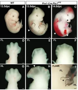
The Prx1-Cretransgene drives Cre recombinase activity in the early limb mesenchyme (Logan et al., 2002). Prx1-Creheterozygotes were mated with mice homozygous for a conditional allele of Ptc1 engineered by flanking exon 3 with loxPsites (Ellis et al., 2003). Heterozygous Prx1-Cre:Ptc1c/wtmice survive to adulthood and are fertile and viable. Homozygous Prx1-Cre:Ptc1c/cembryos survive until 14.0 dpc, allowing analysis of the effects of Ptc1inactivation in the limb mesenchyme during early limb development. Oedema visible from 13.5 dpc (Fig. 1B,C, arrowheads), and vascular haemorrhage evident at 14.0 dpc (Fig. 1C, arrowheads), are likely to be responsible for the lethality at this stage, although this phenotype has yet to be analysed in detail. Loss of tissue integrity in the distal limb often results in blister formation, which is particularly evident in the interdigital region at 13.5 dpc (arrowhead, Fig. 1H). At these later stages we have also detected defects in chondrogenesis, and characterisation of this phenotype will be
presented elsewhere.
D
E
V
E
LO
P
M
E
N
Prx1-Cre:Ptc1c/chindlimbs display polydactyly (Fig. 1H) with expansion of anterior tissue reminiscent of the Gli3Xt/Xtlimb and other Hh gain-of-function mouse models (Hill et al., 2003). Unexpectedly, the forelimb primarily displays an opposing phenotype, with 76% of 13.5 dpc Prx1-Cre:Ptc1c/cforelimbs analysed showing evidence of fewer digit primordia (oligodactyly; Fig. 1E; n=41). A minority (24%) of 13.5 dpc Prx1-Cre:Ptc1c/cforelimbs are polydactylous (Fig. 1F), but most retain the symmetry of shape of the oligodactylous forelimb, rather than the anterior expansion observed in the hindlimb (Fig. 1; compare F to H). Mesenchymal overgrowth visible in the dorsal cranium, maxilla and flank (star, Fig. 1B,C,I) correlates with additional sites of Prx1-Cre activity (see below).
We sought to define the spatiotemporal boundaries of Prx1-Cre activity relative to both Ptc1 excision and subsequent activation of the Hh pathway in the Prx1-Cre:Ptc1c/climbs. Deletion of exon 3 of Ptc1results in the upregulation of a transcript that encodes a non-functional protein incapable of dampening pathway activity via negative autoregulation. However, similar to wild-type Ptc1and Gli1transcripts (Goodrich et al., 1996; Marigo et al., 1996b), this truncated transcript acts as a robust marker of pathway activation. Using a probe capable of detecting both wild-type and exon 3-deleted Ptc1, whole-mount in situ hybridisation revealed wild-type
Ptc1expression primarily in a restricted domain in the limb bud and in the vibrissae at 12.5 dpc (Fig. 2A). Upon Ptc1 inactivation, expression was visible throughout the limb mesenchyme (Fig. 2B), and across the embryo in a pattern correlating closely with sites of Prx1-Cre activity (Fig. 2, compare B with D), as visualised by alkaline phosphatase expression in the Z/AP reporter assay (Lobe et al., 1999). In addition to the limb, these sites include the mesenchyme of the flank, the distal maxilla and the dorsal cranium, although expression of Ptc1appears to be less robust in this latter region (Fig. 2B,D; Fig. S1B,E in the supplementary material) (see also Logan et al., 2002). The extent of Hh pathway activation revealed by Ptc1was confirmed by Gli1 expression (Fig. 2C). A probe directed specifically to the deleted exon 3 of Ptc1showed no hybridisation to regions of the embryo expressing Prx1-Cre outside the normal domains of Ptc1expression (data not shown), confirming that the ectopic Ptc1transcript is the non-functional deleted form.
The precise timing of Ptc1inactivation was determined by RT-PCR of a region spanning exon 3. Detection of only the exon 3-deleted Ptc1 allele in three out of four 9.5 dpc Prx1-Cre:Ptc1c/cforelimb samples indicates, within the limits of detection by RT-PCR, complete excision of exon 3 before 9.5 dpc (Fig. 2E). Wild-type Ptc1was still detectable in the Prx1-Cre:Ptc1c/chindlimb at 10.5 dpc, but not at 11.5 dpc, indicating that exon 3 deletion approaches completion in the hindlimb between 10.5 dpc and 11.5 dpc (Fig. 2F). The relationship between Ptc1exon 3 excision and Hh pathway activation was assessed by quantitative real-time PCR for Ptc1 and Gli1transcripts. These data confirm that earlier excision in the forelimb (Fig. 2E,F) results in earlier and higher upregulation of Ptc1 and Gli1 in the forelimb compared with in the hindlimb (Fig. 2G,H). This provides a likely cause for the phenotypic differences observed between these limbs. A similar trend in expression of a second pathway inhibitor, patched 2 (Ptc2; also known as Ptch2), was also observed (Fig. 2I), although upregulation in both Prx1-Cre:Ptc1c/cforelimbs and hindlimbs did not reach significance until 11.5 dpc. Because we observed no obvious attenuation of Hh signalling at the stages analysed, it is unlikely that upregulated Ptc2 is acting redundantly to significantly dampen pathway activity in this context. Taken together, these data confirm that excision ofPtc1and subsequent upregulation of the Hh pathway correlate closely with sites of Prx1-Cre expression, and demonstrate a delay in these events in the hindlimb compared with in the forelimb.
The Gli3 protein gradient is disrupted in Prx1-Cre:Ptc1c/climbs
Based on the negative regulation of both Gli3transcript and Gli3R production by Shh signalling (Marigo et al., 1996b; Wang et al., 2000), constitutive Hh pathway activation should result in decreased levels of Gli3R. Anterior and posterior halves of 11.5 dpc limbs were assayed for Gli3FL (190 kDa) and Gli3R (83 kDa) by western blot. In control limbs, the anteroposterior gradient of Gli3R (Fig. 2J) was consistent with published results (Chen et al., 2004; Litingtung et al., 2002; Wang et al., 2000). By contrast, this Gli3R gradient was lost in both the Prx1-Cre:Ptc1c/cforelimb and hindlimb, with Gli3R levels being markedly reduced, particularly in the anterior (Fig. 2J). However, no clear increase in Gli3FL levels was observed in either limb.
Ptc1inactivation in the hindlimb produces a molecular profile similar to that ofGli3Xt/Xt
[image:3.612.50.301.57.347.2]The effect of Ptc1 inactivation on the expression of key limb patterning genes was investigated by whole-mount in situ hybridisation. In the hindlimb, changes in gene expression were most obvious at 11.5 dpc (Fig. 3A-L⬘). However, expression of Fgf4, Fig. 1. Prx1-Cre:Ptc1c/cembryos display variable polydactyly/
oligodactyly, and do not survive past 14.0 dpc.Wild-type (A,D,G)
and Prx1-Cre:Ptc1c/c(B,C,E,F,H,I) embryos at 13.5 and 14.0 dpc. At
13.5-14.0 dpc, Prx1-Cre:Ptc1c/cembryos display overgrowth of the cranial mesenchyme, the inter-limb flank region and the maxilla (stars, B,C,I). By 14.0 dpc, embryos display blistering (also in the distal limb at 13.5 dpc, arrowhead, H), and vascular haemorrhage into oedemic areas (arrowheads, C; oedema also visible at 13.5 dpc, arrowhead, B). The majority (76%) of 13.5 dpc Prx1-Cre:Ptc1c/cforelimbs are oligodactylous (E), while 24% are polydactylous (F). All Prx1-Cre:Ptc1c/chindlimbs display asymmetrical polydactyly (H). Scale bars: 500μM in A-C; 200μM in D-I.
D
E
V
E
LO
P
M
E
N
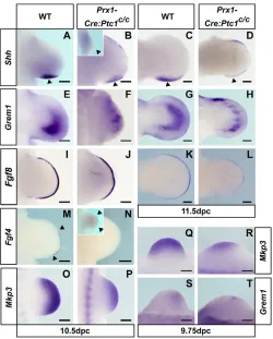
Grem1, Hand2, Hoxd13, Bmp2, Bmp4 and Fgf8 was altered as early as 10.5 dpc (see Fig. S2A-G⬘ in the supplementary material). Additionally, at this stage ectopic Ptc1 expression was weakly detectable in the anterior limb (Fig. S1B in the supplementary material), although the upregulation of Ptc1and Gli1detected by quantitative real-time PCR was not statistically significant at this point (Fig. 2G,H). From 11.5 dpc, Prx1-Cre:Ptc1c/c hindlimb buds displayed a small anterior domain of ectopic Shhexpression, the hallmark of a group of polydactylous mouse mutants that includes Gli3Xt/Xt(Hill, 2007) (arrowhead, Fig. 3A⬘). The anterior mesenchyme of the Prx1-Cre:Ptc1c/chindlimb also displayed expansion of Grem1, Hand2, Hoxd11-Hoxd13 and Jag1at 11.5 dpc (Fig. 3B-E⬘; see also Fig. S2L-M⬘ in the supplementary material). Expression of the anteriorly restricted gene Pax9was abrogated in the mutant hindlimb, reflecting loss of Gli3R (Fig. 3F,F⬘), whereas the domain of Gli3 expression appeared relatively unchanged although slightly expanded proximally (Fig. 3G,G⬘). In addition to gene expansion in the mesenchyme, the anterior AER of the Prx1-Cre:Ptc1c/chindlimb displayed ectopic expression of Bmp2, Bmp4, Fgf4 andFgf8(Fig. 3H-K⬘). Consistent with this, the underlying mesenchyme ectopically expressed the downstream FGF target Mkp3(also known as Dusp6) (Kawakami et al., 2003), suggesting enhanced FGF signalling from
the anterior AER (Fig. 3L,L⬘). In all cases, the anterior AER staining exhibited a sharp boundary, distinct from the anterior tapering normally revealed by Fgf8 expression (Fig. 3K,K⬘, arrowhead). Discrete anterior expression of the above genes in the AER, together with Shh in the underlying mesenchyme, persisted in Prx1-Cre:Ptc1c/c hindlimbs until 12.5 dpc (or 13.5 dpc for Fgf8; Fig. 3M-Q⬘). Overall, the expression patterns of these genes correlate with their behaviour in theGli3Xt/Xtlimb (Bastida et al., 2004; Hill et al., 2007; Litingtung et al., 2002; McGlinn et al., 2005; te Welscher et al., 2002b), indicating that the two models are largely indistinguishable from one another during AP patterning stages.
[image:4.612.51.365.52.478.2]Early Ptc1inactivation in the forelimb results in symmetrical expression of AP patterning genes The molecular profile of the Prx1-Cre:Ptc1c/cforelimb was markedly different from that of the hindlimb. Shh expression was reduced and often lost from the ZPA (arrowheads, Fig. 4B and inset), and ectopic expression was redistributed along the distal anterior margin in 12 out of the 18 forelimbs analysed at 10.5 dpc (Fig. 4B), and in seven out of seven forelimbs analysed at 11.5 dpc (Fig. 4D). In the remaining six (out of 18) Prx1-Cre:Ptc1c/cforelimbs analysed at 10.5 dpc, Shh expression was completely undetectable (Fig. 4B, inset).
Fig. 2. Hh pathway upregulation at sites of
Prx1-Cre activity.(A-C) Whole-mount wild-type
and Prx1-Cre:Ptc1c/cembryos at 12.5 dpc, showing
thatPtc1and Gli1 upregulation in Prx1-Cre:Ptc1c/c embryos correlates closely with activity of Prx1-Cre (D), shown by alkaline phosphatase expression in a 12.0 dpc Prx1-Cre:Ptc1c/wt:Z/AP+/–embryo (compare B and C with D). Scale bars: 500μM. (E,F) RT-PCR of
Ptc1exon 3 from wild-type (WT) and mutant (M) limbs detects only exon 3-deleted Ptc1at 9.5-11.5 dpc in the forelimb (E); the wild-type allele is still detectable in the hindlimb at 9.5-10.5 dpc, but not 11.5 dpc (F). Results are from representative samples of 9.5-11.5 dpc limbs, each consisting of two limbs. Due to variability in budding of the hindlimb at 9.5 dpc (21-29 somites), only one hindlimb pair was obtained from a 9.5 dpc embryo. For other stages, 2 (10.5 dpc), 3 (11.5 dpc) or 4 (9.5 dpc forelimb) samples were analysed. (G-I) Quantification of the delayed upregulation of Ptc1, Gli1andPtc2in hindlimbs versus forelimbs (*P<0.05,**P<0.01). The single 9.5 dpc hindlimb sample was excluded from this analysis, and results from individual limb pairs were pooled for statistical analysis. (J) Western blot showing that Gli3R (83 kDa) is present in an AP gradient across the wild-type hindlimb, but is reduced in anterior Prx1-Cre:Ptc1c/cforelimbs and hindlimbs. No significant increase in levels of Gli3FL (190 kDa) is evident. α-tubulin (55 kDa) serves as a loading control.
D
E
V
E
LO
P
M
E
N
Although Grem1 expression was detected in the anterior of most Prx1-Cre:Ptc1c/cforelimbs at 10.5-11.5 dpc, the size of the domain was reduced and showed a striking discontinuity (Fig. 4E-H; 4/8 at 10.5 dpc, 6/6 at 11.5 dpc). Grem1 expression was undetectable in two out of eight mutant forelimbs, and its expression domain was equivalent in size to that seen in wild type in a further two limbs at 10.5 dpc (data not shown). In general, the proportion of 10.5 dpc forelimbs in which Grem1expression was reduced correlates loosely with the prevalent oligodactylous phenotype (75%). In accordance with the overall reduction in Grem1expression, Fgf4expression was not detected in the AER in 10 out of 14 forelimbs at 10.5 dpc (Fig. 4N), whereas in the remaining four limbs, Fgf4expression was shifted anteriorly to the central AER (Fig. 4N, inset). No Fgf4 expression was observed from 11.5 dpc onwards in wild-type or Prx1-Cre:Ptc1c/c forelimbs (see Fig. S3J,J⬘in the supplementary material).
Coupled with the loss ofFgf4from the AER in the majority of Prx1-Cre:Ptc1c/cforelimbs was a mild decrease in the length of the AER, as shown by Fgf8expression (Fig. 4I-L). A reduced domain of Mkp3expression was detected in the Prx1-Cre:Ptc1c/c forelimb at 10.5 dpc (Fig. 4O-P). Together with a similar reduction in other FGF targets, including Spry1 and Spry4 (Minowada et al., 1999), at 10.5 dpc (Fig. S3E-F⬘ in the supplementary material), this suggests a decreased response to FGF signalling from the AER. BothMkp3andGrem1expression were also downregulated at 9.75 dpc (Fig. 4Q-T), and overall these data support an early disruption of the Shh/Grem1/FGF loop in the mutant forelimb.
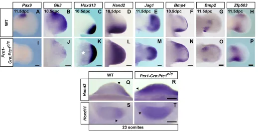
[image:5.612.46.349.56.413.2]A striking phenomenon evident in the expression of virtually all of the genes examined in the Prx1-Cre:Ptc1c/cforelimb from 10.5 dpc was symmetry about the AP midline. Loss of Pax9, an anterior marker in the 11.5 dpc limb (McGlinn et al., 2005), suggests a loss of anterior identity (Fig. 5A,I). Gli3expression was reduced, although still visible in the anterior limb at 10.5 dpc (Fig. 5B,J), with remaining Gli3 expression appearing symmetrical by 11.5 dpc (see Fig. S3K-K⬘in the supplementary material). In addition, those genes normally restricted to the posterior limb were expressed in the anterior Prx1-Cre:Ptc1c/c forelimb (Fig. 5C-G,K-O). Expression of genes such as Fgf4 (4/14; Fig. 4N, inset), Hoxd11-Hoxd13(Fig. 5C,K; see also Fig. S3M-O⬘in the supplementary material) and Hand2(Fig. 5D,L), showed continuous expansion into the anterior. However, this was distinct from the expansion in the hindlimb as the expression domain was now symmetrical and, in the case of the Hoxd genes and Fgf4, was shifted centrally. Expression of Bmp2and Bmp4 tended to occur as two distinct domains in the anterior and posterior mutant forelimb (Fig. 5F,G,N,O), which would suggest duplication of the posterior wild-type mesenchymal expression. This was further reinforced by the expression of the transcriptional repressor Zfp503, which we have previously shown to be downregulated in the anterior of both Gli3Xt/Xtand Prx1-Cre:Ptc1c/chindlimbs (McGlinn et al., 2008; McGlinn et al., 2005). The distinct proximal domain of Zfp503expression visible at the autopod/zeugopod boundary of the posterior limb was duplicated in the anterior Prx1-Cre:Ptc1c/cforelimb, whereas the more distal anterior autopod expression was lost (Fig. 5H,P). Fig. 3. Gene expression in Prx1-Cre:Ptc1c/chindlimbs.
Whole-mount in situ hybridisation of wild-type and Prx1-Cre:Ptc1c/chindlimbs for the indicated genes.
(A-E⬘) Expression of Shh, Grem1, Hand2, Hoxd13, and
Jag1is expanded into the anterior 11.5 dpc Prx1-Cre:Ptc1c/chindlimb. (F-G⬘) Expression of Pax9is abrogated in the Prx1-Cre:Ptc1c/chindlimb (F,F⬘) and Gli3is relatively unchanged but slightly expanded proximally (G,G⬘). (H-K⬘)Bmp2, Bmp4, Fgf4 and Fgf8 are ectopically expressed in the anterior Prx1-Cre:Ptc1c/chindlimb AER (arrowheads, H⬘-K⬘). (L,L⬘) Mkp3 is expanded in the anterior mesenchyme of the Prx1-Cre:Ptc1c/chindlimb at this stage. (M-Q⬘) The anterior AER region displays ectopic expression of Shh(underlying mesenchyme;
M,M⬘, arrowhead), Bmp2, Bmp4andFgf4 at 12.5 dpc, and Fgf8at 13.5 dpc (N-Q⬘, arrowheads). Scale bars: 200μM.
D
E
V
E
LO
P
M
E
N
Importantly, no such distinct anterior mesenchymal duplication of Bmp2, Bmp4orZfp503expression was observed in the Prx1-Cre:Ptc1c/chindlimb (Fig. 3H⬘-I⬘) (see also McGlinn et al., 2008), which clearly demonstrates the dichotomy between the molecular profiles of the forelimb and hindlimb. Taken together, these data suggest that the anterior Prx1-Cre:Ptc1c/cforelimb has undergone posterior re-specification.
To investigate pre-patterning and the generation of asymmetry in the forelimb, expression of Hand2was examined at 9.25 dpc (23 somites), prior to the establishment of the ZPA at around 9.5 dpc, and was found to be expanded into the anterior of the Prx1-Cre:Ptc1c/c forelimb (Fig. 5Q,R). Mice ectopically expressing
Hoxd11-Hoxd13across the forelimb bud displayed symmetrical limbs similar to the Prx1-Cre:Ptc1c/cforelimb phenotype (Zakany et al., 2004). We therefore investigated 5⬘Hoxd gene expression in the early Prx1-Cre:Ptc1c/c forelimb, prior to the onset of Shh
expression. Hoxd11was ectopically expressed across the limb bud at 9.25 dpc (23 somites; Fig. 5S,T). Upregulation of Hoxd13 in Prx1-Cre:Ptc1c/cforelimbs was also observed at the 27 somite stage (data not shown). Thus, the precocious 5⬘Hoxd activity following the loss of Ptc1in the Prx1-Cre:Ptc1c/cforelimb provides a likely mechanism for the symmetrical redistribution of Shh, Hand2and other patterning genes in these limbs.
We next sought to determine whether the changes in gene expression we observed in Prx1-Cre:Ptc1c/climbs were associated with a disruption in the apoptosis/proliferation balance. TUNEL analysis of 10.5 dpc limbs revealed no increase in apoptosis in Prx1-Cre:Ptc1c/climbs compared with that observed in wild type (Fig. 6A-D). This is consistent with a reduction in the Msx1and Msx2 anterior expression domain at 10.5 and 11.5 dpc (see Fig. S3B-C⬘ ,H-I⬘in the supplementary material), and suggests that neither Cre-mediated toxicity nor early apoptosis plays a major role in the forelimb phenotype. Some evidence of enhanced apoptosis in the central mesenchyme was detected at 12.5 dpc (data not shown), but as this later apoptosis occurs in both Prx1:Ptc1c/cforelimbs and hindlimbs, it is unlikely to specifically affect early forelimb patterning events. At 11.5 dpc, a time when the size difference between mutant and wild-type limbs generally becomes obvious, proliferation levels were significantly increased in Prx1-Cre:Ptc1c/c hindlimbs (P=0.001; Fig. 6E-H,I). Although a downward trend in proliferation was detected in the 11.5 dpc forelimb, no statistically significant change was observed (Fig. 6I). Hence neither altered apoptosis nor proliferation is likely to be the major determinant of the oligodactyly observed in Prx1-Cre:Ptc1c/cforelimbs.
Prx1-Cre:Ptc1c/climbs display reduced interdigital apoptosis and delayed AER regression
We next analysed the contribution of sustained Hh pathway activation to the relatively late process of digit separation, beyond the termination of Shh signalling in the wild-type limb. Results were identical for both limbs, and hindlimb data are presented here. Reduction of Bmp2and Msx1 expression in the interdigital mesenchyme (IDM) of 12.5 dpc Prx1-Cre:Ptc1c/climbs (Fig. 6J-K,N-O) suggests that this tissue is not programmed to participate in the apoptotic pathway. Other BMP pathway members were also downregulated in the IDM at 12.5-13.5 dpc (see Fig. S4A-N⬘in the supplementary material). Concomitantly, Fgf8 expression persisted above the IDM of Prx1-Cre:Ptc1c/chindlimbs at 13.5 dpc, when the AER is regressing in wild-type limbs (Fig. 6L,P). TUNEL analysis also revealed that apoptosis of the IDM was markedly reduced in Prx1-Cre:Ptc1c/c limbs relative to that observed in wild type (star, Fig. 6M,Q), even taking into account altered tissue integrity in some interdigital spaces. These data indicate that, in addition to early patterning changes, constitutive Hh signalling during digit morphogenesis inhibits the crucial process of apoptosis that is required for correct digit separation.
DISCUSSION
We have demonstrated for the first time the effects of unchecked ligand-independent upregulation of the Hh pathway specifically in the early limb mesenchyme by conditional ablation of functional Ptc1. Strikingly, we observe variable decrease or increase in digit number in the forelimb and hindlimb, respectively, together with posterior re-specification of the anterior forelimb. To our knowledge, this forelimb phenotype is unique amongst Hh gain-of-function mouse models, and we propose that this is primarily due to the timing and level of Hh pathway activation. Early inactivation of Ptc1in the Prx1-Cre:Ptc1c/cforelimb reveals a key role for Hh Fig. 4. ZPA/AER interactions are disrupted in Prx1-Cre:Ptc1c/c
forelimbs.Whole-mount in situ hybridisation of wild-type and
Prx1-Cre:Ptc1c/cforelimbs for the indicated genes. (A-D) At 10.5 dpc,Shh
expression in Prx1-Cre:Ptc1c/cforelimbs is redistributed from the ZPA (marked by arrowheads, A-D) to the anterior margin, with reduced (B) or completely undetectable (B, inset) ZPA expression; compare with wild type in A. At 11.5 dpc, Shhexpression is reduced in the ZPA of Prx1-Cre:Ptc1c/cforelimbs but is present in the anterior margin (D). (E-H)Grem1expression is anteriorly expanded in Prx1-Cre:Ptc1c/c forelimbs but is reduced and discontinuous at 10.5 dpc (F) and 11.5 dpc (H). (I-N) The Fgf8 expression domain is slightly shortened at 10.5 dpc (J) and 11.5 dpc (L) in Prx1-Cre:Ptc1c/cforelimbs, and Fgf4 expression is lost in most 10.5 dpc Prx1-Cre:Ptc1c/cforelimbs (N), but is shifted centrally in a subset (approx. 28%) of limbs (N, inset). (O-T)Mkp3 expression is reduced at 10.5 dpc (O-P), and Mkp3and
Grem1are reduced at 9.75 dpc (27 somites; Q-T) in Prx1-Cre:Ptc1c/c
forelimbs. Scale bars: 200μM.
D
E
V
E
LO
P
M
E
N
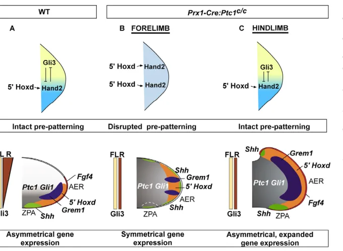
[image:6.612.50.300.57.368.2]pathway repression in early pre-patterning events prior to Shh expression in the ZPA, and results in perturbation of gene expression networks responsible for subsequent patterning (Fig. 7).
Ptc1excision activates the Hh pathway
Prx1-Cre is expressed in limb mesenchyme as well as in restricted domains in other regions of the mouse embryo. Hh pathway activation in Prx1-Cre:Ptc1c/cembryos, as determined byPtc1 andGli1expression, was observed in domains that closely mirror sites of Prx1-Cre expression. This unexpected pathway activation outside the normal domains of detectable Ptc1 expression is consistent with a model in which Hh signalling in these areas is normally inhibited by low-level Ptc1 transcription, below the limits of in situ hybridisation detection. Upon removal of this functional Ptc1 transcript, the pathway is activated inappropriately in a more widespread domain than anticipated. Our results, together with those from studies showing de-repression of Ptc1-lacZexpression in early Ptc1 null embryos (Goodrich et al., 1997; Milenkovic et al., 1999), suggest that much of the embryonic mesenchyme is competent to respond to Hh signalling. This indicates that, in addition to maintaining the fine balance of signalling mediated by Hh ligands, negative regulation by Ptc1 in the absence of ligand is essential to maintain pathway repression in otherwise signalling-competent tissue. The finding that an expanded Shhdomain is likely to underpin the drastic phenotype difference between the bat forelimb and hindlimb, suggests a precedent for evolutionary variation in the spatial restriction of Shh signalling as a means of controlling limb patterning to generate diversity (Hockman et al., 2008).
We determined the precise timing of Ptc1 excision in Prx1-Cre:Ptc1c/c limbs and found that, consistent with Prx1-Cre expression (Logan et al., 2002), there is a delay in excision in the hindlimb compared with in the forelimb, and this translates to a corresponding delay in pathway activation. The mutant hindlimb is therefore exposed to wild-type Ptc1 transcript for a considerable time after the establishment of Shhexpression. By contrast, Ptc1 excision is virtually complete in most forelimbs prior to normal Shh expression, and this appears to lead to defects in the establishment of the ZPA. Our data and published results show that expression of Prx1-Cre throughout the forelimb mesenchyme at 9.5 dpc varies between individuals (Logan et al., 2002). Although this variability could explain the occurrence of polydactyly in a minority of forelimbs, we are not able to confirm this with our model.
Early expression cascades are perturbed in the symmetrical Prx1-Cre:Ptc1c/cforelimb
[image:7.612.50.568.57.320.2]In contrast to the asymmetry in Prx1-Cre:Ptc1c/chindlimbs, mutant forelimbs appear largely morphologically and molecularly symmetrical about the midline (Fig. 7B). Although embryonic lethality prevents unequivocal determination of digit identity in the Prx1-Cre:Ptc1c/c forelimbs, anterior expansion of Hoxd11 and Hoxd12implies that digit 1 identity is lost, as Hoxd11and Hoxd12 expression in the wild-type limb is excluded from the digit 1 primordium (Kmita et al., 2005; Nelson et al., 1996). Importantly, this distinguishes the oligodactyly of the Prx1-Cre:Ptc1c/cforelimb from that produced by Shh inactivation, in which digit 1 identity is preserved in the absence of all other digits (Chiang et al., 2001). The lack of extensive ectopic anterior cell death at 10.5 dpc, as seen in Fig. 5. Loss of asymmetry in Prx1-Cre:Ptc1c/cforelimbs. (A-T) Whole-mount in situ hybridisation of wild-type and Prx1-Cre:Ptc1c/cforelimbs for the indicated genes.Pax9 (11.5 dpc) andGli3 (10.5 dpc) are anteriorly reduced in Prx1-Cre:Ptc1c/cforelimbs (I,J) compared with in wild type (A,B).
Hoxd13 and Hand2are anteriorly expanded at 10.5 dpc in Prx1-Cre:Ptc1c/cforelimbs (K,L) compared with in wild type (C,D). Hoxd13is also
generally expanded in the more proximal mesenchyme in Prx1-Cre:Ptc1c/cforelimbs (star, K). The posterior domains of Jag1, Bmp4, Bmp2and
Zfp503are duplicated in the anterior Prx1-Cre:Ptc1c/cforelimb (M-P; compare with wild type, E-H). Hand2 and Hoxd11are anteriorly expanded in
Prx1-Cre:Ptc1c/cforelimbs at 9.25 dpc compared with in wild type (23 somites; Q-T). Scale bars: 200μM.
D
E
V
E
LO
P
M
E
N
the posteriorised limbs of Msx1/2mouse mutants (Lallemand et al., 2005), indicates that the symmetrical Prx1-Cre:Ptc1c/cforelimb phenotype is unlikely to reflect a loss of anterior tissue. This, and the expression of normally posteriorly restricted genes in the anterior limb, suggest that the symmetrical forelimb results from re-specification of anterior tissue to a posterior identity.
Limb asymmetry and establishment of the ZPA is determined by interplay between 5⬘Hoxd genes, Gli3and Hand2in the early limb bud (Tarchini et al., 2006; te Welscher et al., 2002a; Zakany et al., 2004). Our data show that early inactivation of Ptc1in the forelimb disturbs these early patterning events, as judged by the anterior expansion of Hoxd11 and Hand2 expression at 9.25 dpc (23 somites), and ofHoxd13 slightly later (27 somites). By contrast, there is no change in expression of these genes between wild-type and Prx1-Cre:Ptc1c/chindlimbs at the equivalent stage. In a previous study, inactivation of Shhusing Prx1-Cre produced a hypomorphic phenotype (Lewis et al., 2001), suggesting that this driver does not inactivate gene expression in all cells prior to Shhexpression. Indeed, it is likely that a small fraction of cells escape Ptc1 excision prior to the normal onset ofShh pathway activation in the Prx1-Cre:Ptc1c/cforelimb. However, our data indicate that a high enough level of Ptc1inactivation is achieved to drive downstream signalling to a sufficient threshold to perturb pre-patterning.
Following early expansion of Hoxd11and Hand2expression across the entire Prx1-Cre:Ptc1c/cforelimb paddle, Shhexpressing cells are visible around the distal limb periphery. Similar changes in Shh expression are induced by ectopic anterior expression of Hand2 (McFadden et al., 2002), and 5⬘Hoxd genes (Zakany et al., 2004). In these cases, limbs show a loss of asymmetry and a conversion of anterior digits to posterior identities (McFadden et al., 2002; Zakany et al., 2004). The ectopic Hoxd11-Hoxd13 expression in the latter model also produces variable oligodactyly (Zakany et al., 2004), which further implicates early ectopic 5⬘Hoxd gene expression in the generation of the Prx1-Cre:Ptc1c/cforelimb phenotype. Although the limb symmetry in these other mouse models is likely to be caused by the distally expanded Shh, this is unlikely to be a major contributing
factor in the Prx1-Cre:Ptc1c/cforelimb phenotype, because this limb is characterised by widespread high-level ectopic Hh pathway activation. It is more likely that signalling events downstream of the constitutively active Hh pathway underpin the forelimb symmetry, although a contribution of properties intrinsic to the redistributed Shh-expressing cells and their descendants cannot be ruled out.
The Shh/Grem1/FGF signalling loop is disrupted in Prx1-Cre:Ptc1c/cforelimbs
To date, activation of Hh signalling in both mouse models and human dysmorphologies has generally been associated with extra digits (Hill et al., 2003). Although we observe this phenotype in the Prx1-Cre:Ptc1c/chindlimbs, the forelimbs predominantly display an oligodactylous phenotype with an average of 3-4 digits. Analysis of Gli3 processing at 11.5 dpc revealed that the Gli3FL:Gli3R gradient is abolished in both forelimbs and hindlimbs in a similar manner. In both cases there is a decrease in Gli3R levels in the anterior limb, consistent with inhibition of Gli3 cleavage by Shh signalling (Wang et al., 2000), although a clear increase in Gli3FL was not detectable by western blotting. These data suggest that the forelimb-specific phenotype is not due to differences in Gli3 cleavage.
[image:8.612.49.431.55.319.2]Early reduced expression of Mkp3and other targets of AER-FGF signalling suggests that the mesenchymal response to FGF signalling is attenuated specifically in the Prx1-Cre:Ptc1c/c forelimb. This reduced response corresponds to a lack of Fgf4expression in most limbs, and to a relatively subtle shortening of the Fgf8 expression domain. However, mice lacking Fgf4do not display limb defects (Moon et al., 2000; Sun et al., 2000), and recent studies demonstrate conclusively that of all the AER FGFs, Fgf8is sufficient for limb patterning (Mariani et al., 2008). Thus, the reduction in Fgf4 expression is unlikely to be the major cause of thePrx1-Cre:Ptc1c/c forelimb phenotype, but it is nevertheless indicative of perturbation of the Shh/Grem1/FGF loop (Verheyden and Sun, 2008). Accordingly, we detected evidence of reduced Grem1 expression in the Prx1-Cre:Ptc1c/cforelimb very early after limb budding. Although Shh normally maintains expression of Grem1in the limb, Shh-expressing Fig. 6. Apoptosis and proliferation in Prx1-Cre:Ptc1c/climbs.
(A-D) Apoptosis is unchanged in 10.5 dpc Prx1-Cre:Ptc1c/climbs from that in
wild type, as assessed by TUNEL. (E-I) At 11.5 dpc, proliferation measured by BrdU incorporation was significantly increased in Prx1-Cre:Ptc1c/chindlimbs but not forelimbs
(***P=0.001). (J-K,N-O) Expression of
Bmp2and Msx1is reduced in the IDM at 12.5 dpc in Prx1-Cre:Ptc1c/climbs compared with in wild type. (L,P) At 13.5 dpc, Fgf8persists above the interdigits in Prx1-Cre:Ptc1c/climbs. (M,Q) The IDM of sectioned 13.5 dpc
Prx1-Cre:Ptc1c/climbs contains fewer apoptotic cells than wild type, as assessed by TUNEL (stars). Scale bars: 500μM in Q; 200μM in all other panels.
D
E
V
E
LO
P
M
E
N
cells and their descendants cannot express Grem1, a fact attributed to very high levels of autocrine Shh signalling (Nissim et al., 2006; Scherz et al., 2004). Early high-level autocrine activation of the Hh pathway across the Prx1-Cre;Ptc1c/cforelimb might therefore mediate an early reduced capacity for cells to express Grem1, thus disturbing the Shh/Grem1/FGF loop. We favour this over a more direct effect on the later FGF/Grem1 inhibitory loop (Verheyden and Sun, 2008), as the early reduced expression domain of Mkp3 at 9.75 dpc is inconsistent with the high levels of FGF signalling required to repress Grem1. Given that a primary feature of the Prx1-Cre:Ptc1c/cforelimb is the widespread upregulation of 5⬘Hoxd genes, it may be that high-level constitutive Hh pathway activity is acting through this early ectopic Hox gene expression to downregulate Grem1. This leads to the intriguing but speculative hypothesis that high levels of 5⬘Hoxd genes contribute to the refractoriness toGrem1expression inShh lineage cells of the wild-type limb.
Like the Prx1-Cre:Ptc1c/c forelimb, Grem1 null limbs are characterised by fewer digits, but they also display reduced Shh signalling and increased apoptosis (Khokha et al., 2003; Michos et al., 2004). By contrast, we see no evidence of enhanced early forelimb-specific apoptosis in the Prx1-Cre;Ptc1c/c model. Furthermore, we demonstrate a reduction in the anterior expression domain of Msx1 andMsx2,which are thought to mediate apoptosis downstream of both Shh/Gli3R and BMP signalling in the limb (Lallemand et al., 2009; Pizette et al., 2001). Although reduced Msx gene expression is not consistent with the observed expanded anterior expression of Bmp2 and Bmp4, it supports a recent suggestion that anterior Msx gene expression might be mediated predominantly by Gli3R (Lallemand et al., 2009). This is further supported by the selective maintenance of Msx gene expression in the most anterior region of the limbs of Bmpr1a conditional-knockout mice (Ovchinnikov et al., 2006). Conversely, despite the duplication of both Bmp2and Bmp4expression domains, a decrease in overall BMP signalling in the Prx1-Cre:Ptc1c/cforelimb cannot be ruled out. However, this is less likely in those limbs lacking Fgf4, as it has been shown that inhibition of BMP signalling allows FGF expression in the AER (Capdevila et al., 1999; Pizette and Niswander, 1999; Zuniga et al., 1999).
While much effort has focused on the mechanisms underlying the positive potentiation of the posteriorly biased expression of Shh, our data highlight a previously unappreciated requirement for Ptc1-mediated negative control over the precise timing and levels of Shh signalling in the developing limb. Importantly, the Prx1-Cre:Ptc1c/c forelimb phenotype suggests that failure to prevent activation of this pathway throughout the early pre-patterning stages of limb budding results in perturbations to key gene expression cascades responsible for defining limb asymmetry and digit number.
Acknowledgements
We thank C. Tabin, P. Koopman, M. Little and D. Ovchinnikov for kindly providing probes with permission from cited sources, M. Logan for the Prx1-Cremouse line, and B. Wang for the Gli3 antibody. We also thank C. C. Hui, R. Villani and T. Ellis for helpful discussion, and A. Hardacre, J. Conway and T. Davidson for assistance with mouse husbandry. This work was supported by the National Health and Medical Research Council of Australia (NHMRC). N.C.B. was a recipient of an Australian Postgraduate Award, and C.W. is an NHMRC Senior Research Fellow.
Supplementary material
Supplementary material for this article is available at http://dev.biologists.org/cgi/content/full/136/20/3515/DC1
References
Bastida, M. F., Delgado, M. D., Wang, B., Fallon, J. F., Fernandez-Teran, M. and Ros, M. A.(2004). Levels of Gli3 repressor correlate with Bmp4 expression and apoptosis during limb development. Dev. Dyn. 231, 148-160.
Benazet, J. D., Bischofberger, M., Tiecke, E., Goncalves, A., Martin, J. F., Zuniga, A., Naef, F. and Zeller, R.(2009). A self-regulatory system of interlinked signaling feedback loops controls mouse limb patterning. Science 323, 1050-1053.
Bruce, S. J., Gardiner, B. B., Burke, L. J., Gongora, M. M., Grimmond, S. M. and Perkins, A. C.(2007). Dynamic transcription programs during ES cell differentiation towards mesoderm in serum versus serum-freeBMP4 culture.
BMC Genomics8, 365.
Capdevila, J., Tsukui, T., Rodriquez Esteban, C., Zappavigna, V. and Izpisua Belmonte, J. C.(1999). Control of vertebrate limb outgrowth by the proximal factor Meis2 and distal antagonism of BMPs by Gremlin. Mol. Cell4, 839-849.
Chen, W., John, J., Lin, C. and Chang, C.(2002). Molecular cloning and developmental expression of zinc finger transcription factor MTF-1 gene in zebrafish, Danio rerio. Biochem. Biophys. Res. Commun. 291, 798-805.
[image:9.612.58.398.56.305.2]Chen, Y. and Struhl, G.(1996). Dual roles for patched in sequestering and transducing Hedgehog. Cell87, 553-563.
Fig. 7. A summary of the gene expression changes in Prx1-Cre:Ptc1c/climbs. Interplay
between 5⬘Hoxd genes, Gli3andHand2
establishes limb asymmetry in wild-type (WT) limbs primarily by determining correct formation of the posterior Shh-expressing ZPA (A). Constitutive Hh signalling in Prx1-Cre:Ptc1c/climbs occurs prior to (forelimb, B)
and subsequent to (hindlimb, C) early patterning. Early and strong Hh signalling in
Prx1-Cre:Ptc1c/cforelimbs disrupts the
pre-pattern and generates mirror-image symmetry of gene expression (B). Pre-patterning is intact inPrx1-Cre:Ptc1c/chindlimbs, which
predominantly display anterior expansion of genes (C).
D
E
V
E
LO
P
M
E
N
Chen, Y., Knezevic, V., Ervin, V., Hutson, R., Ward, Y. and Mackem, S.(2004). Direct interaction with Hoxd proteins reverses Gli3-repressor function to promote digit formation downstream of Shh. Development131, 2339-2347.
Chiang, C., Litingtung, Y., Harris, M. P., Simandl, B. K., Li, Y., Beachy, P. A. and Fallon, J. F.(2001). Manifestation of the limb prepattern: limb development in the absence of sonic hedgehog function. Dev. Biol. 236, 421-435.
Ellis, T., Smythe, I., Riley, E., Graham, S., Elliot, K., Narang, M., Kay, G., Wicking, C. and Wainwright, B.(2003). Patched1 conditional null allele in mice. Genesis36, 158-161.
Fowles, L. F., Bennetts, J. S., Berkman, J. L., Williams, E., Koopman, P., Teasdale, R. D. and Wicking, C.(2003). Genomic screen for genes involved in mammalian craniofacial development. Genesis35, 73-87.
Goodrich, L. V., Johnson, R. L., Milenkovic, L., McMahon, J. A. and Scott, M. P.(1996). Conservation of the hedgehog/patched signaling pathway from flies to mice: induction of a mouse patched gene by Hedgehog. Genes Dev. 10, 301-312.
Goodrich, L. V., Milenkovic, L., Higgins, K. M. and Scott, M. P.(1997). Altered neural cell fates and medulloblastoma in mouse patched mutants. Science277, 1109-1113.
Hahn, H., Christiansen, J., Wicking, C., Zaphiropoulos, P. G., Chidambaram, A., Gerrard, B., Vorechovsky, I., Bale, A. E., Toftgard, R., Dean, M. et al.
(1996). A mammalian patched homolog is expressed in target tissues of sonic hedgehog and maps to a region associated with developmental abnormalities. J. Biol. Chem. 271, 12125-12128.
Hill, P., Wang, B. and Ruther, U.(2007). The molecular basis of Pallister-Hall associated polydactyly. Hum. Mol. Genet. 16, 2089-2096.
Hill, P., Gotz, K. and Ruther, U.(2009). A SHH-independent regulation of Gli3 is a significant determinant of anteroposterior patterning of the limb bud. Dev. Biol. 328, 506-516.
Hill, R. E.(2007). How to make a zone of polarizing activity: insights into limb development via the abnormality preaxial polydactyly. Dev. Growth Differ. 49, 439-448.
Hill, R. E., Heaney, S. J. and Lettice, L. A.(2003). Sonic hedgehog: restricted expression and limb dysmorphologies. J. Anat. 202, 13-20.
Hockman, D., Cretekos, C. J., Mason, M. K., Behringer, R. R., Jacobs, D. S. and Illing, N.(2008). A second wave of Sonic hedgehog expression during the development of the bat limb. Proc. Natl. Acad. Sci. USA 105, 16982-16987.
Hui, C. C. and Joyner, A. L.(1993). A mouse model of greig
cephalopolysyndactyly syndrome: the extra-toesJmutation contains an intragenic deletion of the Gli3 gene. Nat. Genet. 3, 241-246.
Ingham, P. W. and McMahon, A. P.(2001). Hedgehog signaling in animal development: paradigms and principles. Genes Dev. 15, 3059-3087.
Kawakami, Y., Rodriguez-Leon, J., Koth, C. M., Buscher, D., Itoh, T., Raya, A., Ng, J. K., Esteban, C. R., Takahashi, S., Henrique, D. et al.(2003). MKP3 mediates the cellular response to FGF8 signalling in the vertebrate limb. Nat. Cell Biol. 5, 513-519.
Khokha, M. K., Hsu, D., Brunet, L. J., Dionne, M. S. and Harland, R. M.
(2003). Gremlin is the BMP antagonist required for maintenance of Shh and Fgf signals during limb patterning. Nat. Genet. 34, 303-307.
Kmita, M., Tarchini, B., Zakany, J., Logan, M., Tabin, C. J. and Duboule, D.
(2005). Early developmental arrest of mammalian limbs lacking HoxA/HoxD gene function. Nature435, 1113-1116.
Lallemand, Y., Nicola, M. A., Ramos, C., Bach, A., Cloment, C. S. and Robert, B.(2005). Analysis of Msx1; Msx2 double mutants reveals multiple roles for Msx genes in limb development. Development132, 3003-3014.
Lallemand, Y., Bensoussan, V., Cloment, C. S. and Robert, B.(2009). Msx genes are important apoptosis effectors downstream of the Shh/Gli3 pathway in the limb. Dev. Biol. 331, 189-198.
Lewis, P. M., Dunn, M. P., McMahon, J. A., Logan, M., Martin, J. F., St-Jacques, B. and McMahon, A. P.(2001). Cholesterol modification of sonic hedgehog is required for long-range signaling activity and effective modulation of signaling by Ptc1. Cell105, 599-612.
Litingtung, Y., Dahn, R. D., Li, Y., Fallon, J. F. and Chiang, C.(2002). Shh and Gli3 are dispensable for limb skeleton formation but regulate digit number and identity. Nature418, 979-983.
Lobe, C. G., Koop, K. E., Kreppner, W., Lomeli, H., Gertsenstein, M. and Nagy, A.(1999). Z/AP, a double reporter for cre-mediated recombination. Dev. Biol. 208, 281-292.
Logan, M., Martin, J., Nagy, A., Lobe, C., Olson, E. N. and Tabin, C.(2002). Expression of Cre recombinase in the developing mouse limb bud driven by a Prx1 enhancer. Genesis33, 77-80.
Mariani, F. V., Ahn, C. P. and Martin, G. R.(2008). Genetic evidence that FGFs have an instructive role in limb proximal-distal patterning. Nature453, 401-405.
Marigo, V., Davey, R. A., Zuo, Y., Cunningham, J. M. and Tabin, C. J.(1996a). Biochemical evidence that patched is the Hedgehog receptor. Nature384, 176-179.
Marigo, V., Johnson, R. L., Vortkamp, A. and Tabin, C. J.(1996b). Sonic hedgehog differentially regulates expression of GLI and GLI3 during limb development. Dev. Biol. 180, 273-283.
McFadden, D. G., McAnally, J., Richardson, J. A., Charite, J. and Olson, E. N.
(2002). Misexpression of dHAND induces ectopic digits in the developing limb bud in the absence of direct DNA binding. Development129, 3077-3088.
McGlinn, E. and Tabin, C. J.(2006). Mechanistic insight into how Shh patterns the vertebrate limb. Curr. Opin. Genet. Dev. 16, 426-432.
McGlinn, E., van Bueren, K. L., Fiorenza, S., Mo, R., Poh, A. M., Forrest, A., Soares, M. B., Bonaldo Mde, F., Grimmond, S., Hui, C. C. et al.(2005). Pax9 and Jagged1 act downstream of Gli3 in vertebrate limb development. Mech. Dev. 122, 1218-1233.
McGlinn, E., Richman, J. M., Metzis, V., Town, L., Butterfield, N. C., Wainwright, B. J. and Wicking, C.(2008). Expression of the NET family member Zfp503 is regulated by hedgehog and BMP signaling in the limb. Dev. Dyn. 237, 1172-1182.
Michos, O., Panman, L., Vintersten, K., Beier, K., Zeller, R. and Zuniga, A.
(2004). Gremlin-mediated BMP antagonism induces the epithelial-mesenchymal feedback signaling controlling metanephric kidney and limb organogenesis.
Development131, 3401-3410.
Milenkovic, L., Goodrich, L. V., Higgins, K. M. and Scott, M. P.(1999). Mouse patched1 controls body size determination and limb patterning. Development 126, 4431-4440.
Minowada, G., Jarvis, L. A., Chi, C. L., Neubuser, A., Sun, X., Hacohen, N., Krasnow, M. A. and Martin, G. R.(1999). Vertebrate Sprouty genes are induced by FGF signaling and can cause chondrodysplasia when overexpressed.
Development126, 4465-4475.
Moon, A. M., Boulet, A. M. and Capecchi, M. R.(2000). Normal limb development in conditional mutants of Fgf4. Development127, 989-996.
Nelson, C. E., Morgan, B. A., Burke, A. C., Laufer, E., DiMambro, E., Murtaugh, L. C., Gonzales, E., Tessarollo, L., Parada, L. F. and Tabin, C.
(1996). Analysis of Hox gene expression in the chick limb bud. Development 122, 1449-1466.
Nissim, S., Hasso, S. M., Fallon, J. F. and Tabin, C. J.(2006). Regulation of Gremlin expression in the posterior limb bud. Dev. Biol. 299, 12-21.
Ovchinnikov, D. A., Selever, J., Wang, Y., Chen, Y. T., Mishina, Y., Martin, J. F. and Behringer, R. R.(2006). BMP receptor type IA in limb bud mesenchyme regulates distal outgrowth and patterning. Dev. Biol. 295, 103-115.
Pizette, S. and Niswander, L.(1999). BMPs negatively regulate structure and function of the limb apical ectodermal ridge. Development126, 883-894.
Pizette, S., Abate-Shen, C. and Niswander, L.(2001). BMP controls proximodistal outgrowth, via induction of the apical ectodermal ridge, and dorsoventral patterning in the vertebrate limb. Development128, 4463-4474.
Riddle, R. D., Johnson, R. L., Laufer, E. and Tabin, C.(1993). Sonic hedgehog mediates the polarizing activity of the ZPA. Cell75, 1401-1416.
Roelink, H., Augsburger, A., Heemskerk, J., Korzh, V., Norlin, S., Ruiz i Altaba, A., Tanabe, Y., Placzek, M., Edlund, T., Jessell, T. M. et al.(1994). Floor plate and motor neuron induction by vhh-1, a vertebrate homolog of hedgehog expressed by the notochord. Cell76, 761-775.
Scherz, P. J., Harfe, B. D., McMahon, A. P. and Tabin, C. J.(2004). The limb bud Shh-Fgf feedback loop is terminated by expansion of former ZPA cells. Science 305, 396-399.
Sun, X., Lewandoski, M., Meyers, E. N., Liu, Y. H., Maxson, R. E., Jr and Martin, G. R.(2000). Conditional inactivation of Fgf4 reveals complexity of signalling during limb bud development. Nat. Genet. 25, 83-86.
Tarchini, B., Duboule, D. and Kmita, M.(2006). Regulatory constraints in the evolution of the tetrapod limb anterior-posterior polarity. Nature443, 985-988.
te Welscher, P., Fernandez-Teran, M., Ros, M. A. and Zeller, R.(2002a). Mutual genetic antagonism involving GLI3 and dHAND prepatterns the vertebrate limb bud mesenchyme prior to SHH signaling. Genes Dev. 16, 421-426.
te Welscher, P., Zuniga, A., Kuijper, S., Drenth, T., Goedemans, H. J., Meijlink, F. and Zeller, R.(2002b). Progression of vertebrate limb development through SHH-mediated counteraction of GLI3. Science298, 827-830.
Verheyden, J. M. and Sun, X.(2008). An Fgf/Gremlin inhibitory feedback loop triggers termination of limb bud outgrowth. Nature454, 638-641.
Wang, B., Fallon, J. F. and Beachy, P. A.(2000). Hedgehog-regulated processing of Gli3 produces an anterior/posterior repressor gradient in the developing vertebrate limb. Cell100, 423-434.
Wang, C., Pan, Y. and Wang, B.(2007a). A hypermorphic mouse Gli3 allele results in a polydactylous limb phenotype. Dev. Dyn. 236, 769-776.
Wang, C., Ruther, U. and Wang, B.(2007b). The Shh-independent activator function of the full-length Gli3 protein and its role in vertebrate limb digit patterning. Dev. Biol. 305, 460-469.
Zakany, J., Kmita, M. and Duboule, D.(2004). A dual role for Hox genes in limb anterior-posterior asymmetry. Science304, 1669-1672.
Zuniga, A., Haramis, A. P., McMahon, A. P. and Zeller, R.(1999). Signal relay by BMP antagonism controls the SHH/FGF4 feedback loop in vertebrate limb buds.
Nature401, 598-602.