N A N O E X P R E S S
Open Access
Antitumor Activity of Alloy and
Core-Shell-Type Bimetallic AgAu Nanoparticles
Igor Shmarakov
1*, Iuliia Mukha
2, Nadiia Vityuk
2, Vira Borschovetska
1, Nelya Zhyshchynska
1, Galyna Grodzyuk
3,4and Anna Eremenko
2Abstract
Nanoparticles (NPs) of noble metals, namely gold and silver, remain promising anticancer agents capable of enhancing current surgery- and chemotherapeutic-based approaches in cancer treatment. Bimetallic AgAu composition can be used as a more effective agent due to the synergetic effect. Among the physicochemical parameters affecting gold and silver nanoparticle biological activity, a primary concern relates to their size, shape, composition, charge, etc. However, the impact of metal components/composition as well as metal topological distribution within NPs is incompletely characterized and remains to be further elucidated and clarified. In the present work, we tested a series of colloidal solutions of AgAu NPs of alloy and core-shell type for an antitumor activity depending on metal molar ratios (Ag:Au = 1:1; 1:3; 3:1) and topological distribution of gold and silver within NPs (AucoreAgshell; AgcoreAushell).
The efficacy at which an administration of the gold and silver NPs inhibits mouse Lewis lung carcinoma (LLC) growth in vivo was compared. The data suggest that in vivo antitumor activity of the studied NPs strongly depends on gold and silver interaction arising from their ordered topological distribution. NPs with Ag core covered by Au shell were the most effective among the NPs tested towards LLC tumor growth and metastasizing inhibition. Our data show that among the NPs tested in this study, AgcoreAushellNPs may serve as a suitable anticancerous prototype.
Keywords:Gold, Silver, Nanoparticles, Anticancer activity, Oxidative stress
PACS:78.67.Bf (Nanocrystals, nanoparticles, and nanoclusters), 87.85.Rs (Nanotechnologies-applications), 87.85.em
(Tissue damage)
Background
Rapid advances in nanomedicine have generated an in-creasing number of potential diagnostic and therapeutic applications of nanoparticles in recent years [1, 2]. Among the different types of nanoparticles, metal nanoparticles including gold and silver have widespread use across the biomedical sciences [3, 4]. Nanoparticles (NPs) based on noble metals remain promising antican-cer agents capable of enhancing existing surgery and chemotherapeutic-based approaches in cancer treat-ment. These NPs are being extensively used in a diag-nostics and anticancer therapy due to intrinsic noble metal properties including ultramicroscopic size, large surface area-to-volume ratio, and consequently their
electronic and optical properties as well as high reactivity and stability [4]. In vitro and in vivo studies demonstrated that gold and silver NPs have strong antitumor activities [3, 5–7]. Bimetallic AgAu NPs are advantageous because they combine and resemble the unique properties of both gold and silver in one nanoparticle that are distinctly different from those inherent to individual components [1, 8]. Such physicochemical parameters as size, shape, composition, and charge greatly influence biological ac-tivity of NPs [9–12]. These properties of bimetallic AgAu NPs have been also shown to be advantageous in a development of aptamer-based biosensors [13]. How-ever, many aspects of gold and silver NP-based systems remain unexplored, limiting the efficiency of their ap-plication. For instance, the impact of metal composition as well as metal topological distribution within NPs is incompletely characterized and remains to be further elucidated and clarified. An inclusion of different * Correspondence:[email protected]
1Department of Biochemistry and Biotechnology, Yuriy Fedkovych Chernivtsi
National University, Kotsuybynskyiy St., 2, Chernivtsi 58012, Ukraine Full list of author information is available at the end of the article
metallic compositions has been shown to significantly affect reactive oxygen species (ROS) formation and has been suggested to be a major cause of different cellular disruptions and cell death enhancement observed in cells exposed to nanoparticles [12]. Our previous study demonstrated that combination of both gold and silver in one bimetallic NP confers advantageous biological activities and low toxicity in vivo [14]. In the present work, we test the ability of various bimetallic AgAu NPs to inhibit the metastatic tumor growth in vivo, employing mouse Lewis lung carcinoma (LLC) as a model. Aiming at expanding our knowledge on the AgAu nanoparticles biological activity, NPs were tested for an antitumor activity depending on metal molar ratios (Ag:Au = 1:1; 1:3; 3:1) and topological dis-tribution of gold and silver within NPs (AucoreAgshell; AgcoreAushell). The data, revealed in the present study, emphasize that both parameters, metal molar ratio as well as metal topological distribution within bimetallic gold and silver nanoparticles, are critical features of NPs and can affect their antitumor activity. Among the NPs tested in this study, AgcoreAushell NPs may serve as the most suitable prototype for the develop-ment of an anticarcinomatous agent.
Methods
Nanoparticle Synthesis and Characterization
Colloidal solutions of bimetallic silver and gold NPs were obtained via chemical reduction of silver nitrate and tetrachlorauratic acid (AgNO3and HAuCl4, Merck, Germany) with amino acid tryptophan (Trp, SC12-20120713, China). The components interacted in a molar ratio Trp:M = 2:1. The concentration of metal in the resulting solution was C(M) = 10-4M followed by concentrating it to 10−3M. Bimetallic NPs of alloy type were obtained via simultaneous reduction of metal ions using initial metal molar ratios of Ag:Au = 3:1, 1:1, and 1:3, indicated as AgAu(3:1), AgAu(1:1), and AgAu(1:3) correspondingly. For the preparation of NPs of coshell type with different topology, metal ions were re-duced sequentially using the molar ratio Ag:Au = 1:1 and were indicated as AucoreAgshelland AgcoreAushell, For all colloids, the initial solutions of tryptophan were adjusted to pH = 10. Thus, amino acid was used in an anionic state that exists in alkaline medium. The pH control of the medium was performed using a pH meter I-160MI. As a working electrode, a glass electrode was used. Silver chloride electrode served as a reference one. Metals were injected into boiling solution of Trp followed by con-tinuous heating of the reaction mixture.
The absorption spectra of the colloidal solutions of AgAu NPs were recorded in the UV-visible region with a spectrophotometer Lambda 35 (Perkin-Elmer, USA) in 1-cm quartz cells.
The size and morphology of nanoparticles were charac-terized with a transmission electron microscope (TEM) JEM-1230 (JEOL, Japan) with an accelerating voltage of 80 kV. One microliter of colloid was placed on carbon-coated copper grids and dried at room temperature. For the calculation of particle size distribution, the program ImageJ was used.
Scanning electron microscopy (SEM) was performed using a Tescan Mira 3 LMU microscope with an acceler-ating voltage of 5–20 kV. Energy-dispersive X-ray (EDX) spectra were taken using a built-in Oxford X-max 80 mm2setup.
The particle size distribution function was studied by a laser correlation spectrometer Zetasizer Nano S (Malvern, UK) equipped with a correlator (multicomputing corre-lator type 7032 ce) by a method based on the scattering of light on any micro-objects. The information signal from the random movement of nanoparticles was analyzed by multichannel spectrum analyzer and colorimeters; 1 ml of studied suspension was placed in a cylindrical optical glass cell with a diameter of 10 mm, which was located in a thermostated sample holder of a laser correlation spec-trometer. Registration and statistical processing of the scattered laser light at 173° from the suspension (helium-neon laser LGN-111 was used with power output of 25 mW and wavelength of 633 nm) was performed three times during 120 s at 25 °C. The resulting autocorrelation function was treated with standard computer programs PCS–Size mode v 1.61.
Animal Husbandry and NP Administration
where Ag:Au = 1:1) at a dose of 500 μg/kg/day for 12 days. Control group of mice received an intra-peritoneal injection of 100 μL of saline. General mor-phological parameters (animal weight, primary tumor size) and survival rate were monitored daily.
Twenty-four hours after the last NP injection (day 18 after tumor cells inoculation), the mice underwent eu-thanasia. At the time of sacrifice, mice were weighed, blood was taken from the inferior vena cava, and liver, lungs, and primary tumor were immediately removed. Blood serum was collected after centrifugation at 1500×g for 10 min. To isolate primary peripheral blood lymphocytes, the blood was first layered onto Ficoll-Paque (1.0777 g/ml; Pharmacia) and sedimented at 1000×g for 20 min at 21 °C. Mononuclear cells were harvested from the interface and pelleted three times in phosphate-buffered saline (PBS) without Ca2+ and Mg2+ to remove platelets (first at 650×gthen twice at 235×g).
NP Antitumor Activity Analysis
Antitumor and antimetastatic activities of the NPs were evaluated on day 18 after tumor transplantation based on morphological parameters of primary tumor volume and number and size of lung metastases [15]. Primary tumor diameter was measured three times per week starting from day 5 after tumor inoculation. Tumor vol-ume (V) was calculated by the formulaV= 0.52d3, where
d is the tumor diameter [15]. Anticancer activity was evaluated as tumor growth inhibition (TGI) rate calcu-lated by the formula
TGI¼V0−V1 V0 ⋅
100%;
whereV0andV1are average tumor volumes in animals of a control and experimental group respectively.
The number and size of lung metastases were rou-tinely analyzed using binocular microscope and milli-meter scale. Lung metastasis inhibition (MI) index was calculated by a formula
MI¼A0⋅B0−A1⋅B1 A0⋅B0 ⋅
100%;
whereA0andA1are metastasis frequencies in a control and experimental group and B0 and B1 are the average number of metastases in lungs of animals in a control and experimental group, respectively.
Subsequently, lung metastasis growth inhibition (MGI) index was calculated by a formula
MGI¼S0−S1
S0
100%;
whereS0andS1are the average metastasis size in a con-trol and experimental group, respectively.
Serum Lactate Dehydrogenase Activity
As a functional indicator for the monitoring of the activity of tumor growth and tissue infiltration rate, blood serum lactate dehydrogenase (LDH, EC 1.1.1.27) test was used. Lactate dehydrogenase enzymatic activity (IU/l) was de-termined in blood serum using commercially available kits (Felicit Diagnostics, Ukraine) according to manu-facturer’s protocol.
DNA Comet Assay
A single-cell DNA electrophoresis (DNA comet assay) was employed in order to assess NPs cytotoxicity towards mouse lymphocytes in vivo. Comet assay was performed exactly as described in [16]. Comets were visualized using a light microscope at ×400 magnification, and images were captured with a digital camera. Typically, 100 sequentially observed cells were analyzed per sample. Comet tail moment as a product of the tail length and the fraction of total DNA in the tail was then calculated automatic-ally using TriTek CometScoreTM software. Results were expressed as a percentage distribution of comets with a certain tail moment.
Oxidative Damage Product Measurement
The degree of oxidative modification of hepatic and pri-mary tumor tissue proteins was assessed as previously described [14] through assessment of the levels of pro-tein carbonylation and of propro-tein sulfhydryl groups. Lipid peroxidation in hepatic and primary tumor tissue was determined by assessing the level of thiobarbituric acid-reactive substances (TBRAS).
Statistical Analysis
All data are presented as means ± S.D. Statistical com-parisons involving treatment and control groups were first analyzed by a one-way ANOVA followed by mul-tiple comparisons employing Tukey’s HSD post hoc test.P values less than 0.05 were considered to be sta-tistically significant.
Results and Discussion Bimetallic Nanoparticles
occur, donor-acceptor complex formation and metal re-duction through indole ring. Thus, one molecule of Trp can serve as a bridge between NPs causing their aggrega-tion. Reduction of the noble metal with tryptophan leads to the formation of large aggregates of NPs. To avoid such process, an excess of amino acid was used.
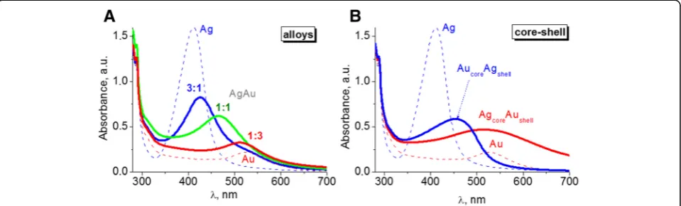
Colloidal solutions of obtained NPs had a bright color due to the localized surface plasmon resonance (LSPR) band absorption. Only one LSPR band is characteristic for bimetallic alloy NPs formed when Ag+ and [AuCl4]− ions are simultaneously reduced. Its maximum is located between the maxima inherent to the LSPR bands of indi-vidual metals (Fig. 1a) and is shifted from silver to gold with decreasing Ag:Au molar ratio. Namely, band max-ima of AgAu NPs were observed at 427 nm for the ratio of 3:1, 466 nm for 1:1, and 511 nm for 1:3, between LSPR bands of individual metals obtained in the same experimental conditions at 411 nm for nanosized silver and 526 nm for gold.
According to SEM images, the average size of AgAu NPs of alloy type was of 7–10 nm (Fig. 2a). We used elemental analysis to check the content of metals in ob-tained NPs. For the sample AgAu(1:1) studied with SEM/EDX, the Ag:Au ratio corresponds to a given ex-perimental quantity. Namely, the average value shown in atomic % ratio was Ag:Auaverage= 1 (Fig. 2a, inset). Also, we fixed amount of carbon that was added to the mix-ture in the form of tryptophan. The experimental molar ratio Trp:M = 2:1 corresponds to atomic ratio C:M = 22:1, where C—carbon and M—metal. SEM/EDX showed that in the area of nanoparticles, carbon quantity exceeds a given number by several times, suggesting the accumula-tion of amino acid and its fragments around NPs, and thus forming an organic stabilizing shell.
Bimetallic core-shell NPs were synthesized by sequen-tial reduction of metal ions. The disappearance of LSPR absorption band of previously formed monometallic par-ticles, used as a core, and the growth of LSPR band of
the second metal in the spectra of the same colloid indi-cates the occurrence of new plasmonic metal shell. Such
process is what we observed for both systems—
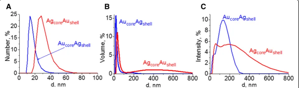
previously formed silver core with a new gold shell and vice versa (Fig. 1b). High-intensive characteristic band of Ag NPs at 411 nm is screened by the absorption of gold nanoparticles at 515 nm. Also, LSPR band of Au NPs at 526 nm is covered by silver absorption with a maximum at 454 nm. Broad LSPR bands as well as their asymmetry indicate polydispersity and association of NPs of core-shell type in colloidal solutions. We suggest that it can be the aggregation of small NPs occurring due to cross-linking of particles through tryptophan during the syn-thesis. Polydispersity in colloids was evidenced by the dynamic light scattering (DLS) data (Fig. 3). Volume and intensity basis suggested the formation of a wide range of structures up to 300 nm in case of AucoreAgshell. And large aggregates up to 800 nm are characteristic for AgcoreAushell. At the same time, there are big fractions of small particles with an average diameter of 15 and 30 nm correspondingly.
The clear difference in topology of core-shell NPs is confirmed by TEM measurements. AucoreAgshellNPs are mainly spherical particles of 10–25 nm (Fig. 4) while their gold core (previously formed monometallic Au NPs) was smaller with the average size of 5–10 nm. Thus, as expected, the growth of the shell is accompanied by an increase of particle size. The same trend is charac-teristic for the system with silver core of 10–25 nm, and twice bigger AgcoreAushell NPs of 25–50 nm, but in this case, the shell is not uniform. According to TEM, the sil-ver cores are mainly cosil-vered by small gold particles. The presence of small Au NPs of 5–10 nm in colloid presum-ably confirms that the formation of gold particles in the presence of tryptophan occurs simultaneously with the gold shell growth around Ag core due to fast reduction of gold with amino acid. Moreover, it causes an aggregation shown by TEM as well as by DLS.
[image:4.595.59.539.558.703.2]Antitumor Activity
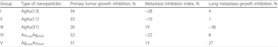
An in vivo antitumor activity of the administered NPs was estimated based on the rate of primary tumor growth inhibition (TGI), lung metastasis growth inhib-ition (MGI), and metastasis inhibinhib-ition (MI) indexes [15]. An inhibitory effect of NPs towards LLC primary tumor growth was observed for mice treated with NPs either with different Ag:Au molar ratio (AgAu(1:3), AgAu(1:1), AgAu(3:1)) or with different topology (AucoreAgshell, AgcoreAushell) (Table 1). Based on a primary tumor growth inhibition rate, all the administered NPs pos-sessed an antitumor activity in vivo. For the majority of AgAu NPs administered, primary tumor growth inhib-ition was more than 30%. The maximum value (34%) of LLC primary tumor inhibition was attributed to NP with metal molar ratio as 1:3, AgAu(1:3) (Table 1).
Despite a primary tumor growth inhibition, the adminis-tered NPs were not efficient in inhibiting of LLC lung me-tastases development. Metastasis inhibition (MI) index was calculated based on a frequency and a number of
metastatic nodes in mouse lungs. During the administra-tion of AgAu(3:1) and AgcoreAushellNPs, MI index was 19 and 13%, respectively, whereas the treatment of mice with AgAu(1:3) (−28%), AucoreAgshell (−22%), and AgAu(1:1) (−10%) was characterized even by stimulation of metasta-sizing (Table 1).
Lung metastases were observed in mice of all the treatment groups. Lung metastasis growth inhibition index ranged from 4% (in AgAu(1:3)-treated group) to 27% (in AgcoreAushell-treated group). Conversely, the treatment with AgAu(3:1) NPs led to an accelerated metastasis growth in mice (MGI =−36%).
Thus, based on the parameters of primary tumor growth inhibition, lung metastasis growth inhibition, and metastasis inhibition index, it can be concluded that the highest antitumor activity was attributed to AgcoreAushell NPs (Table 1).
NPs administration to mice was accompanied with low serum lactate dehydrogenase (LDH) activity, an indi-cator of tumor growth activity and antitumor treatment
Fig. 2SEM images of alloy NPs: sample AgAu(1:1) (a),insetshows atomic ratio of metals Ag:Au and carbon to metal ratio according to SEM/EDX measurements; sample AgAu(1:3) (b), scale 50 nm
[image:5.595.58.540.87.259.2] [image:5.595.58.539.561.702.2]effectiveness [17]. For the mice with LLC that were not treated with NP, the level of serum LDH activity was more than 8500 IU/I (Fig. 5). As seen from the Fig. 5, NP administration led to a reduction of serum LDH activity supporting an observed inhibitory effect of NP administration towards tumor growth. The lowest value of LDH activity was detected in serum of mice treated with AgcoreAushelland AucoreAgshell. At the same time in the groups characterized with comparably higher pri-mary tumor growth inhibition rates, serum LDH activity tended to be significantly lower. However, AgAu(3:1) NP administration had no significant effect on serum LDH activity in mice with LLC. This was consistent given the lowest primary tumor growth inhibition rate in this treatment group (Table 1, Fig. 5).
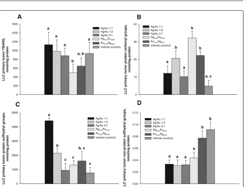
Oxidative stress within the cells is considered as one of the primary mechanisms underlying NP antitumor activity [18–20]. Silver and gold nanoparticles were reported to have an intrinsic property that allows a Fenton-type reaction to produce ROS [21, 22] with a subsequent lipid and protein oxidation, DNA damage, and cell death induction [23]. Several studies under-scored a direct interaction between the ability of NPs to initiate ROS production and the subsequent cell death [2, 23]. Our results are consistent with this paradigm showing that administration of NPs resulted in oxidative stress induction in primary tumor tissue as was evi-denced by accumulation of protein oxidation markers. However, based on the data, it can be concluded that only protein carbonylation may have an impact on pri-mary tumor growth rates and metastasizing.
The analysis of oxidative processes intensity in the primary tumor tissue was performed by determining lipid and protein oxidative damage product accumula-tion. Consistent with morphological parameters of anti-tumor activity, we observed higher levels of oxidative damage product accumulation in primary tumor tissue of mice treated with NPs (Fig. 6). NP treatment did not accelerate lipid oxidation in the primary tumor (Fig. 6A), whereas led to higher protein oxidation (Fig. 6B). The administration of NPs with well-defined topology of metal components (either AgcoreAushell or AucoreAgshell) was the most sufficient in inducing pro-tein carbonylation in LLC primary tumor tissue. Con-versely, the use of NPs with different metal molar ratios (AgAu(1:1) and AgAu(3:1)) did not result in higher tumor protein carbonylation and did not affect or even cause elevation of primary tumor protein sulfhydryl groups (Fig. 6C). Unexpectedly, the action of NPs in case of AgAu(1:1), AgAu(1:3), and AucoreAgshell was accompanied with even significantly higher levels of protein thiol groups. However, the administration of NPs, except for the AucoreAgshell NPs, resulted in significantly lower levels of nonprotein thiol groups in primary tumor tissue (Fig. 6D).
One of the major limitations of any substances with antitumor activity is a side effect expressed in cytotox-icity towards untransformed cells and tissues [20]. Since both therapeutic and toxic effects of nanoparticles need to be studied simultaneously, we have analyzed the ability of NPs to induce hepatic lipid and protein oxidation as well as lymphocyte DNA stability.
[image:6.595.58.539.88.209.2]Fig. 4The TEM images of bimetallic NPs of core-shell type: AucoreAgshell(a) and AgcoreAushell(b). Theinsetsshow previously formed monometallic NPs applied as a core. Scale 100 nm
Table 1The indexes of antitumor activity of gold/silver bimetallic nanoparticles
Group Type of nanoparticles Primary tumor growth inhibition, % Metastasis inhibition index, % Lung metastasis growth inhibition, %
I AgAu(1:3) 34 −28 4
II AgAu(1:1) 33 −10 1
III AgAu(3:1) 26 19 −36
IV AucoreAgshell 32 −22 8
[image:6.595.56.540.648.731.2]l/ UI , yti vi t c a H D L d o ol B 0 500 1000 1500 2000 4000 6000 8000 10000 12000 Ag/Au 1:1 Ag/Au 1:3 Ag/Au 3:1 AgcoreAushell AucoreAgshell Vehicle a b c d d
Fig. 5Blood serum lactate dehydrogenase (LDH) activity in mice with LLC following NP administration. LDH activity was assessed in serum of mice on day 18 after tumor cell inoculation and after daily intra-peritoneal injections of NPs for 12 days at a dose 500μg/kg/day. Values marked withdifferent letters(a,b,c,d) are statistically different,P< 0.05. All values are given as the mean ± 1 S.D.,n= 5 for each group
, S R A B T r o m ut yr a mi r p C L L ni et or p g m/l o m n 0 200 400 600 800 1000 1200 1400 1600 Ag/Au 1:1 Ag/Au 1:3 Ag/Au 3:1 AgcoreAushell
AucoreAgshell
Vehicle (control) a a a a b a, b
A
, s p u or g l y n o br a c ni et or p r o m ut yr a mi r p C L L ni et or p g m/l o m n 0 10 20 30 40 Ag/Au 1:1 Ag/Au 1:3 Ag/Au 3:1 AgcoreAushellAucoreAgshell
Vehicle (control) a a b b b a, c
B
, s p u or g l yr d y hfl u s ni et or p r o m ut yr a mi r p C L L ni et or p g m/l o m n 0 1000 2000 3000 4000 5000 Ag/Au 1:1 Ag/Au 1:3 Ag/Au 3:1 AgcoreAushellAucoreAgshell
Vehicle (control) a b b, c c c c
C
, s p u or g l yr d y hfl u s ni et or p-n o n r o m ut yr a mi r p C L L ni et or p g m/l o m n 0.00 0.02 0.04 0.06 0.08 0.10 0.12 Ag/Au 1:1 Ag/Au 1:3 Ag/Au 3:1 AgcoreAushellAucoreAgshell
Vehicle (control)
a a a a
b b
D
[image:7.595.61.539.88.276.2] [image:7.595.61.540.314.681.2]Fig. 7Tail moment distribution of primary lymphocyte DNA comets of mice with LLC following bimetallic NP administration. DNA comet assay was performed employing primary lymphocytes isolated from mice with LLC following AgAu(3:1) (a) and AgcoreAushell(b) bimetallic NP administration;
X-axis—the length of tail moment,μm;Y-axis—percentage of the comets
, S R A B T ci t a p e H ni et or p g m/l o m n 0 100 200 300 400 500 600 Ag/Au 1:1 Ag/Au 1:3 Ag/Au 3:1 AgcoreAushell
AucoreAgshell
Vehicle (control) a a a a a b
A
, s p u or g l y n o br a c ni et or p ci t a p e H ni et or p g m/l o m n 0 20 40 60 80 100 120 140 Ag/Au 1:1 Ag/Au 1:3 Ag/Au 3:1 AgcoreAushell AucoreAgshellVehicle (control) a a a b b c
B
, s p u or g l yr d y hfl u s ni et or p ci t a p e H ni et or p g m/l o m n 0 10000 20000 30000 40000 Ag/Au 1:1 Ag/Au 1:3 Ag/Au 3:1 AgcoreAushellAucoreAgshell
Vehicle (control) a b a c c a
C
, s p u or g l yr d y hfl u s ni et or p-n o n ci t a p e H ni et or p g m/l o m n 0.00 0.05 0.10 0.15 0.20 0.25 Ag/Au 1:1 Ag/Au 1:3 Ag/Au 3:1 AgcoreAushellAucoreAgshell
Vehicle (control) a a a b b b
D
[image:8.595.59.539.89.264.2] [image:8.595.58.539.303.685.2]Our attention was predominantly focused on Agcore Aushell NPs as these showed the highest antitumor ac-tivity. AgcoreAushell NPs upon their administration to mice with LLC possessed low in vivo genotoxicity to-wards untransformed cells as evidenced by single-cell DNA comet analysis of primary mouse lymphocytes (Fig. 7). The analysis of comet moment distribution re-vealed that up to 70% of cell population was character-ized with intact integrated nuclear DNA after NPs administration. The percentage of cells with a damaged DNA (tail moment more than 150 μm) was only 10% (Fig. 7). Conversely, in a group of mice treated with AgAu(3:1) NPs, only 59% of the cells had intact nuclear DNA; 25% of the comets analyzed in this group had tail moment more than 150 μm, suggesting higher geno-toxicity of these NPs towards lymphocytes.
All of the NPs administered in the present study did not lead to an increased hepatic lipid oxidation (Fig. 8A). After AgcoreAushell and AucoreAgshell NP treatment, the livers retained even higher protein sulfhydryl group levels (Fig. 8C), lower level of protein carbonylation (Fig. 8B) compared to the same parameters in the livers of untreated mice with LLC. These data suggest lower levels of oxidative damage in the livers of mice with LLC receiving NPs.
The data generated in the current study allow for con-cluding the presence of antitumor activity of bimetallic gold and silver NPs. Among the tested samples, and es-pecially among the NPs with the same metal ratio of Ag:Au = 1:1 (alloy and core-shell type), NPs that con-sisted of Ag core covered by Au shell were the most ac-tive towards LLC tumor growth and metastasizing inhibition and possessed the lowest toxicity. Thus, the studied biological activity was the highest in case of or-dered topological distribution of metal compounds within AgcoreAushell NPs, suggesting that in vivo antitu-mor activity of the studied NPs is strongly dependent on gold and silver interaction arising from their ordered topological distribution.
Conclusions
Based on the parameters of primary tumor growth in-hibition, lung metastasis growth inin-hibition, and metas-tasis inhibition index, the highest antitumor activity was attributed to AgcoreAushell NPs among the studied series of bimetallic nanoparticles with different metal content and topology. This was also confirmed by low serum lactate dehydrogenase activity compared to un-treated mice with LLC and higher tumor tissue protein carbonylation. AgcoreAushellNPs showed low genotoxicity towards mouse lymphocytes as evidenced by DNA comet analysis and did not affect hepatic lipid and protein oxida-tion. Our data suggest that AgcoreAushell NPs used in this study may serve as a suitable prototype to develop anticar-cinomatous agent and anticancer drug vehicle.
Abbreviations
DLS:Dynamic light scattering; LDH: Lactate dehydrogenase; LLC: Lewis lung carcinoma; LSPR: Localized surface plasmon resonance; MGI: Metastasis growth inhibition; MI: Metastasis inhibition; NPs: Nanoparticles; ROS: Reactive oxygen species; TGI: Tumor growth inhibition; Trp: Tryptophan
Authors’Contributions
AE and IS supervised this work and helped in the analysis and interpretation of the data. IM, NV, and GG carried out the synthesis of nanoparticles and their following characterization and analysis. VB and NZ performed the studies of antitumor activity and biochemical measurements. IS and IM worked on the drafting and revisions of the manuscript. All the authors have read and approved the final manuscript.
Competing Interests
The authors declare that they have no competing interests.
Publisher’s Note
Springer Nature remains neutral with regard to jurisdictional claims in published maps and institutional affiliations.
Author details
1Department of Biochemistry and Biotechnology, Yuriy Fedkovych Chernivtsi
National University, Kotsuybynskyiy St., 2, Chernivtsi 58012, Ukraine. 2Laboratory of Photonics of Nanosized Oxide Systems, Chuiko Institute of
Surface Chemistry, National Academy of Sciences of Ukraine, General Naumov St., 17, Kyiv 03164, Ukraine.3L.V. Pisarzhevskii Institute of the Physical Chemistry, National Academy of Sciences of Ukraine, Kyiv, Ukraine. 4NanoMedTech LLC, Kyiv, Ukraine.
Received: 17 December 2016 Accepted: 26 April 2017
References
1. Arvizo RR, Bhattacharyya S, Kudgus RA, Giri K, Bhattacharya R, Mukherjee P (2012) Intrinsic therapeutic applications of noble metal nanoparticles: past, present and future. Chem Soc Rev 41(7):2943–2970
2. Liu G, Li Q, Ni W, Zhang N, Zheng X, Wang Y et al (2015) Cytotoxicity of various types of gold-mesoporous silica nanoparticles in human breast cancer cells. Int J Nanomedicine 10:6075–6087
3. Bhattacharya R, Mukherjee P (2008) Biological properties of“naked”metal nanoparticles. Adv Drug Deliv Rev 60(11):1289–1306
4. Gheorghe DE, Cui L, Karmonik C, Brazdeikis A, Penaloza JM, Young JK et al (2011) Gold-silver alloy nanoshells: a new candidate for nanotherapeutics and diagnostics. Nanoscale Res Lett 6:554
5. Loutfy SA, Al-Ansary NA, Abdel-Ghani NT, Hamed AR, Mohamed MB, Craik JD et al (2015) Anti-proliferative activities of metallic nanoparticles in an in vitro breast cancer model. Asian Pac J Cancer Prev 16(14):6039–6046 6. Mukherjee P, Bhattacharya R, Wang P, Wang L, Basu S, Nagy JA et al
(2005) Antiangiogenic properties of gold nanoparticles. Clin Cancer Res 11(9):3530–3534
7. Chakraborty B, Pal R, Ali M, Singh LM, Shahidur Rahman D, Kumar Ghosh S et al (2016) Immunomodulatory properties of silver nanoparticles contribute to anticancer strategy for murine fibrosarcoma. Cell Mol Immunol 13(2):191–205
8. Holden MS, Nick KE, Hall M, Milligan JR, Chen Q, Perry CC (2014) Synthesis and catalytic activity of pluronic stabilized silver-gold bimetallic nanoparticles. RSC Adv 4(94):52279–52288
9. Hu M, Chen J, Li ZY, Au L, Hartland GV, Li X et al (2006) Gold nanostructures: engineering their plasmonic properties for biomedical applications. Chem Soc Rev 35(11):1084–1094
10. Millstone JE, Hurst SJ, Metraux GS, Cutler JI, Mirkin CA (2009) Colloidal gold and silver triangular nanoprisms. Small (Weinheim an der Bergstrasse, Germany) 5(6):646–664
11. Samal AK, Polavarapu L, Rodal-Cedeira S, Liz-Marzan LM, Perez-Juste J, Pastoriza-Santos I (2013) Size tunable Au@Ag core-shell nanoparticles: synthesis and surface-enhanced Raman scattering properties. Langmuir 29(48):15076–15082
13. Hamidi-Asl E, Dardenne F, Pilehvar S, Blust R, De Wael K (2016) Unique properties of core shell Ag@Au nanoparticles for the aptasensing of bacterial cells. Chemosensors 4(3):16
14. Shmarakov IO, Mukha IP, Karavan VV, Chunikhin OY, Marchenko MM, Smirnova NP, et al. Tryptophan-assisted synthesis reduces bimetallic gold/ silver nanoparticle cytotoxicity and improves biological activity. Nanobiomedicine. 2014;1(6). doi:10.5772/59684.
15. Pyaskovskaya ON, Kolesnik DL, Fedorchuk AG, Prokhorova IV, Solyanik GI (2016) 2-Deoxy-D-glucose enhances dichloroacetate antitumor action against Lewis lung carcinoma. Exp Oncol 38(3):176–180
16. Kamins’kyi VO, Lutsyk MD, Stoika RS (2005) Comet assay of DNA fragmentation: modification of silver staining for obtaining permanent preparations. Ukrains’kyi Biokhimichnyi Zhurnal (1999) 77(6):105–108 17. Faloppi L, Del Prete M, Casadei Gardini A, Santini D, Silvestris N, Bianconi M
et al (2016) The correlation between LDH serum levels and clinical outcome in advanced biliary tract cancer patients treated with first line chemotherapy. Scientific Reports 6:24136
18. Manke A, Wang L, Rojanasakul Y (2013) Mechanisms of nanoparticle-induced oxidative stress and toxicity. Biomed Res Int 2013:942916
19. Pal R, Chakraborty B, Nath A, Singh LM, Ali M, Rahman DS et al (2016) Noble metal nanoparticle-induced oxidative stress modulates tumor associated macrophages (TAMs) from an M2 to M1 phenotype: an in vitro approach. Int Immunopharmacol 38:332–341
20. Yamada M, Foote M, Prow TW (2015) Therapeutic gold, silver, and platinum nanoparticles. Wiley Interdisciplinary Rev Nanomed Nanobiotechnol 7(3):428–445 21. Sang Y, Zhang L, Li YF, Chen LQ, Xu JL, Huang CZ (2010) A visual detection of hydrogen peroxide on the basis of Fenton reaction with gold nanoparticles. Anal Chim Acta 659(1–2):224–228
22. Navalon S, de Miguel M, Martin R, Alvaro M, Garcia H (2011) Enhancement of the catalytic activity of supported gold nanoparticles for the Fenton reaction by light. J Am Chem Soc 133(7):2218–2226
23. Dutta D, Sahoo AK, Chattopadhyay A, Ghosh SS (2016) Bimetallic silver nanoparticle-gold nanocluster embedded composite nanoparticles for cancer theranostics. J Mater Chem B 4(4):793–800
Submit your manuscript to a
journal and benefi t from:
7Convenient online submission 7Rigorous peer review
7Immediate publication on acceptance 7Open access: articles freely available online 7High visibility within the fi eld
7Retaining the copyright to your article