Copyright © 1998, American Society for Microbiology. All Rights Reserved.
Evaluation of Three Commercial Enzyme-Linked Immunosorbent
Assays for Diagnosis of Chagas’ Disease
WALTER M. R. OELEMANN,1* MARIA DA GLO´ RIA M. TEIXEIRA,1GIOVANI C. VERI´SSIMO DA COSTA,1
JOSE´ BORGES-PEREIRA,2JOSE´ ADAIL F. DE CASTRO,3JOSE´ RODRIGUES COURA,2
ANDJOSE´ MAURO PERALTA1
Institute of Microbiology, Federal University of Rio de Janeiro,1and Department of Tropical Medicine,
Oswaldo Cruz Institute, FIOCRUZ,2Rio de Janeiro, and Department of Parasitology,
Federal University of Piauı´, Teresina,3Brazil
Received 23 February 1998/Returned for modification 6 April 1998/Accepted 2 June 1998
Chagas’ disease is a common cause of morbidity in Latin American countries. In Brazil, naturally occurring transmission of its etiologic agent, Trypanosoma cruzi, has been almost completely abolished through effective control programs aimed at the triatomid insect vector. Thus, transfusion of blood from infected donors has become the major route for contracting Chagas’ disease due to the socioeconomically motivated migration of residents from areas where the disease is endemic to the larger urban centers. Therefore, the employment of screening tests is mandatory for all blood banks throughout the country. We compared the diagnostic perfor-mances of three commercially available screening assays used in routine testing in Brazilian blood banks: the Abbott Chagas antibody enzyme immunoassay (Abbott Laborato´rios do Brasil, Sa˜o Paulo), the BIOELISA-CRUZI kit (Biolab-Me´rieux, Rio de Janeiro, Brazil), and the BIOZIMA Chagas kit (Polychaco S.A.I.C., Buenos Aires, Argentina). The evaluation was performed with sera obtained from chagasic patients and healthy residents of four different areas in Brazil where Chagas’ disease is either endemic or emergent and where clinical manifestations of the disease and circulating parasite strains vary. The results obtained with each kit were compared to matched in-house enzyme-linked immunosorbent assay and immunofluorescence assay data obtained for each sample. Depending on the area under investigation, the three commercial kits produced specificity values between 93.3 and 100.0%, sensitivity values between 97.7 and 100%, and accuracies ranging from 93.6 to 100.0%.
The protozoan parasite Trypanosoma cruzi is the etiologic agent of Chagas’ disease, which is endemic throughout Latin America and which is a major cause of morbidity and death in the affected countries. According to World Health Organiza-tion estimates (31), 16 to 18 million people are infected by the parasite and about 50,000 chagasic patients die each year from the disease. In Brazil, the area in which the disease is endemic extends over 17 states in the northeastern, southeastern, south-ern, and central western regions (21), but successful vector control programs have abolished almost completely the natural transmission of T. cruzi by its reduviid insect vector. Recent studies reported few chagasic patients younger than 12 years in the state of Minas Gerais (8, 20). Apart from vectorial trans-mission, Chagas’ disease can be contracted either orally (39), congenitally (23), or by transfusion of blood from an infected donor (38). Due to socioeconomic factors, the migration of infected people from the areas in which the disease is endemic to the urban centers is very frequent, and blood transfusion has become the principal way of infection, accounting for an esti-mated 20,000 new cases per year in Brazil, a country with five to six million blood transfusions per year (21). Therefore, ef-ficient donor screening is very important in order to identify and discard contaminated blood without negatively affecting the country’s blood supply.
T. cruzi infection is lifelong, and after a short and mostly
asymptomatic acute phase, during which the parasites can be detected in blood smears, patients enter the indeterminate
phase of the disease, which is marked by an extremely low parasitemia and no sequelae. This stage can last for 10 to 30 years, after which a significant percentage of patients develop the chronic manifestations of Chagas’ disease (cardiopathy, megacolon, and/or megaesophagus). While traditional meth-ods of parasite detection (hemoculture and xenodiagnosis) are time-consuming and of low sensitivity, PCR amplification of nuclear (32, 40) or kinetoplast (3, 43) DNA was shown to be very sensitive (10, 46). However, at present, PCR is not feasible for blood bank screening, and the best way of diagnosing an indeterminate or chronic T. cruzi infection is the serologic detection of antibodies directed against the parasite. Serologic assays include the indirect immunofluorescence assay (IFA), indirect hemagglutination, complement fixation, the radioim-munoprecipitation assay, the enzyme-linked immunosorbent assay (ELISA), and Western blots. Antigen preparations em-ployed in these tests range from crude parasite extracts and subcellular fractions to cloned antigens and synthetic peptides (24, 27–30, 34–36, 41, 44, 45). Some of these tests are available commercially, while others are in-house assays being used only in research settings. In Brazilian blood banks today, the screen-ing of donors for Chagas’ disease by at least two tests based on different methodologies is obligatory. Although IFAs and hemagglutination often lead to false-positive or -negative test results due to subjective interpretation, both assays are still widely used in blood bank screening and epidemiolog-ical surveys, and the results are generally confirmed by an ELISA.
T. cruzi is polymorphic, and different parasite strains
circu-late in different areas (21). While to date no definite correla-tion between infecting strain and clinical manifestacorrela-tion has been demonstrated, survey studies in regions in which the
* Corresponding author. Mailing address: Departamento de Imuno-logia, Instituto de MicrobioImuno-logia, UFRJ-CCS, Ilha do Funda˜o, 21941-590 Rio de Janeiro, Brazil. Phone: 0055-21-270 0990. Fax: 0055-21-560 8028. E-mail: IMIMWAL@MICROBIO.UFRJ.BR.
2423
on May 15, 2020 by guest
http://jcm.asm.org/
disease is endemic show differences in antibody titers found in the patients and in the degree of the clinical manifestations in the chronic phase of the disease (21). Since the infected donor populations encountered in the large urban centers of Brazil migrated from many different regions of the country in which the disease is endemic, in this study, we compared the perfor-mances of three commercial enzyme immunoassays (EIAs) by using panels of sera obtained from patients and healthy resi-dents of four Brazilian areas where Chagas’ disease is either endemic or emergent.
MATERIALS AND METHODS
Study population and description of areas in which Chagas’ disease is en-demic.Sera were obtained from patients and healthy residents from the follow-ing areas: the state of Minas Gerais in the south-central region of Brazil (mu-nicipality of Virgem da Lapa, n5261; 12.6% seroprevalence), where the cardiac and digestive forms of the disease are common (4, 5); the hinterlands of the northeastern states of Paraı´ba (n5466; 9.5% seroprevalence) and Piauı´ (n5 253; 5.9% seroprevalence), where the indeterminate form of the disease is common (7, 16, 17); and the Amazon state in the north of Brazil (municipality of Barcellos, n585; 13.2% seroprevalence), where Chagas’ disease is emergent (15, 18, 19).
The municipalities and regions situated in the states of Minas Gerais, Paraı´ba, and Piauı´ are part of the dry hinterlands with sparse vegetation and rainless periods lasting from 1 to 3 years. On the other hand, the study area located in the
Amazon state is part of the rain forest. The chagasic patients in the areas under investigation became infected mainly by vectorial transmission, with the predom-inant vectors being the triatomid bugs Panstrongylus megistus and Triatoma
in-festans (Minas Gerais) (6), Triatoma brasiliensis (Paraı´ba and Piauı´) (16), Tria-toma pseudomaculata (Paraı´ba) (16), and Rhodnius brethesi (Amazon) (19).
Professional occupations in these regions are agriculture and stock raising (Mi-nas Gerais, Paraı´ba, and Piauı´) and gathering of palm fibers (Amazon). Illiteracy reaches levels between 30 and 40%. Most members of the study population were less than 30 years or more than 50 years of age. The intermediate age group (30 to 50 years old) is underrepresented in these areas as a conse-quence of the migration to the cities of Manaus, Salvador, Sa˜o Paulo, and Rio de Janeiro.
Indirect immunofluorescence.All sera were tested at a final dilution of 1/40 in in-house tests according to the method of Camargo (12) with T. cruzi Y epimas-tigotes as antigen and fluorescein isothiocyanate-conjugated goat anti-human immunoglobulin G (IgG) (Cappel Biomedical Inc., Malvern, Pa.).
In-house ELISA.The cytosolic fraction of T. cruzi Y epimastigotes was used as antigen. Briefly, Nunc microtiter plates were sensitized overnight at 4°C with 100 ml of antigen solution in 0.05 M sodium bicarbonate buffer (pH 9.6) at a con-centration of 200 ng/ml. Sera were diluted 1/200 in phosphate-buffered saline– Tween 20 (PBST) (0.3%)–fetal calf serum (5.0%), and 100ml of the mixture was added to the wells. After 30 min at 37°C, the plates were washed eight times with PBST, anti-human IgG-peroxidase conjugate (Cappel Biomedical Inc.) was add-ed to the wells at a dilution of 1/10,000 in PBST-fetal calf serum, and the wells were incubated at 37°C for 30 min. After eight additional washes, the immune complexes were developed with tetramethylbenzidine-H2O2(Sigma), and the
absorbances were read at 450 nm. Cutoff values were calculated by dividing the difference of the average absorbances of two positive and three negative controls by three.
To compare the results of ELISAs performed on different days, the results were expressed as ratios by dividing the absorbance values of each plate by the cutoff value obtained for the same plate. A sample was considered positive if the ratio was equal to or greater than 1.1, negative if the ratio was equal to or smaller than 0.9, and indeterminate if the ratio was between 0.9 and 1.1. After the results of the in-house ELISA were compared to those previously obtained by IFA, the sera were classified as either positive, negative, or discrepant. Sera with repeat-edly indeterminate ELISA results were a priori considered discrepant. The results are shown in Table 1.
[image:2.612.49.292.90.194.2]Commercial EIAs.Three commercial EIAs were evaluated in this study: the Abbott Chagas antibody EIA (Abbott Laborato´rios do Brasil, Sa˜n Paulo), the BIOELISACRUZI kit (Biolab-Me´rieux, Rio de Janeiro, Brazil), and the BIOZIMA Chagas kit (Polychaco S.A.I.C., Buenos Aires, Argentina). Each EIA was carried out strictly according to the instructions provided by the manufac-turer. Calculations of the cutoff values and evaluation of the test results were performed as described in the respective sections of each manual.
TABLE 1. Consensus classifications of sera from four regions of Brazil by in-house ELISAs and IFAs
Source of sera
No. of sera
col-lected
No. of sera classified as: Positive
by both assays
Negative by both
assays
Positive or neg-ative
Discrepant (assay results
differed) Minas Gerais 261 180 81 261 0 Paraı´ba 466 135 305 440 26 Piauı´ 253 202 44 246 7
Amazon 85 3 75 78 7
Total 1,065 520 505 1,025 40
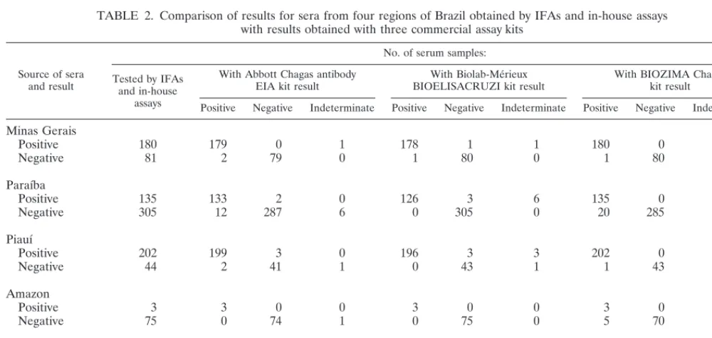
TABLE 2. Comparison of results for sera from four regions of Brazil obtained by IFAs and in-house assays with results obtained with three commercial assay kits
Source of sera and result
No. of serum samples:
Tested by IFAs and in-house
assays
With Abbott Chagas antibody
EIA kit result BIOELISACRUZI kit resultWith Biolab-Me´rieux With BIOZIMA Chagaskit result
Positive Negative Indeterminate Positive Negative Indeterminate Positive Negative Indeterminate
Minas Gerais
Positive 180 179 0 1 178 1 1 180 0 0
Negative 81 2 79 0 1 80 0 1 80 0
Paraı´ba
Positive 135 133 2 0 126 3 6 135 0 0
Negative 305 12 287 6 0 305 0 20 285 0
Piauı´
Positive 202 199 3 0 196 3 3 202 0 0
Negative 44 2 41 1 0 43 1 1 43 0
Amazon
Positive 3 3 0 0 3 0 0 3 0 0
Negative 75 0 74 1 0 75 0 5 70 0
Total for all sera
Positive 520 514 5 1 503 7 10 520 0 0
Negative 505 16 481 8 1 503 1 27 478 0
on May 15, 2020 by guest
http://jcm.asm.org/
[image:2.612.49.549.488.726.2]RESULTS
Evaluation of the EIAs.The results of the evaluation of the
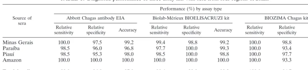
three kits are presented in Tables 2 and 3 for each study area and for the study population as a whole. Due to the lack of a serologic “gold standard” for the indeterminate and chronic phases of Chagas’ disease (see also Discussion), the sera em-ployed in the evaluation were characterized by matched IFA and in-house ELISA results. Of a total of 1,025 sera, 520 were consensus positive and 505 were consensus negative. Perfor-mances of the commercial tests were expressed as relative sensitivity, relative specificity, and accuracy (11).
Looking at the results obtained for each study area and for the population as a whole, all three kits performed comparably (Table 3). Considering the entire study population, the BIO-ELISACRUZI kit had the highest relative specificity and was the most accurate test, whereas the BIOZIMA Chagas kit showed the highest relative sensitivity.
With respect to the IFA and in-house ELISA consensus classifications, 63 sera of a total of 1,025 (6.1%) gave either a discrepant or indeterminate result with at least one of the kits evaluated in this study (some of the 76 discordant results shown in Table 2 appeared in the same sample). Due to the lack of a gray-zone definition for the BIOZIMA Chagas kit, indeterminate results were observed only for the Abbott Cha-gas antibody EIA (9 of 1,025; 0.9%) and the Biolab-Me´rieux BIOELISACRUZI kit (11 of 1,025; 1.1%).
One of the four sera that tested indeterminate with the BIOELISACRUZI kit was a consensus-negative serum from Piauı´ that tested positive with the two other kits. The remain-ing 10 sera (1 from Minas Gerais, 6 from Paraı´ba, and 3 from Piauı´) were consensus positive and were classified as such by both the Abbott Chagas antibody EIA and BIOZIMA Chagas kit.
On the other hand, the BIOELISACRUZI kit diagnoses were in agreement with the consensus on all nine sera which gave indeterminate results with the Abbott Chagas antibody EIA (six sera from Paraı´ba and one each from Minas Gerais, Piauı´, and Amazon), whereas the BIOZIMA Chagas kit clas-sified as positive three consensus-negative sera of the six from Paraı´ba.
The BIOELISACRUZI kit showed the highest relative spec-ificity, with only 1 of 505 (0.02%; from the panel of Minas Gerais sera) consensus-negative sera diagnosed as positive. This serum was classified as negative by the two other kits. The Abbott Chagas antibody EIA and the BIOZIMA Chagas kit showed much lower relative specificities, with 16 (3.2%) and 27 (5.3%) positive results for consensus-negative sera, respec-tively. From the 16 consensus-negative sera that were positive in the Abbott test, 12 (75%) were from the Paraı´ba panel and
2 each were from Minas Gerais and Piauı´. Six of these Paraı´ba sera and one of the Piauı´ sera also tested positive with the BIOZIMA Chagas kit. Additionally, the latter test gave posi-tive results with another group of 20 consensus-negaposi-tive sera (14 from Paraı´ba, 5 from the Amazon, and 1 from Minas Gerais), which were all diagnosed as negative by the Abbott and Biolab-Me´rieux EIAs.
The BIOZIMA Chagas EIA did not yield any negative result for consensus-positive sera and, therefore, was the most sen-sitive test in this study. On the other hand, the Abbott Chagas antibody EIA gave negative results for five (1.0%) consensus-positive sera, three of which were from Piauı´ and two of which were from Paraı´ba. The BIOELISACRUZI kit yielded nega-tive results for seven (1.3%) consensus-posinega-tive sera. Of these, three sera were from Piauı´, three sera were from Paraı´ba, and one serum was from Minas Gerais. Two of the three sera from Piauı´ also tested negative in the Abbott Chagas antibody EIA. However, the remaining five sera were classified as positive by the other two tests. Taken together, these results clearly indi-cate that sera from patients residing in the states of Paraı´ba and Piauı´ have to be considered problematic for routine serol-ogy testing (see also Discussion).
DISCUSSION
In the present study, we compared the performances of the Abbott Chagas antibody EIA, the Biolab-Me´rieux BIOELISA-CRUZI kit, and the BIOZIMA Chagas kit, which are routinely employed in Brazilian blood banks for the detection of anti-bodies against T. cruzi.
[image:3.612.49.549.80.194.2]The BIOZIMA Chagas and BIOELISACRUZI kits are 96-well ELISAs, while the Abbott Chagas antibody EIA employs coated beads as the solid matrix. The Brazilian prices (in U.S. dollars) for a single test are $2.17, $1.59, and $3.51, respec-tively. The total test incubation times varied from 50 to 120 min, with the BIOZIMA Chagas kit being the fastest, provid-ing a result by visual readprovid-ing after a little over 1 h. In addition, it was the most easily performed, with controls, conjugate, and substrate supplied in dropper bottles as ready-to-use solutions. All three assays gave satisfactory results with sera which were obtained in four Brazilian areas and classified as either consensus positive or negative by matched in-house IFA and in-house ELISA results. Relative assay sensitivities and speci-ficities varied depending on the area under investigation (Ta-ble 3) and ranged for the total population from 98.6 to 100% and 94.7 to 99.8%, respectively. The observed area-dependent differences may in part be attributed to the disproportional fractions of positive and negative sera obtained in each area (e.g., 3 positive versus 75 negative sera from the Amazon and 202 positive versus 44 negative sera from Piauı´ [Table 2]).
TABLE 3. Diagnostic performance of three assay kits with sera from four regions of Brazila
Source of sera
Performance (%) by assay type
Abbott Chagas antibody EIA Biolab-Me´rieux BIOELISACRUZI kit BIOZIMA Chagas kit Relative
sensitivity specificityRelative Accuracy sensitivityRelative specificityRelative Accuracy sensitivityRelative specificityRelative Accuracy
Minas Gerais 100.0 97.5 99.2 99.4 98.8 99.2 100.0 98.8 99.6
Paraı´ba 98.5 96.0 96.8 97.7 100.0 99.3 100.0 93.4 95.5
Piauı´ 98.5 95.3 98.0 98.5 100.0 98.8 100.0 97.7 99.6
Amazon 100.0 100.0 100.0 100.0 100.0 100.0 100.0 93.3 93.6
Total for all sera 99.0 96.8 97.9 98.6 99.8 99.2 100.0 94.7 97.4
aIndeterminate results have been omitted.
on May 15, 2020 by guest
http://jcm.asm.org/
However, for the total population, we employed 520 (50.7%) consensus-positive and 505 (49.3%) consensus-negative sera. Consequently, assay performances calculated for the four pan-els as a whole should reflect interassay differences more pre-cisely.
The ELISAs yielded conflicting results for a number of sera, but the same sera were not necessarily problematic for each of the three kits evaluated in this study. Thus, for 21 of 520 (4.0%) positive and 42 of 505 (8.3%) negative sera, the results obtained with at least one of the three kits were not in agree-ment with the consensus. These findings corroborate the re-sults obtained by others (1, 26). A chemiluminescent ELISA for the diagnosis of active infection by T. cruzi (1) was evalu-ated with sera which yielded inconclusive results in eight con-ventional serologic tests. Depending on the combination of test results, the percentage of inconclusive results varied between 18 and 78%. In another study (26), the Abbott Chagas anti-body EIA, the Biolab-Me´rieux BIOELISACRUZI kit, and the Chagas IgG ELISA (Gull Laboratories, Salt Lake City, Utah) were evaluated with 60 sera obtained at a blood bank. The authors defined a combined assay performance in which a serum was considered positive if at least two of the three ELISAs to be evaluated plus a confirmatory IFA were positive. Using the combined assay performance results as the gold standard, ELISA sensitivities were reported to be 100% and specificities varied from 87 to 97%. Carvalho et al. (13) compared the performances of an in-house recombinant-an-tigen ELISA and four commercial ELISAs (Abbott, Biolab-Me´rieux, Gull Laboratories, and Ortho Diagnostic, Buenos Aires, Argentina) with sera obtained in Virgem da Lapa, Mi-nas Gerais, and at the state blood bank of Sa˜o Paulo. The au-thors report for the commercial tests specificities ranging from 95.0 to 98.0% and sensitivities from 99.0 to 100.0%.
The observed variation of sera that were problematic for a given assay is not surprising since the antigen preparations employed in each of the evaluated kits are obtained by differ-ent procedures. Furthermore, the T. cruzi Y epimastigotes are cultivated according to different protocols in various culture media. As previously reported (37), extraction procedures in-fluence drastically the epitopes retained on antigenic mole-cules. Furthermore, binding of these molecules to solid sur-faces hides or exposes epitopes that have different affinities for both specific and nonspecific antibodies present in the sera, thus accounting for conflicting results.
The Abbott Chagas antibody EIA was also evaluated in two studies published earlier (9, 33). Pan et al. (33) reported a sensitivity of 93.48% and a specificity of 99.48% with 1,392 sera from Brazil and Argentina which had been previously charac-terized by a commercial indirect hemagglutination assay.
Brashear et al. (9) used the Abbott Chagas antibody EIA to screen 13,309 sera from a potentially high-risk U.S. donor population and calculated a specificity of 99.98% and positive and negative predictive values of 81.25 and 99.99%, respec-tively.
The sera employed in our study were obtained in Minas Gerais, Paraı´ba, Piauı´, and the Amazon, regions where disease manifestation, circulating parasite strains, and parasitemia vary (6, 7, 16, 18, 19). As a consequence of the sampling technique, in which houses and dwellings were first investigated for the presence of triatomid bugs and then, in a second step, blood samples were drawn from the residents and their neighbors (Minas Gerais, Piauı´, and Paraı´ba), the panels we used did not reflect the overall prevalences of T. cruzi infection described in serologic surveys for the populations in the study areas. How-ever, in the Amazon region, triatomid bugs are not found in houses, and people get infected while working in the rain forest.
The Amazon panel utilized in this study consists of a small part of the samples obtained during the serologic survey (19).
In the particular case of Chagas’ disease, no serologic gold standard for the definition of the disease status exists, since detection of T. cruzi-specific antibodies depends on the pa-tient’s immune status and since cross-reactivity of T. cruzi an-tigens with antibodies raised against other coendemic parasites (Leishmania and Trypanosoma rangeli) is frequent (2, 25, 42). Nevertheless, despite its drawbacks, IFA is the most commonly used serologic test for Chagas’ disease and, as a result, is widely accepted as the gold standard (22). Therefore, as a first step we determined the status of the sera according to the results obtained in an in-house IFA and an in-house ELISA (Table 1). The sera were considered either positive or negative if IFA and ELISA results were concordant and indeterminate if the two results were discrepant. However, while no indeterminate se-rum was found in the panel from Minas Gerais, 26 (5.6%) of the 466 sera from Paraı´ba were found to be indeterminate, as were 7 (2.8%) of the 253 sera from Piauı´ and 7 (8.2%) of the 85 sera from the Amazon. These findings can be explained by the epidemiological characteristics and circulating parasite strains in the different areas. In Virgem da Lapa, Minas Gerais, the cardiac and digestive forms of Chagas’ disease are fre-quent, and the circulating T. cruzi strains generally cause a high-titer immune response in the patients (6). Furthermore, this area is not one in which Leishmania spp. (5), which can cause false-positive results in Chagas’ disease serology (14), is endemic. On the other hand, in the states of Paraı´ba and Piauı´, the indeterminate form of the disease predominates, and pa-tients show mostly moderate or weak immune responses to the infection (7). Also, in these areas leishmaniasis is frequent. As far as the Amazon is concerned, cross-reactions with
Leishma-nia spp. may account for the high seroprevalence reported for
this region (15, 19), and infections with the nonpathogenic parasite T. rangeli have been demonstrated (18). Taken to-gether, these facts are likely to account for the indeterminate classification of some sera by our in-house tests. In addition, we cannot rule out the possibility that some of the discrepan-cies observed between the in-house consensus results and those obtained with the three kits were due to a misclassifica-tion of the sera by our in-house assays. However, the use of in-house tests for the characterization of serum panels and subsequent evaluation of a commercial kit has been reported by others (35).
This study shows that the Abbott Chagas antibody EIA, the Biolab-Me´rieux BIOELISACRUZI kit, and the BIOZIMA Chagas test are well suited for the detection of IgG antibodies against T. cruzi. Nevertheless, when used for routine diagnoses and blood bank screening, problems can occur if the patients or donors come from areas in which the epidemiology of Cha-gas’ disease is complex. Therefore, confirmatory tests with higher specificities need to be developed, and good candidates for such tests are those that include a combination of T. cruzi-specific cloned antigens and/or synthetic peptides (13, 28, 35, 36).
ACKNOWLEDGMENTS
This work was supported in part by Conselho Nacional de Desen-volvimento Cientı´fico e Tecnolo´gico (CNPq), Financiadora de Estudos e Projetos (FINEP), Coordenac¸a˜o de Aperfeic¸oamento de Pessoal de Nı´vel Superior (CAPES), and Conselho de Ensino para Graduados de Universidade Federal do Rio de Janeiro (CEPG-UFRJ).
We are deeply indebted to Carmen Nogueira from the blood bank of the University Hospital Clementino Fraga Filho, Rio de Janeiro, Brazil, for permission to use the Commander Dynamic Incubator and Quantum II reader with the Abbott Chagas antibody EIA and to Carlos A. B. de Souza for excellent technical assistance.
on May 15, 2020 by guest
http://jcm.asm.org/
REFERENCES
1. Almeida, I. C., D. T. Covas, L. M. T. Soussumi, and L. R. Travassos. 1997. A highly sensitive and specific chemiluminescent enzyme-linked immunosor-bent assay for diagnosis of active Trypanosoma cruzi infection. Transfusion
37:850–857.
2. Araujo, F. G. 1986. Analysis of Trypanosoma cruzi antigens bound by specific antibodies and by antibodies to related trypanosomatids. Infect. Immun. 53: 179–185.
3. Avila, H. A., D. S. Sigman, L. M. Cohen, R. C. Millikan, and L. Simpson. 1991. Polymerase chain reaction amplification of Trypanosoma cruzi kineto-plast minicircle DNA isolated from whole blood lysates: diagnosis of chronic Chagas’ disease. Mol. Biochem. Parasitol. 48:211–222.
4. Borges-Pereira, J. 1997. Doenc¸a de Chagas humana: estudo da infecc¸a˜o croˆnica, morbidade e mortalidade em Virgem da Lapa, MG, Brasil (1976– 1996). Ph.D. thesis. Fundac¸a˜o Oswaldo Cruz, Rio de Janeiro, Brazil. 5. Borges-Pereira, J. 1998. Personal communication.
6. Borges-Pereira, J., and J. R. Coura. 1986. Morbidade da doenc¸a de Chagas. Estudo seccional em uma a´rea endeˆmica, Virgem da Lapa, Minas Gerais. Rev. Soc. Bras. Med. Trop. 19:139–148.
7. Borges-Pereira, J., and J. R. Coura. 1987. Morbidade da doenc¸a de Chagas em populac¸o˜es urbanas do Serta˜o da Paraı´ba. Rev. Soc. Bras. Med. Trop. 20: 101–107.
8. Borges-Pereira, J., R. C. R. Santos, E. R. S. Lemos, M. L.-S. Cruz, C. E. A.
Subia, H. P. F. Willcox, and R. V. Cunha. 1989. Infecc¸a˜o chaga´sica em menores de treze anos no municı´pio de Virgem da Lapa, Minas Gerais. Estudo longitudinal no perı´odo de seis anos. Rev. Soc. Bras. Med. Trop.
22(Suppl. II):124.
9. Brashear, R. J., M. A. Winkler, J. D. Schur, H. Lee, J. D. Burczak, H. J. Hall,
and A. A. Pan.1995. Detection of antibodies to Trypanosoma cruzi in the southwestern and western United States. I. Evaluation of the sensitivity and specificity of an enzyme immunoassay for detecting antibodies to T. cruzi. Transfusion 35:213–218.
10. Britto, C., M. A. Cardoso, C. M. Monteiro Vanni, A. Hasslocher-Moreno,
S. S. Xavier, W. Oelemann, A. Santoro, C. Pirmez, C. M. Morel, and P. Wincker.1995. Polymerase chain reaction detection of Trypanosoma cruzi in human blood samples as a tool for diagnosis and treatment evaluation. Parasitology 110:241–247.
11. Buck, A. A., and J. J. Gart. 1966. Comparison of screening tests and refer-ence tests in epidemiologic studies. I. Indices of agreement and their relation to prevalence. Am. J. Epidemiol. 83:586–592.
12. Camargo, M. E. 1966. Fluorescent antibody test for the serodiagnosis of American trypanosomiasis. Technical modification employing preserved cul-ture forms of Trypanosoma cruzi in a slide test. Rev. Inst. Med. Trop. Sa˜o Paulo 8:227–234.
13. Carvalho, M. R., M. A. Krieger, E. C. Almeida, W. Oelemann, M. A.
Shika-nai-Yasuda, A. W. Ferreira, J. Borges-Pereira, A. Sa´ez-Alque´zar, P. E. Dorl-hiac-Llacer, D. F. Chamone, and S. Goldenberg. 1993. Chagas’ disease diagnosis: evaluation of several tests in blood bank screening. Transfusion
33:830–834.
14. Chiller, T. M., M. A. Samudio, and G. Zouler. 1990. IgG antibody reactivity with Trypanosoma cruzi and Leishmania antigens in sera of patients with Chagas’ disease and leishmaniasis. Am. J. Trop. Med. Hyg. 43:650–656. 15. Coura, J. R., T. V. Barrett, and M. A. Naranjo. 1994. Ataque de populac¸o˜es
humanas por triatomı´neos silvestres no Amazonas: uma nova forma de transmissa˜o da infecc¸a˜o chaga´sica? Rev. Soc. Bras. Med. Trop. 27:251–253. 16. Coura, J. R., J. Borges-Pereira, F. I. Alves Filho, J. A. F. de Castro, R. V. da
Cunha, W. Costa, and A. C. V. Junqueira.1996. Morbidade da doenc¸a de Chagas em a´reas do Serta˜o da Paraı´ba e da Caatinga do Piauı´. Rev. Soc. Bras. Med. Trop. 29:197–205.
17. Coura, J. R., L. L. de Abreu, L. E. G. Dubois, F. C. Lima, E. de Arruda, Jr.,
H. P. F. Willcox, N. Annunziato, and W. Petana.1984. Morbidade da doenc¸a de Chagas. II. Estudos seccionais em quatro a´reas de campo no Brasil. Mem. Inst. Oswaldo Cruz 79:101–124.
18. Coura, J. R., O. Fernandes, M. Arboleda, T. V. Barrett, N. Carrara, W.
Degrave, and D. Campbell.1996. Human infection by Trypanosoma rangeli in the Brazilian Amazon. Trans. R. Soc. Trop. Med. Hyg. 90:278–279. 19. Coura, J. R., H. P. F. Willcox, M. Arboleda Naranjo, O. Fernandes, and
D. D. de Paiva. 1995. Chagas’ disease in the Brazilian Amazon. III. A cross-sectional study (1). Rev. Inst. Med. Trop. Sa˜o Paulo 37:415–420. 20. Dias, J. C. P. 1987. Control of Chagas’ disease in Brazil. Parasitol. Today 3:
336–341.
21. Dias, J. C. P. 1992. Epidemiology of Chagas’ disease, p. 49–80. In S. Wendel, Z. Brener, M. E. Camargo, and A. Rassi (ed.), Chagas’ disease (American trypanosomiasis): its impact on transfusion and clinical medicine. Interna-tional Society of Blood Transfusion, Sa˜o Paulo, Brazil.
22. Ferreira, A. W., and S. L. Moraes de Avila. 1995. Laboratory diagnosis of Chagas’ heart disease. Sa˜o Paulo Med. J./RPM 113:767–771.
23. Freilij, H., and J. Altcheh. 1995. Congenital Chagas’ disease: diagnostic and clinical aspects. Clin. Infect. Dis. 21:551–555.
24. Godsel, L. M., R. S. Tibbetts, C. L. Olson, B. M. Chaudoir, and D. M.
Eng-man.1995. Utility of recombinant flagellar calcium-binding protein for
se-rodiagnosis of Trypanosoma cruzi infection. J. Clin. Microbiol. 33:2082–2085. 25. Guhl, F., L. Hudson, C. J. Marinkelle, C. A. Jaramillo, and D. Bridge. 1987. Clinical Trypanosoma rangeli infection as complication of Chagas’ disease. Parasitology 94:475–484.
26. Hamerschlak, N., J. Pasternak, V. Amato Neto, M. B. de Carvalho, C. S.
Guerra, A. L. Coscina, O. C. Ferreira, J. Rosenblit, and L. N. Szterling.1997. Chagas’ disease: an algorithm for donor screening and positive donor coun-seling. Rev. Soc. Bras. Med. Trop. 30:205–209.
27. Knecher, L. M., L. F. Rojkin, G. A. Capriotti, and L. E. Lorenzo. 1993. Chagas’ disease screening in blood bank employing enzyme immunoassay. Int. J. Parasitol. 24:207–211.
28. Krieger, M. A., E. C. Almeida, W. Oelemann, J. J. Lafaille, J.
Borges-Pereira, H. Krieger, M. R. Carvalho, and S. Goldenberg. 1992. Use of recombinant antigens for the accurate immunodiagnosis of Chagas’ disease. Am. J. Trop. Med. Hyg. 46:427–434.
29. Levin, M. J., J. F. da Silveira, A. C. C. Frasch, M. E. Camargo, S. Lafon,
W. M. Degrave, and R. Rangel-Alda˜o.1991. Recombinant Trypanosoma cruzi antigens and Chagas’ disease diagnosis: analysis of a workshop. FEMS Mi-crobiol. Immunol. 89:11–20.
30. Mendes, R. P., S. Hoshino-Shimizu, A. M. M. da Silva, I. Mota, R. A. G.
Heredia, A. O. Luquetti, and P. G. Leser.1997. Serological diagnosis of Chagas’ disease: a potential confirmatory assay using preserved protein an-tigens of Trypanosoma cruzi. J. Clin. Microbiol. 35:1829–1834.
31. Moncayo, A. 1993. Chagas’ disease, p. 62–75. In Tropical disease research: eleventh programme report. World Health Organization, Geneva, Switzer-land.
32. Moser, D. R., L. V. Kirchhoff, and J. E. Donelson. 1989. Detection of
Trypanosoma cruzi by DNA amplification using the polymerase chain
reac-tion. J. Clin. Microbiol. 27:1477–1482.
33. Pan, A. A., G. B. Rosenberg, M. K. Hurley, G. J. H. Schock, V. P. Chu, and
A. Aiyappa.1992. Clinical evaluation of an EIA for the sensitive and specific detection of serum antibody to Trypanosoma cruzi (Chagas’ disease). J. In-fect. Dis. 165:585–588.
34. Paranhos-Bacalla, G. S., M. R. M. Santos, P. C. Cotrim, A. Rassi, M. Jolivet,
M. E. Camargo, and J. F. da Silveira.1994. Detection of antibodies in sera from Chagas’ disease patients using a Trypanosoma cruzi immunodominant recombinant antigen. Parasite Immunol. 16:165–169.
35. Pastini, A. C., S. R. Iglesias, V. C. Carricarte, M. E. Guerin, D. O. Sa´nchez,
and A. C. C. Frasch.1994. Immunoassay with recombinant Trypanosoma
cruzi antigens potentially useful for screening donated blood and diagnosing
Chagas’ disease. Clin. Chem. 40:1893–1894.
36. Peralta, J. M., M. D. G. M. Teixeira, W. G. Shreffler, J. B. Pereira, J. M.
Burns, Jr., P. R. Sleath, and S. G. Reed.1994. Serodiagnosis of Chagas’ disease by enzyme-linked immunosorbent assay using two synthetic peptides as antigens. J. Clin. Microbiol. 32:971–974.
37. Schechter, M., and N. Nogueira. 1988. Variations induced by different meth-odologies in Trypanosoma cruzi surface antigen profiles. Mol. Biochem. Parasitol. 29:37–46.
38. Schmun˜is, G. A. 1991. Trypanosoma cruzi, the etiologic agent of Chagas’ disease: status in the blood supply in endemic and nonendemic countries. Transfusion 31:547–557.
39. Shikanai-Yasuda, M. A., C. Brisola Marcondes, L. A. Guedes, G. S. Siqueira,
A. A. Barone, J. C. P. Dias, V. Amato Neto, J. E. Tolezano, B. A. Peres, E. R. Arruda, Jr., M. H. Lopes, M. Shiroma, and E. Chapadeiro.1991. Possible oral transmission of acute Chagas’ disease in Brazil. Rev. Inst. Med. Trop. Sa˜o Paulo 33:351–357.
40. Silber, A. M., J. Bu´a, B. M. Porcel, E. L. Segura, and A. M. Ruiz. 1997.
Trypanosoma cruzi: specific detection of parasites by PCR in infected
hu-mans and vectors using a set of primers (BP1/BP2) targeted to a nuclear DNA sequence. Exp. Parasitol. 85:225–232.
41. Solana, M. E., A. M. Katzin, E. S. Umezawa, and C. S. Miatello. 1995. High specificity of Trypanosoma cruzi epimastigote ribonucleoprotein as antigen in serodiagnosis of Chagas’ disease. J. Clin. Microbiol. 33:1456–1460. 42. Sousa, O. E., and J. M. Johnson. 1973. Prevalence of Trypanosoma cruzi and
Trypanosoma rangeli in the Republic of Panama. Am. J. Trop. Med. Hyg. 22:
18–23.
43. Sturm, N. R., W. Degrave, C. M. Morel, and L. Simpson. 1989. Sensitive detection and schizodeme classification of Trypanosoma cruzi cells by ampli-fication of kinetoplast minicircle DNA sequences: use in the diagnosis of Chagas’ disease. Mol. Biochem. Parasitol. 33:205–214.
44. Teixeira, M. G. M., J. Borges-Pereira, E. Natizert, M. L. N. X. Souza, and
J. M. Peralta. 1994. Development and evaluation of an enzyme linked immunotransfer blot technique for serodiagnosis of Chagas’ disease. Trop. Med. Parasitol. 45:308–312.
45. Vergara, U., C. Veloso, A. Gonzalez, and M. Lorca. 1992. Evaluation of an enzyme-linked immunosorbent assay for the diagnosis of Chagas’ disease using synthetic peptides. Am. J. Trop. Med. Hyg. 46:39–43.
46. Wincker, P., C. Britto, J. Borges-Pereira, M. A. Cardoso, W. Oelemann, and
C. M. Morel.1994. Use of a simplified polymerase chain reaction procedure to detect Trypanosoma cruzi in blood samples from chronic chagasic patients in a rural endemic area. Am. J. Trop. Med. Hyg. 51:771–777.