Direct effects of leptin on brown and white
adipose tissue.
C A Siegrist-Kaiser, … , J Zapf, C A Meier
J Clin Invest.
1997;
100(11)
:2858-2864.
https://doi.org/10.1172/JCI119834
.
Leptin is thought to exert its actions on energy homeostasis through the long form of the
leptin receptor (OB-Rb), which is present in the hypothalamus and in certain peripheral
organs, including adipose tissue. In this study, we examined whether leptin has direct
effects on the function of brown and white adipose tissue (BAT and WAT, respectively) at
the metabolic and molecular levels. The chronic peripheral intravenous administration of
leptin in vivo for 4 d resulted in a 1.6-fold increase in the in vivo glucose utilization index of
BAT, whereas no significant change was found after intracerebroventricular administration
compared with pair-fed control rats, compatible with a direct effect of leptin on BAT. The
effect of leptin on WAT fat pads from lean Zucker Fa/ fa rats was assessed ex vivo, where a
9- and 16-fold increase in the rate of lipolysis was observed after 2 h of exposure to 0.1 and
10 nM leptin, respectively. In contrast, no increase in lipolysis was observed in the fat pads
from obese fa/fa rats, which harbor an inactivating mutation in the OB-Rb. At the level of
gene expression, leptin treatment for 24 h increased malic enzyme and lipoprotein lipase
RNA 1.8+/-0.17 and 1.9+/-0.14-fold, respectively, while aP2 mRNA levels were unaltered in
primary cultures of brown adipocytes from lean Fa/fa rats. Importantly, however, no
significant […]
Research Article
Find the latest version:
J. Clin. Invest.
© The American Society for Clinical Investigation, Inc. 0021-9738/97/12/2858/07 $2.00
Volume 100, Number 11, December 1997, 2858–2864 http://www.jci.org
Direct Effects of Leptin on Brown and White Adipose Tissue
Catherine A. Siegrist-Kaiser,* Véronique Pauli,* Cristiana E. Juge-Aubry,* Olivier Boss,‡ Agnès Pernin,* William W. Chin,§
Isabelle Cusin,i Françoise Rohner-Jeanrenaud,i Albert G. Burger,* Jürgen Zapf,¶ and Christoph A. Meier*§**‡‡
*Unité de Thyroïde, Division d’Endocrinologie et Diabétologie, Hôpital Universitaire de Genève, ‡Biochimie Médicale, Centre Médical
Universitaire, Université de Genève, CH-1211 Geneva 14, Switzerland; §Division of Genetics, Brigham and Women’s Hospital, Boston,
Massachusetts 02115; iLaboratoires des Recherches Métaboliques, Faculté de Médecine, Université de Genève, CH-1211 Geneva 14, Switzerland; ¶Division of Endocrinology and Diabetes, University Hospital Zürich, CH-8091 Zurich, Switzerland; **Clinique de
Médecine II, Département de Médecine, Hôpital Universitaire de Genève, CH-1211 Geneva 14, Switzerland; and ‡‡Endocrine Division,
Massachusetts General Hospital, Boston, Massachusetts 02114
Abstract
Leptin is thought to exert its actions on energy homeostasis
through the long form of the leptin receptor (OB-Rb), which
is present in the hypothalamus and in certain peripheral
or-gans, including adipose tissue. In this study, we examined
whether leptin has direct effects on the function of brown
and white adipose tissue (BAT and WAT, respectively) at the
metabolic and molecular levels. The chronic peripheral
in-travenous administration of leptin in vivo for 4 d resulted in
a 1.6-fold increase in the in vivo glucose utilization index of
BAT, whereas no significant change was found after
intra-cerebroventricular administration compared with pair-fed
control rats, compatible with a direct effect of leptin on BAT.
The effect of leptin on WAT fat pads from lean Zucker
Fa/
fa
rats was assessed ex vivo, where a 9- and 16-fold increase
in the rate of lipolysis was observed after 2 h of exposure to
0.1 and 10 nM leptin, respectively. In contrast, no increase
in lipolysis was observed in the fat pads from obese
fa/fa
rats, which harbor an inactivating mutation in the OB-Rb.
At the level of gene expression, leptin treatment for 24 h
in-creased malic enzyme and lipoprotein lipase RNA 1.8
6
0.17
and 1.9
6
0.14-fold, respectively, while aP2 mRNA levels
were unaltered in primary cultures of brown adipocytes
from lean
Fa/fa
rats. Importantly, however, no significant
effect of leptin was observed on these genes in brown
adipo-cytes from obese
fa/fa
animals. The presence of OB-Rb
re-ceptors in adipose tissue was substantiated by the detection
of its transcripts by RT-PCR, and leptin treatment in vivo
and in vitro activated the specific STATs implicated in the
signaling pathway of the OB-Rb.
Taken together, our data strongly suggest that leptin has
direct effects on BAT and WAT, resulting in the activation
of the Jak/STAT pathway and the increased expression of
certain target genes, which may partially account for the
ob-served increase in glucose utilization and lipolysis in
leptin-treated adipose tissue. (
J. Clin. Invest.
1997. 100:2858–2864.)
Key words: leptin
•adipocytes
•lipolysis
•Jak/STAT
Introduction
Adipocyte differentiation and metabolism are regulated by a variety of endocrine and paracrine factors, including certain cytokines, such as IL-6 and TNF-a (1–3). The ob gene product (leptin) was initially cloned in ob/ob mice and shown to be a 167–amino acid cytokine-like peptide, secreted by adipocytes in a regulated fashion (4, 5). Subsequent physiological studies revealed that leptin was able to inhibit food intake and en-hance energy expenditure by acting at the hypothalamic level, providing a feedback signal reflecting the nutritional status of the periphery (6–8). The gene encoding a leptin receptor (OB-R)1
was cloned from hypothalamic cDNA, and several differen-tially expressed splice variants were detected in various other tissues (9–12). While most of these variants are currently thought to be involved in leptin binding and transport, the in-tracellular domain of the long form of the OB-R (OB-Rb) is homologous to the IL-6/gp130 receptor family (13). Several studies performed in vivo and in vitro demonstrated the capa-bility of the OB-Rb to activate the Janus kinase (Jak)/signal transducers and activators of transcription (STAT) pathway, resulting in the nuclear translocation of specific STATs, such as STAT 1, 3, 5, and 6 (12–16). Although the OB-Rb is highly expressed in the hypothalamus and the lymphoid system, the presence of small amounts of its mRNA was also demon-strated in other organs, including pancreatic b cells and brown and white adipose tissue (BAT and WAT, respectively) (12, 17, 18). Recently, direct functional effects of leptin on some of these nonhypothalamic target organs were described, such as on hematopoietic cells, the endocrine pancreas, pituitary, and ovary (17, 19–23). However, no data are currently available on potential direct paracrine or autocrine effects of leptin on adi-pose tissue, although several studies have provided some evi-dence compatible with such an hypothesis (24–28).
In this study, we examined the effect of leptin on brown and white adipocytes in vivo, ex vivo, and in primary cultures to demonstrate direct actions of leptin on metabolism and gene expression. In addition, we provide evidence that leptin admin-istration in vivo and in vitro activates the Jak/STAT pathway in adipocytes, resulting in the nuclear translocation of STAT1. Address correspondence to Dr. Christoph A. Meier, Division of
Ge-netics, Thorn 10, Department of Medicine, Brigham and Women’s Hospital, 20 Shattuck Street, Boston, MA 02115. Phone: 617-732-5857; FAX: 617-732-5123.
Received for publication 24 March 1997 and accepted in revised form 10 October 1997.
Methods
Measurement of glucose utilization in BAT in vivo. Freely moving adult male rats were infused for 4 d with vehicle (0.1 M Tris-HCl, pH 9.0) or leptin (12 mg/d) administered into the lateral cerebral ventri-cle. Another group of rats was given either vehicle (Ringer solution) or 1 mg/d of recombinant mouse leptin (Novartis, Basel, Switzerland) for 4 d into the femoral vein via a custom-made device including a swivel mounted on a counterbalanced arm, supporting the catheters and allowing the animals to move freely. To eliminate the confound-ing variable of leptin-induced hypophagia, all vehicle-infused rats were pair-fed to the amount of food consumed by leptin-infused ani-mals. At the end of the treatment period, the animals were anesthe-tized with 60 mg/kg sodium pentobarbital intraperitoneally and pre-pared for euglycemic-hyperinsulinemic clamps associated with the labeled 2-deoxyglucose technique to measure the insulin-stimulated glucose utilization index in BAT in vivo. This technique was de-scribed previously and validated (29–31).
Measurement of lipolysis in WAT fat pads ex vivo. Epididymal fat pads were removed and pooled from Sprague-Dawley (140–160 g) or lean (Fa/fa, 170–190 g) or obese (fa/fa, 205–230 g) Zucker rats, and the amount of glycerol released into the medium was determined as described (32). Briefly, after a preincubation period of 1 h, the pooled fat pads were incubated at 378C for different time intervals in the presence of Krebs-Ringer-Hepes buffer containing dialyzed human serum albumin (30 mg/ml), 2 mg/ml glucose, as well as epinephrine (0.5–5,000 nM) or leptin (0.1–1,000 nM), as appropriate. After the incu-bation period, glycerol release into the medium was determined and the results were expressed as micromoles of glycerol released per 100 mg of adipose tissue per hour. The fat pads from the leptin-resistant obese fa/fa rats served as negative controls for these experiments.
Primary culture of BAT and WAT. 3-wk-old male lean Zucker
Fa/fa rats were killed and interscapular BAT as well as inguinal WAT were immediately removed. For both tissues, precursor cells were iso-lated as reported elsewhere and suspended in a (1:1 vol/vol) mixture of Dulbecco’s modified Eagle’s medium and Ham’s F-12, supple-mented with 16 mM biotin, 18 mM pantothenic acid, 5 mM glutamine, 16 mM glucose, 15 mM Hepes, penicillin 50 IU/ml, streptomycin 50 mg/ml, 100 mM ascorbate, and 10% fetal calf serum (complete me-dium) (33). The medium was changed on day 1 and replaced with a differentiation medium containing 510 nM insulin, 100 nM dexa-methasone, 0.2 nM L-triiodothyronine, and 10 mg/ml transferrin in complete medium (34). The medium was changed every 2 d until the cells were fully differentiated after 14 d, as ascertained by the presence of lipid accumulation and the expression of uncoupling protein-1 (UCP-1) mRNA. The brown adipocytes from the leptin-resistant obese fa/fa rats served as negative controls for these experi-ments.
Analysis of gene expression in brown adipocytes. Fully differenti-ated brown adipocytes were tredifferenti-ated for 24 h with 1 mM BRL49653 (kind gift of Dr. W. Wahli, Lausanne, Switzerland), 625 nM leptin, or a combination of both. Total RNA was isolated by guanidinium isothio-cyanate extraction (35). Cells were lysed in a 4 M guanidinium thiocy-anate solution containing 5 mM sodium citrate, pH 7.0, 0.5% sarcosyl, and 0.1 M b-mercaptoethanol and ultracentrifuged overnight through a CsCl step gradient. RNA (15 mg/sample) was denatured and subjected to electrophoresis on a 1% agarose-formaldehyde gel before blotting and ultraviolet cross-linking onto a nylon membrane (NYTRAN 13 N; Schleicher & Schuell, Dassel, Germany).
The probes for malic enzyme (EcoRI insert from pME6, kindly provided by Dr. V. Nikodem, Bethesda, MD), lipoprotein lipase (EcoRI insert from pGEM2, kindly provided by Dr. M. Schotz, Los Angeles, CA), and aP2 (PstI insert from pAL422, kindly provided by Dr. D. Lane, Baltimore, MD) were labeled with 50 mCi [a-32P]dCTP (Rediprime random primer labeling kit; Amersham, Buchs, Switzerland) to a spe-cific activity of at least 2 3 109 cpm/mg (36, 37). Hybridization was carried out for 2 h with 2 ng/ml of labeled probe, using the Rapid-Hyb buffer (Amersham). Membranes were washed, with the last step
being 0.13 SSC, 0.1% SDS at 658C for 15 min. The autoradiograms were quantified by laser scanning densitometry (Molecular Dynamics, Sunnyvale, CA). The results were corrected for 18S and 28S ribo-somal RNA on the gel as quantitated by scanning densitometry.
Reverse transcription PCR (RT-PCR) assay for the detection of OB-Rb.RNA was prepared from the hypothalamus, WAT, and BAT, as well as cultured brown and white adipocytes as described above. 5 mg of total RNA was used for reverse transcription with Moloney murine leukemia virus reverse transcriptase (2 3 40 min, 378C), primed by oligo-dT18. Primers for PCR amplification were syn-thesized according to the published sequence for the long form of the rat OB-R (OB-Rb, GenBank accession No. D85558), and the cDNA was amplified using the sense primer OB1F (nucleotides 2541–2564: 59-TATGTCATTGTACCGATAATTATT-39) and the reverse primer OB1R (nucleotides 2895–2913: 59 -CAGAGAAGTTAGCACT-GTT-39). All PCR reactions were carried out in standard buffer [10 mM KCl, 20 mM Tris-HCl, pH 8, 0.1% Triton X-100, 6 mM (NH4)2SO4, 0.2 mM dNTP, 2 mM MgCl2, 0.5 mM of each primer, 2.5 U/100 ml of na-tive Pfu (Stratagene, Basel, Switzerland)], for . 40 cycles with the fol-lowing temperatures: denaturation 30 s at 928C, annealing 30 s at 508C, and elongation for 90 s at 728C. The PCR products were analyzed on a 2% agarose gel and stained with ethidium bromide. The PCR prod-ucts obtained from WAT and BAT and culture were sequenced by direct sequencing using standard protocols for the ABI373A auto-mated sequencer with primers OB1F and OB1R.
Preparation of nuclear extracts. Lean male Fa/fa Zucker rats were injected intravenously with either vehicle or 500 mg of leptin in Tris 0.1 M, pH 8.1. After 60 and 90 min, the animals were killed and BAT was removed. Nuclear extractswere prepared as described elsewhere (15). Briefly, tissues were homogenized in 10 mM KCl, 1.5 mM MgCl2, 10 mM Hepes, pH 7.9, 1 mM DTT, 1 mM Na3VO4, 10 mg/ml leupeptin, 0.3 TIU/ml aprotinin, 1 mM pepstatin A, and 1 mM PMSF. The homogenate was centrifuged at 2,000 g for 10 min and the pellet was washed with the same buffer. The washed pellet was resuspended in a buffer containing 420 mM NaCl, 10 mM KCl, 20 mM Hepes, pH 7.9, 20% glycerol, 1 mM DTT, 1 mM Na3VO4, and the protease inhibitors specified above. Nuclei were extracted for 30 min at 48C on a shaking rotor and centrifuged for 10 min at 16,000 g. Supernatants were di-luted in a buffer containing 10 mM KCl, 20 mM Hepes, pH 7.9, 20% glycerol, 1 mM DTT, 1 mM Na3VO4, and the protease inhibitors spec-ified above. After additional centrifugation, the supernatants were concentrated on a Microcon-50 ultrafiltration column (Amicon, Wal-lisellen, Switzerland).
Nuclear extracts from primary brown or white adipocyte cultures were prepared as follows (38). Cells were cultured in 6-cm petri dishes as described above. The cells were placed in serum-free me-dium for 4 h before stimulation with 0.2 mM leptin for 15 or 30 min as indicated. Cells were washed with 2 ml of cold PBS and scraped off the dish in 1 ml of PBS. Two plates were pooled for nuclear protein extraction. Cells were pelleted by centrifugation at 5,200 g for 15 s. The pellet was resuspended in 400 ml of hypotonic buffer (10 mM Hepes, pH 7.9, 10 mM KCl, 0.1 mM EDTA, 0.1 mM EGTA, 1 mM DTT, 1 mM PMSF, and 0.5 mM Na3VO4) by gentle pipetting. The cells were left to swell for 15 min on ice, after which 25 ml of 1% NP-40 was added and the tubes were vigorously vortexed. After spinning at 15,800 g for 30 s, the pellet was resuspended by mild vortexing in 50 ml of cold hypertonic buffer (20 mM Hepes, pH 7.9, 0.4 M NaCl, 1 mM EDTA, 1 mM EGTA, 20% glycerol, 1 mM DTT, 1 mM PMSF, and 0.5 mM Na3VO4). After 15 min of incubation, the tubes were centri-fuged for 5 min at 15,800 g at 48C. Supernatants were aliquoted and stored at 2708C. The protein concentration in the nuclear extracts was determined by the Bradford assay (BioRad, Glattbrugg, Switzer-land).
nuclear extracts from BAT, was incubated with 25,000 cpm of the la-beled M67-SIE, and 2 mg of poly [d(I-C)] in EMSA binding buffer (50 mM KCl, 20 mM Hepes, 20% glycerol, 0.05% NP-40, 10 mM b-mercaptoethanol, pH 7.5) with a final volume of 25 ml. Incubation was performed at room temperature for 20 min and the samples were analyzed on a 5% polyacrylamide gel in 0.53 TBE (44.5 mM Tris, 44.5 mM boric acid, 1 mM EDTA, pH 8) for 75 min at 300 V. Gels were dried and then autoradiographed.
For the supershift assay, either 0.3 or 0.75 mg of a mouse mono-clonal IgG1 anti-STAT1 antibody (Santa Cruz Biotechnology, Basel, Switzerland) or of an anti-STAT3 antibody, respectively, was added to the EMSA incubation mixture for 15 min before the addition of the labeled oligonucleotide.
Statistical analysis. Results are expressed as the mean6SEM and the unpaired Student’s t test was used to assess statistical significance.
P . 0.05 was considered not significant (NS).
Others. All experimental animal protocols were approved by the Review Board for Animal Experimentation of the Canton of Geneva, Switzerland.
Results
[image:4.612.57.555.287.609.2]Leptin enhances glucose utilization of BAT in vivo.To assess direct peripheral effects of leptin on BAT, male rats were given leptin either intracerebroventricularly or intravenously for 4 d, and euglycemic-hyperinsulinemic clamps were per-formed to determine the insulin-stimulated glucose utilization index. As shown in Fig. 1 A, the glucose utilization index in BAT remained unchanged in rats treated with leptin intracere-broventricularly, as compared with pair-fed controls (195623 vs. 174611 ng/min/mg tissue, NS). In contrast, when leptin was administered intravenously, the insulin-stimulated glucose uti-lization index increased 1.6-fold to 320645 ng/min/mg tissue, compared with 196614 ng/min/mg tissue in pair-fed controls (P , 0.05). Hence, the peripheral, but not central, administra-tion of leptin enhances glucose utilizaadministra-tion in BAT compared with pair-fed control rats, compatible with a direct peripheral effect of leptin on this tissue.
Leptin increases lipolysis in WAT fat pads ex vivo.The ef-fect of leptin on the basal lipolytic rate of epididymal fat pads was assessed ex vivo. As shown in Fig. 1, B and C, leptin in-creases glycerol release in a dose- and time-dependent manner in fat pads from Sprague-Dawley rats, where leptin is a strong stimulator of lipolysis. On a molar basis, epinephrine stimu-lated lipolysis about fourfold more potently than leptin. To as-certain that this effect is indeed mediated by leptin and the OB-Rb, a similar experiment was performed in lean (Fa/fa) and obese (fa/fa) Zucker rats. As shown in Fig. 1 D, epineph-rine was able to enhance the glycerol release in the fat pads from Fa/fa and fa/fa animals, as expected, whereas only the leptin-sensitive Fa/fa animals responded to leptin in a dose-dependent manner.
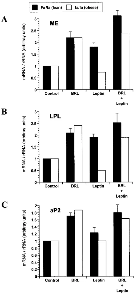
Effects of leptin treatment on gene expression in brown adi-pocytes. Primary cultures of in vitro differentiated brown adi-pocytes from genetically lean Fa/fa or obese fa/fa rats were treated for 24 h with either 1 mM of the thiazolidinedione BRL49653, 625 nM of leptin, or both. Analysis of the extracted total RNA by Northern blotting after treatment with BRL49653 revealed as expected an about twofold increase in the mRNA for malic enzyme, lipoprotein lipase, and the fatty acid–bind-ing protein, aP2, in brown adipocytes derived from lean rats (Fig. 2) (40–42). Interestingly, treatment with leptin resulted in a similar increase in malic enzyme and lipoprotein lipase mRNA (1.860.17 and 1.960.14-fold, respectively; P , 0.01 compared with control), while the expression of the aP2 gene remained unaltered. The combined treatment with leptin and BRL49653 resulted in a partially additive effect on malic enzyme expres-sion (3.160.22-fold, P , 0.05 compared with BRL alone). In contrast, the brown adipocyte cultures from obese Zucker fa/ fa rats harboring a partially inactivating mutation in the OB-Rb did not respond to leptin (0.73-fold stimulation), although the thiazolidinedione BRL49653 induced gene expression as expected (2.2-fold) (43–45).
Adipose tissue expresses OB-Rb. The presence of small amounts of OB-Rb in adipose tissue has been reported previ-ously (12). Using total RNA extracted from rat hypothalamus, BAT, and WAT, as well as from primary brown and white adi-pocyte cultures, we performed RT-PCR to demonstrate the presence of OB-Rb transcripts. The amplified products de-rived from native and cultured adipocytes were sequenced, confirming the presence of the correct PCR product (data not shown). With this assay, OB-Rb mRNA could be detected in tissue samples as well as cultures derived from BAT (Fig. 3). Lean Fa/fa rats as well as obese fa/fa animals expressed OB-Rb as expected, since the fa missense mutation alters intracellular trafficking, rather than expression of the OB-R (43). Similar results were obtained for native and cultured WAT (data not shown).
Leptin activates STATs in brown and white adipocytes.
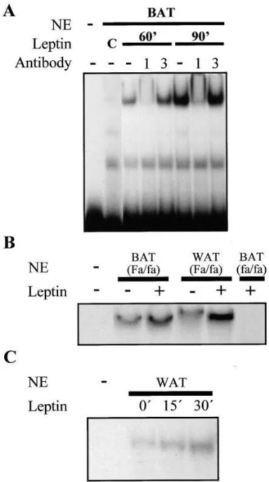
[image:5.612.315.531.58.526.2]To determine whether leptin is capable of activating the Jak/ STAT pathway in adipocytes, as previously reported for the hypothalamus, we examined whether leptin would induce nu-clear STAT translocation in vivo and in cultured primary adi-pocytes (15). Nuclear extracts were prepared from BAT 60 and 90 min after the intravenous injection of Fa/fa rats with 500 mg of leptin. The extracts were assayed for the presence of specific STATs by means of an EMSA. Leptin strongly in-duced the translocation of STAT1, as demonstrated by binding to the M67-SIE DNA element and supershifting with an anti-STAT1, but not anti-STAT3, antibody (Fig. 4 A). In contrast,
Figure 2. Effect of leptin on gene expression in primary cultures of brown adipocytes. Northern blot of total RNA isolated from brown adipocytes after treatment for 24 h with vehicle (Control), 1 mM BRL49653 (BRL), and/or 625 nM leptin. Equal amounts of RNA (15 mg) were loaded on each lane and, after transfer, the membrane was hybridized with the probe for malic enzyme. The experiments were repeated at least three times in adipocytes derived from Fa/fa
rats. The results were normalized to the 18S and 28S rRNA bands and expressed as the mean6SEM. (A) Quantitation of three different experiments examining the effects of BRL and/or leptin on malic en-zyme (ME) mRNA in brown adipocytes from lean Fa/fa rats and of one parallel experiment in obese fa/fa animals (for the Fa/fa animals: control vs. leptin P , 0.01; BRL vs. BRL 1 leptin P , 0.05). (B) Quantitation of three different experiments examining the effects of BRL and/or leptin on lipoprotein lipase (LPL) mRNA in brown adi-pocytes from lean Fa/fa rats and of one parallel experiment in obese
and in parallel with the data on glucose utilization (Fig. 1 A), no peripheral activation of STATs was observed after the ad-ministration of 12 mg of leptin i.c.v. (data not shown).
To determine whether these effects of leptin were direct, primary brown and white adipocyte cultures were treated with leptin and the nuclear extracts assayed for STAT activity. Lep-tin-inducible binding of STATs in the nuclear extracts from brown and white adipocytes increased 1.8- and 2.7-fold, re-spectively, after 15 min on the M67-SIF element, which binds STAT1 and 3 (Fig. 4 B). The shift in mobility of the band ob-served after treatment of white adipocytes with leptin suggests the possible transition from hetero- to homodimeric com-plexes or vice versa. However, no activation of STATs was ob-served in primary cultures of BAT from obese fa/fa rats, com-patible with the genetic leptin resistance of these animals (Fig. 4 B, right-most lane). Interestingly, even basal STAT activity was absent in the fa/fa adipocytes, suggesting either that the basal signal observed in Fa/fa cells is due to the paracrine ef-fects of leptin secreted in culture, or, alternatively, due to a general desensitization of STAT signaling in fa/fa adipocytes. Time course experiments performed in WAT demonstrated a rapid increase in STAT translocation at 15 min (1.7-fold), which increased further after 30 min (3.1-fold; Fig. 4 C).
Discussion
[image:6.612.62.555.57.195.2]In this paper, we demonstrate that leptin has direct effects on BAT and WAT at a metabolic and molecular level. This con-clusion is supported by several independent lines of evidence: (a) peripheral, but not central, administration of leptin in-creases the insulin-stimulated utilization of glucose in BAT when compared with pair-fed control rats; (b) leptin at concen-trations as low as 0.1 nM stimulates basal lipolysis in white fat pads ex vivo in a time- and dose-dependent manner, and this effect is absent in fat pads from obese fa/fa rats which are known to be deficient in functional OB-R; (c) leptin induces the expression of malic enzyme and lipoprotein lipase in pri-mary cultures of brown adipocytes; (d) BAT, WAT, and pri-mary adipocyte cultures all express OB-Rb mRNA; and (e) treatment of brown and white adipocytes with leptin in vivo or in vitro induces the nuclear translocation of STATs, compati-ble with the currently known mechanism of action of leptin through the OB-R. Taken together, our results suggest that
Figure 3. Detection of OB-Rb by RT-PCR in hypothalamus (Hypoth) as well as tissue (T) and primary cultures (C) from BAT from lean Fa/fa and obese fa/fa rats. The PCR products were run on an ethidium bromide– stained 2% agarose gel. The products were of the expected size (371 bp) and the specificity of the amplification was further assessed by direct sequencing of the PCR products derived from adipose tissue and culture. No band was present in the control reactions (2) where the reverse transcriptase was omitted.
[image:6.612.57.249.265.609.2]leptin has direct auto- or paracrine effects on adipocytes, which possibly contribute to the weight- and fat-reducing ac-tivity of leptin.
While the hypothalamus was the first target organ de-scribed for leptin, recent evidence at the level of mRNA and cellular function suggests that peripheral organs also express OB-Rb and are responsive to leptin (12, 17, 22). Specifically, several recent reports demonstrated an inhibitory effect of lep-tin on the release of insulin from pancreatic b cells, and an-other paper reported decreased synergism of insulin-like growth factor I on the follicle-stimulating hormone–induced estradiol synthesis when leptin was present (17, 19, 20). In ad-dition, leptin was shown to increase the pituitary secretion of luteinizing hormone, independent of any confounding hypo-thalamic effects (22). We now show that leptin increases glu-cose utilization in BAT in vivo and the rate of lipolysis in white fat pads ex vivo. This latter result is compatible with the recent observations of Shimabukuro et al. that leptin treatment in vivo dramatically reduces tissue triglyceride stores compared with pair-fed controls, already suggesting a role for leptin be-yond its appetite-reducing properties (25, 26). In addition, the same group has demonstrated increased lipolysis and the in-creased expression of lipolytic enzymes in primary cultures of pancreatic islets, analogous to our findings in white fat pads (28). However, other groups have failed to detect a significant effect of leptin on basal lipolysis in cultured adipocytes in vitro (27). Our positive results in this study are likely due to the use of intact fat pads ex vivo, since the model of isolated adipo-cytes used in the negative reports was shown previously to be less responsive to other lipolytic stimuli, such as epinephrine (Zapf, J., unpublished data). The observation that already 0.1 nM of leptin is able to stimulate basal lipolysis and that fat pads from Zucker fa/fa rats, which are known to harbor a par-tially inactivating mutation in the OB-Rb gene, are unrespon-sive to leptin suggests that this effect is specific and of potential of physiological relevance. However, the possibility cannot be excluded that the adipocytes from fa/fa animals have acquired postreceptor defects related to their obese and insulin-resis-tant state, contributing to their unresponsiveness to the lipoly-tic action of leptin. Nevertheless, the observation that leptin fails to activate STATs in brown adipocytes derived from the leptin-resistant fa/fa animals is compatible with the concept that the observed direct effects of leptin might be mediated through the OB-R.
Leptin also enhances the mRNA expression of malic en-zyme and lipoprotein lipase, but not of the fatty acid–binding protein aP2, in brown adipocytes from lean Fa/fa rats, but not in brown adipocytes derived from obese Zucker fa/fa rats. The only gene shown previously to be regulated directly by leptin in a preadipocyte cell line is the acetyl-CoA carboxylase gene, which is suppressed in the presence of leptin (24). Since the malic enzyme and lipoprotein lipase genes are both regulated by the peroxisome proliferator-activated receptors (PPARs), it is conceivable that leptin activates this nuclear receptor pathway. However, the fact that leptin does not significantly increase aP2 mRNA, which represents another PPAR-regu-lated gene, suggests that any putative functional interaction between the OB-R and PPAR signaling pathways is more complex. Since leptin not only decreases food intake, but also increases energy expenditure, we examined its effect on the expression of UCP-1 in brown adipocyte cultures. However, we were unable to detect any chances in the levels of UCP-1
mRNA after leptin treatment, and hence it remains to be seen whether the direct effects of leptin on BAT are implicated in regulating thermogenesis (data not shown).
The levels of OB-Rb in all target organs other than the hy-pothalamus and the hematopoietic system are very low, re-quiring RT-PCR or RNase protection assays for the detection of transcripts. In this study, we were able to detect OB-Rb mRNA in BAT, WAT, and primary adipocyte cultures, which is compatible with previous reports demonstrating the pres-ence of OB-Rb mRNA in adipose tissue by RNase protection (12, 18). However, other than the hypothalamus, no target tissue for leptin was hitherto shown to respond by nuclear STAT translocation (15). The present experiments demon-strate that the peripheral, but not central, administration of leptin in vivo activates STAT1 in BAT. Experiments with pri-mary cultures of brown and white adipocytes suggest that this effect of leptin on adipocytes is indeed direct and does not in-volve the hypothalamus. However, the exact nature of the STATs required for transducing the leptin signal is still contro-versial and may depend on the cellular context and the con-comitant presence of other stimuli in vivo. For example, a pre-vious in vivo study implicated only STAT3 in hypothalamic leptin signaling, whereas experiments involving transfected cells have provided evidence for the activation of either STAT3, 5, 6, STAT1, 3, 5, or only STAT1 and 3, depending on the cellular model (12–15).
In summary, we have shown that physiological levels of leptin have direct and specific effect on the metabolism and gene expression of brown and white adipocytes. These obser-vations suggest that leptin may exert its weight-reducing action not only through an endocrine, hypothalamic mode of action, but also through an auto- or paracrine pathway.
Acknowledgments
We are grateful to Dr. D. Lane for providing the aP2 probe, to Dr. M. Schotz for the lipoprotein lipase probe, and Dr. V. Nikodem for the malic enzyme cDNA. Leptin was kindly provided by Novartis. The expert technical assistance of Mrs. Mireille de Meyer and Mrs. Mar-jana Paponja is gratefully acknowledged.
C.A. Meier is supported by a Fellowship from the Howard Hughes Medical Institute. The work was supported by the Swiss Na-tional Science Foundation (grants 32-37536.93 to A.G. Burger and C.A. Meier, and 32-046808.96 to J. Zapf) and also in part through a grant-in-aid from Boehringer-Ingelheim International Inc. (to C.A. Meier).
References
1. Spiegelman, B.M., and J.S. Flier. 1996. Adipogenesis and obesity: round-ing out the big picture. Cell. 87:377–389.
2. Stouthard, J.M.L., R.P.J.O. Elferink, and H.P. Sauerwein. 1996. Interleu-kin-6 enhances glucose transport in 3T3-L1 adipocytes. Biochem. Biophys. Res. Commun. 220:241–245.
3. Peraldi, P., G.S. Hotamisligil, W.A. Buurman, M.F. White, and B.M. Spiegelman. 1996. Tumor necrosis factor (TNF)-a inhibits insulin signaling through stimulation of the p55 TNF receptor and activation of sphingomyeli-nase. J. Biol. Chem. 271:13018–13022.
4. Caro, J.F., M.K. Sinha, J.W. Kolaczynski, P.L. Zhang, and R.V. Consi-dine. 1996. Leptin: the tale of an obesity gene. Diabetes. 45:1455–1462.
5. Zhang, Y., R. Proenca, M. Maffei, M. Barone, L. Leopold, and J.M. Friedman. 1994. Positional cloning of the mouse obese gene and its human ho-mologue. Nature. 372:425–432.
6. Pelleymounter, M.A., M.J. Cullen, M.B. Baker, R. Hecht, D. Winters, T. Boone, and F. Collins. 1995. Effects of the obese gene product on body weight regulation in ob/ob mice. Science. 269:540–543.
Rabinowitz, R.L. Lallone, S.K. Burley, and J.M. Friedman. 1995. Weight-reducing effects of the plasma protein encoded by the obese gene. Science. 269: 543–546.
8. Campfield, L.A., F.J. Smith, Y. Guisez, R. Devos, and P. Burn. 1995. Re-combinant mouse OB protein: evidence for a peripheral signal linking adiposity and central neural networks. Science. 269:546–549.
9. Tartaglia, L.A., M. Dembski, X. Weng, N.H. Deng, J. Culpepper, R. De-vos, G.J. Richards, L.A. Campfield, F.T. Clark, J. Deeds, et al. 1995. Identifica-tion and expression cloning of a leptin receptor, OB-R. Cell. 83:1263–1271.
10. Chen, H., O. Charlat, L.A. Tartaglia, E.A. Woolf, X. Weng, S.J. Ellis, N.D. Lakey, J. Culpepper, K.J. Moore, R.E. Breitbart, et al. 1996. Evidence that the diabetes gene encodes the leptin receptor: identification of a mutation in the leptin receptor gene in db/db mice. Cell. 84:491–495.
11. Lee, G.H., R. Proenca, J.M. Montez, K.M. Carroll, J.G. Darvishzadeh, J.I. Lee, and J.M. Friedman. 1996. Abnormal splicing of the leptin receptor in diabetic mice. Nature. 379:632–635.
12. Ghilardi, N., S. Ziegler, A. Wiestner, R. Stoffel, M.H. Heim, and R.C. Skoda. 1996. Defective STAT signaling by the leptin receptor in diabetic mice. Proc. Natl. Acad. Sci. USA. 93:6231–6235.
13. Baumann, H., K.K. Morella, D.W. White, M. Dembski, P.S. Bailon, H.K. Kim, C.F. Lai, and L.A. Tartaglia. 1996. The full-length leptin receptor has signaling capabilities of interleukin 6-type cytokine receptors. Proc. Natl. Acad. Sci. USA. 93:8374–8378.
14. Rosenblum, C.I., M. Tota, D. Cully, T. Smith, R. Collum, S. Qureshi, J.F. Hess, M.S. Phillips, P.J. Hey, A. Vongs, et al. 1996. Functional STAT 1 and 3 signaling by the leptin receptor (OB-R). Reduced expression of the rat fatty leptin receptor in transfected cells. Endocrinology. 137:5178–5181.
15. Vaisse, C., J.L. Halaas, C.M. Horvath, J.E. Darnell, Jr., M. Stoffel, and J.M. Friedman. 1996. Leptin activation of Stat3 in the hypothalamus of wildtype and ob/ob mice but not db/db mice. Nat. Genet. 14:95–97.
16. White, D.W., K.K. Kuropatwinski, R. Devos, H. Baumann, and L.A. Tartaglia. 1997. Leptin receptor (OB-R) signaling. Cytoplasmic domain muta-tional analysis and evidence for receptor homo-oligomerization. J. Biol. Chem. 272:4065–4071.
17. Emilsson, V., Y.L. Liu, M.A. Cawthorne, N.M. Morton, and M. Daven-port. 1997. Expression of the functional leptin receptor mRNA in pancreatic is-lets and direct inhibitory action of leptin on insulin secretion. Diabetes. 46:313–316. 18. Fei, H., H.J. Okano, C. Li, G.H. Lee, C. Zhao, R. Darnell, and J.M. Friedman. 1997. Anatomic localization of alternatively spliced leptin receptors (Ob-R) in mouse brain and other tissues. Proc. Natl. Acad. Sci. USA. 94:7001– 7005.
19. Zachow, R.J., and D.A. Magoffin. 1997. Direct intraovarian effects of leptin: impairment of the synergistic action of insulin-like growth factor-I on follicle-stimulating hormone-dependent estradiol-17b production by rat ova-rian granulosa cells. Endocrinology. 138:847–850.
20. Kieffer, T.J., R.S. Heller, and J.F. Habener. 1996. Leptin receptors ex-pressed on pancreatic b-cells. Biochem. Biophys. Res. Commun. 224:522–527.
21. Bennett, B.D., G.P. Solar, J.Q. Yuan, J. Mathias, G.R. Thomas, and W. Matthews. 1996. A role for leptin and its cognate receptor in hematopoiesis. Curr. Biol. 6:1170–1180.
22. Yu, W.H., M. Kimura, A. Walczewska, S. Karanth, and S.M. McCann. 1997. Role of leptin in hypothalamic-pituitary function. Proc. Natl. Acad. Sci. USA. 94:1023–1028.
23. Gainsford, T., T.A. Willson, D. Metcalf, E. Handman, C. McFarlane, A. Ng, N.A. Nicola, W.S. Alexander, and D.J. Hilton. 1996. Leptin can induce pro-liferation, differentiation, and functional activation of hemopoietic cells. Proc. Natl. Acad. Sci. USA. 93:14564–14568.
24. Bai, Y., S.Y. Zhang, K.S. Kim, J.K. Lee, and K.H. Kim. 1996. Obese gene expression alters the ability of 30A5 preadipocytes to respond to lipogenic hormones. J. Biol. Chem. 271:13939–13942.
25. Shimabukuro, M., K. Koyama, G. Chen, M.Y. Wang, F. Trieu, Y. Lee, C.B. Newgard, and R.H. Unger. 1997. Direct antidiabetic effect of leptin through triglyceride depletion of tissues. Proc. Natl. Acad. Sci. USA. 94:4637– 4641.
26. Chen, G., K. Koyama, X. Yuan, Y. Lee, Y.T. Zhou, R. O’Doherty, C.B. Newgard, and R.H. Unger. 1996. Disappearance of body fat in normal rats in-duced by adenovirus-mediated leptin gene therapy. Proc. Natl. Acad. Sci. USA.
93:14795–14799.
27. Müller, G., J. Ertl, M. Gerl, and G. Preibisch. 1997. Leptin impairs met-abolic actions of insulin in isolated rat adipocytes. J. Biol. Chem. 272:10585– 10593.
28. Zhou, Y.T., M. Shimabukuro, K. Koyama, Y. Lee, M.Y. Wang, F. Trieu, C.B. Newgard, and R.H. Unger. 1997. Induction by leptin of uncoupling pro-tein-2 and enzymes of fatty acid oxidation. Proc. Natl. Acad. Sci. USA. 94:6386– 6390.
29. Ferre, P., A. Leturque, A.F. Burnol, L. Penicaud, and J. Girard. 1985. A method to quantify glucose utilization in vivo in skeletal muscle and white adi-pose tissue of the anaesthetized rat. Biochem. J. 228:103–110.
30. Terrettaz, J., F. Assimacopoulos-Jeannet, and B. Jeanrenaud. 1986. Se-vere hepatic and peripheral insulin resistance as evidenced by euglycemic clamps in genetically obese fa/fa rats. Endocrinology. 118:674–678.
31. James, D.E., K.M. Burleigh, and E.W. Kraegen. 1986. In vivo glucose metabolism in individual tissues of the rat. Interaction between epinephrine and insulin. J. Biol. Chem. 261:6366–6374.
32. Zapf, J., D. Feuerlein, M. Waldvogel, and E.R. Froesch. 1975. Increased sensitivity of diabetic rat adipose tissue towards the lipolytic action of epineph-rine. Diabetologia. 11:509–516.
33. Rehnmark, S., J. Kopecky, A. Jacobsson, M. Nechad, D. Herron, B.D. Nelson, M.J. Obregon, J. Nedergaard, and B. Cannon. 1989. Brown adipose tis-sue differentiated in vitro can express the gene for the uncoupling protein ther-mogenin: effects of hypothyroidism and norepinephrine. Exp. Cell. Res. 182: 75–83.
34. Shima, A., Y. Shinohara, K. Doi, and H. Terada. 1994. Normal differen-tiation of rat brown adipocytes in primary culture judged by their expressions of uncoupling protein and the physiological isoform of glucose transporter. Bio-chim. Biophys. Acta. 1223:1–8.
35. Ausubel, F.M., R. Brent, R.E. Kingston, D.D. Moore, J.G. Seidman, J.A. Smith, and K. Struhl. 1997. Current Protocols in Molecular Biology. J. Wiley & Sons, Inc., New York.
36. Magnuson, M.A., H. Morioka, M.F. Tecce, and V.M. Nikodem. 1986. Coding nucleotide sequence of rat liver malic enzyme mRNA. J. Biol. Chem. 261:1183–1186.
37. Bernlohr, D.A., C.W. Angus, M.D. Lane, M.A. Bolanowski, and T.J. Kelly. 1984. Expression of specific mRNAs during adipose differentiation: identification of an mRNA encoding a homologue of myelin P2 protein. Proc. Natl. Acad. Sci. USA. 81:5468–5472.
38. Schreiber, E., P. Matthias, M.M. Muller, and W. Schaffner. 1989. Rapid detection of octamer binding proteins with “mini-extracts,” prepared from a small number of cells. Nucl. Acids Res. 17:6419.
39. Wagner, B.J., T.E. Hayes, C.J. Hoban, and B.H. Cochran. 1990. The SIF binding element confers sis/PDGF inducibility onto the c-fos promoter. EMBO (Eur. Mol. Biol. Organ.) J. 9:4477–4484.
40. Castelein, H., T. Gulick, P.E. Declercq, G.P. Mannaerts, D.D. Moore, and M.I. Baes. 1994. The peroxisome proliferator activated receptor regulates malic enzyme gene expression. J. Biol. Chem. 269:26754–26758.
41. Hertz, R., V. Nikodem, A. Ben-Ishai, I. Berman, and J. Bar-Tana. 1996. Thyromimetic mode of action of peroxisome proliferators: activation of “malic” enzyme gene transcription. Biochem. J. 319:241–248.
42. Pearson, S.L., M.A. Cawthorne, J.C. Clapham, S.J. Dunmore, S.D. Holmes, G.B.T. Moore, S.A. Smith, and M. Tadayyon. 1996. The thiazo-lidinedione insulin sensitiser, BRL 49653, increases the expression of PPARg and aP2 in adipose tissue of high-fat-fed rats. Biochem. Biophys. Res. Commun.
229:752–757.
43. Chua, S.C., Jr., D.W. White, X.S. Wu-Peng, S.M. Liu, N. Okada, E.E. Kershaw, W.K. Chung, L. Power-Kehoe, M. Chua, L.A. Tartaglia, and R.L. Leibel. 1996. Phenotype of fatty due to Gln269Pro mutation in the leptin recep-tor (Lepr). Diabetes. 45:1141–1143.
44. Phillips, M.S., Q.Y. Liu, H.A. Hammond, V. Dugan, P.J. Hey, C.T. Cas-key, and J.F. Hess. 1996. Leptin receptor missense mutation in the fatty Zucker rat. Nat. Genet. 13:18–19.