Generation and characterization of mice
deficient in hepsin, a hepatic transmembrane
serine protease.
Q Wu, … , J E Sadler, J Morser
J Clin Invest.
1998;
101(2)
:321-326.
https://doi.org/10.1172/JCI1617
.
Hepsin is a type II transmembrane serine protease highly expressed on the surface of
hepatocytes. The physiological function of hepsin is not known, although in vitro studies
indicate that hepsin plays a role in the initiation of blood coagulation and in hepatocyte
growth. To determine the functional importance of hepsin, we generated hepsin-deficient
mice by homologous recombination. Homozygous hepsin-/- mice were viable and fertile,
and grew normally. In functional assays including tail bleeding time, plasma clotting times,
and tissue factor- or LPS-induced disseminated intravascular coagulation models, no
significant difference was found between hepsin-/- and wild-type litter mates. Liver weight
and serum concentrations of liver-derived proteins or enzymes were similar in hepsin-/- and
wild-type mice. Interestingly, serum concentrations of bone-derived alkaline phosphatase
were approximately twofold higher in hepsin-/- mice of both sexes when compared with
wild-type litter mates. No obvious abnormalities were found in major organs in
hepsin-/-mice in histological examinations. Our results indicate that hepsin is not essential for
embryonic development and normal hemostasis. Hepsin-/- mice will help to evaluate the
long-term effects of hepsin deficiency in these animals.
Research Article
Find the latest version:
J. Clin. Invest.
© The American Society for Clinical Investigation, Inc. 0021-9738/98/01/0321/06 $2.00
Volume 101, Number 2, January 1998, 321–326 http://www.jci.org
Generation and Characterization of Mice Deficient in Hepsin, a Hepatic
Transmembrane Serine Protease
Qingyu Wu,* Dongyin Yu,* Joseph Post,‡ Meredith Halks-Miller,‡ J. Evan Sadler,§i and John Morser*
*Department of Cardiovascular Research and ‡Department of Pharmacology, Berlex Biosciences, Richmond, California 94804; and §Department of Medicine and iDepartment of Biochemistry and Molecular Biophysics, Howard Hughes Medical Institute, Barnes-Jewish
Hospital, Washington University School of Medicine, St. Louis, Missouri 63110
Abstract
Hepsin is a type II transmembrane serine protease highly expressed on the surface of hepatocytes. The physiological function of hepsin is not known, although in vitro studies indicate that hepsin plays a role in the initiation of blood co-agulation and in hepatocyte growth. To determine the func-tional importance of hepsin, we generated hepsin-deficient mice by homologous recombination. Homozygous hepsin2/2
mice were viable and fertile, and grew normally. In func-tional assays including tail bleeding time, plasma clotting times, and tissue factor- or LPS-induced disseminated intra-vascular coagulation models, no significant difference was found between hepsin2/2 and wild-type litter mates. Liver
weight and serum concentrations of liver-derived proteins or enzymes were similar in hepsin2/2 and wild-type mice.
Interestingly, serum concentrations of bone-derived alka-line phosphatase were approximately twofold higher in hepsin2/2 mice of both sexes when compared with wild-type
litter mates. No obvious abnormalities were found in major organs in hepsin2/2 mice in histological examinations. Our
results indicate that hepsin is not essential for embryonic development and normal hemostasis. Hepsin2/2 mice will
help to evaluate the long-term effects of hepsin deficiency in these animals. (J. Clin. Invest. 1998. 101:321–326.) Key words: gene targeting • embryonic stem cells • homologous
recombination • factor VII • hepatocyte
Introduction
Membrane proteases participate in a variety of important biological events, including development, tissue remodeling, wound healing, cytokine processing, and tumor invasion (1–3). Hepsin is a novel serine protease with a transmembrane do-main near the amino terminus (4, 5). This structural feature distinguishes hepsin from most serine proteases of the trypsin superfamily (6, 7). In Drosophila melanogaster, Stubble-stub-bloid protein, a transmembrane serine protease, has a
struc-ture similar to that of hepsin (8). Genetic studies demonstrate that defects in the Stubble-stubbloid gene lead to malformation of legs, wings, and bristles in the fruitfly (9).
Biochemical studies including proteolytic digestion and membrane fractionation show that hepsin is a type II trans-membrane serine protease mainly expressed on the surface of hepatocytes (10–12). Low levels of hepsin mRNA have been detected in kidney, pancreas, lung, and prostate, but the cell types that express hepsin mRNA in these tissues are unknown. In addition to normal tissues, hepsin gene expression was found in several tumor cell lines, including hepatoma cell lines HepG2 and PLC/PRF/5, mammary carcinoma cell lines MCF784 and T470, and epithelioid HeLa S3 carcinoma cells (11, 13). Recently, hepsin was reported to be frequently over-expressed in ovarian carcinomas (14).
The physiological function of hepsin is unknown, although biochemical studies indicate that hepsin may play a role in the initiation of blood coagulation. Kazama et al. reported that re-combinant human hepsin expressed on the surface of BHK cells activated blood coagulation factor VII (FVII)1 (15). The
reaction was highly specific since hepsin did not activate other structurally related proteases such as factors IX and X, pro-thrombin, and protein C under similar experimental condi-tions. Hepsin-mediated FVII activation was shown to be suffi-cient to initiate the coagulation pathway leading to thrombin formation (15).
In addition, hepsin may participate in other biological pro-cesses such as cell growth or tissue remodeling. Torres-Rosado et al. reported that hepsin is required for the growth of human hepatocytes (13). In a cell culture system, addition of a poly-clonal anti-hepsin antibody or hepsin-specific antisense oligo-nucleotides significantly altered the morphology of HepG3 cells and inhibited their growth (13). These results suggest that hepsin functions either directly as a growth factor or indirectly as an enzyme that processes a growth factor(s) for hepato-cytes.
To evaluate the biological importance of hepsin, we have generated hepsin-deficient mice by gene targeting technology. Homozygous hepsin2/2 mice were viable and fertile, and did
not exhibit obvious defects in growth, liver function, and blood coagulation. Our results show that hepsin is not essential for embryonic development, organogenesis, and hemostasis.
Methods
The murine hepsin gene and the targeting vector.Murine hepsin cDNAs were isolated by PCR amplification and screening of murine liver A preliminary report describing a portion of this work has appeared
in abstract form (1997. Thromb. Haemostasis. [Suppl.]:756a). Address correspondence to Qingyu Wu, M.D., Ph.D., Berlex Bio-sciences, 15049 San Pablo Avenue, Richmond, CA 94804. Phone: 510-669-4737; FAX: 510-669-4246; E-mail: Qingyu_Wu@Berlex.com
Received for publication 27 August 1997 and accepted in revised form 11 November 1997.
cDNA libraries (16). The full-length murine hepsin cDNA was z 1.6 kb in length and shared 84% sequence identity with human hepsin cDNA (4). The hepsin cDNAs were used to screen a 129Sv genomic DNA library (Stratagene Inc., La Jolla, CA) to clone the mouse hep-sin gene. To construct a targeting vector, a 4-kb KpnI/BamHI frag-ment in the hepsin gene was replaced by a neomycin (neo) expression cassette driven by the murine phosphoglycerate kinase promoter (17) (see Fig. 1 A). At the 39 end of the vector, a herpes simplex virus thy-midine kinase (tk) gene cassette was included to serve as a negative selection marker (18) (Fig. 1 A).
Targeting of the hepsin gene in mouse embryonic stem (ES) cells.
30 mg of linearized plasmid vector DNA was introduced into ES cells (RW4; Genome Systems, St. Louis, MO) by electroporation using an Electro Cell Manipulator (ECM 600; BTX Inc., San Diego, CA). Trans-fected ES cells (1.5 3 107) were selected in the presence of 250 mg/ml
G418 (Life Technologies, Gaithersburg, MD) and 2 mM ganciclovir (Syntex Lab., Inc., Palo Alto, CA). Stable clones from the selection were screened for homologous recombination by Southern blotting using a genomic probe flanking the hepsin gene deletion (see Fig. 1
A). Additional Southern analysis was performed using a neo probe to exclude the presence of random insertions of the neo gene in the ge-nome of mutant ES cells.
Generation of chimeric mice and germline transmission of the mu-tant allele.Mutant ES clones were microinjected into blastocysts from C57BL/6J mice. Chimeric male offspring with . 50% coat color chimerism were bred with National Institutes of Health (NIH) Swiss females to establish germline transmission of the mutant hepsin al-lele. Genotyping was performed by PCR using 100 ng genomic DNA isolated from tail or ear biopsies. Oligonucleotide primers for both
neo gene (59-CAA GAT GGA TTG CAC GCA GG-39 and 59-GTA AAG CAC GAG GAA GCG G-39) and the hepsin gene (59-ACG GCA CAT CGG GCT TCT TTT-39 and 59-AAT GCG GTC CAC CGG CAG CTT CCT-39) were included in the reactions. The PCR results were confirmed by Southern analysis of EcoRI digested DNA samples using the hepsin genomic probe. All mice in this study were maintained in an animal facility accredited by the Association for As-sessment and Accreditation of Laboratory Animal Care Interna-tional (AAALACI).
Northern analysis of hepsin mRNA expression. Total RNA was extracted from frozen liver tissue using the Ultraspec™ RNA kit (Biotecx Laboratories Inc., Houston, TX). Samples (15 mg) were fractionated on denaturing agarose gels, transferred onto nitrocellu-lose membranes, and hybridized with 32P-labeled mouse hepsin
cDNA probes spanning the entire coding region. To determine mRNA expression of other liver specific genes, the blot was reprobed with a partial mouse prothrombin cDNA that was PCR-amplified based on the published sequence (19).
Hemostasis assays and blood chemistry analysis. Blood samples were drawn from the inferior vena cava into 1/10 volume of 3.8% so-dium citrate. Blood cell counts and hematocrit were analyzed using a hemostasis instrument (9210 CP Baker System, Allentown, PA). Clot-ting assays (activated partial thromboplastin time and prothrombin time [aPTT and PT]) were performed with platelet poor plasma using the MLA Electra 900C programmable coagulometer (Pleasantville, NY). In the tail-bleeding assay, mice were anesthetized with pento-barbital (90 mg/kg). The tip of the mouse tail (1 mm in diameter) was cut. Tails were soaked immediately in 0.9% NaCl saline prewarmed at 378C and the time until bleeding stopped completely was recorded. For blood chemistry analysis, serum samples were prepared from clotted whole blood. Chemistry analysis was performed by Consoli-dated Veterinary Diagnostics, Inc. (West Sacramento, CA). Alkaline phosphatase isoenzymes were analyzed by a quantitative method de-scribed by Koffmann et al. (20).
LPS-induced septic shock and thromboplastin-induced dissemi-nated intravascular coagulation (DIC). In the LPS-induced septic shock model, mice (13–14 wk old) were injected intraperitoneally with 40 mg/kg of LPS from Escherichia coli serotype O111:B4 (Cal-biochem Corp., La Jolla, CA). The mice were monitored for signs of
endotoxemia and lethality four times a day for the first 3 d and peri-odically thereafter. In the thromboplastin-induced DIC model, anes-thetised mice were injected intravenously with human placental thromboplastin (Behring, Marburg, Germany). The linear range of the thromboplastin dose was determined experimentally. The time in minutes was recorded between the injection of thromboplastin and acute death as measured by cessation of breathing or heart beating.
Histological analysis. Mice were killed under CO2 narcosis and
tissues were fixed by perfusion through the heart with an aldehyde fixative consisting of 4% (wt/vol) paraformaldehyde, 3% (wt/vol) su-crose, in 0.1 M phosphate buffer, pH 7.3. Tissues were dehydrated in graded ethanol solutions and embedded in paraffin blocks. Tissue sections were cut at 5-mm thickness, stained with hematoxylin and eosin (H & E), and examined by light microscopy.
Results
Disruption of the hepsin gene in ES cells and generation of
chi-meric mice. An 18-kb DNA fragment that contained the
mouse hepsin gene was isolated from a 129Sv genomic library. The hepsin gene contains at least 12 exons (Fig. 1 A). The serine protease active site residues, His, Asp, and Ser are en-coded by exons 7, 8, and 11, respectively. A replacement tar-geting vector was constructed by deleting the KpnI/BamHI fragment containing exons 5–8 that code for the conserved ac-tivation site and most of the catalytic domain (Fig. 1 A). Over 700 stable ES cell clones transfected with the targeting vector were analyzed by Southern blotting. Seven ES clones were identified in which one copy of the endogenous hepsin gene was disrupted by homologous recombination. Four mutant ES clones were microinjected into blastocysts from C57BL/6J mice to generate chimeric mice. Germline transmission was achieved with nine chimeric mice derived from three indepen-dent cell lines. The results of phenotypical analysis were ob-tained from mice derived from all these three ES cell lines.
Viability, growth, and fertility of homozygous hepsin2/2
mice. Breeding of hepsin1/2 mice produced over 500 offspring.
Genotypes of F2 progeny were determined by PCR analysis and the results were confirmed by Southern blotting in ran-domly selected samples (Fig. 1 B). Wild-type, hepsin1/2 and
hepsin2/2 mice represented 24.3, 50.0, and 25.7% of progeny,
respectively. The observed Mendelian pattern of inheritance indicates that homozygous hepsin deficiency is compatible with normal embryonic development. Male to female ratio was z 1:1 in wild-type, hepsin1/2, and hepsin2/2 mice. The absence
of hepsin mRNA expression in liver samples from hepsin2/2
mice was demonstrated by Northern analysis using cDNA probes spanning the entire coding region (Fig. 1 C). As a con-trol, the expression of prothrombin mRNA in the liver from hepsin2/2 mice was not significantly affected (Fig. 1 C).
When followed to 10 mo, hepsin2/2 mice appeared to
de-velop normally and exhibited similar body weight gain as wild-type and hepsin1/2 litter mates. No obvious physical
ab-normalities were observed in hepsin2/2 mice. In hematologic
examinations, values for red blood cell, white blood cell, and platelet counts, hematocrit and hemoglobin were similar in hepsin2/2 mice and wild-type controls (data not shown). Both
male and female hepsin2/2 mice were fertile and produced
via-ble offspring.
Tail bleeding and plasma clotting assays in hepsin2/2 mice.
tail bleeding time measures both platelet and blood-clotting activities, whereas aPTT and PT assays measure the function of the intrinsic and extrinsic blood coagulation pathways, re-spectively. As shown in Table I, no prolongation in time was detected in hepsin2/2 mice in these three assays, indicating that
hepsin deficiency did not adversely affect hemostasis. These results are consistent with the observation that no spontaneous bleedings occurred in hepsin2/2 mice.
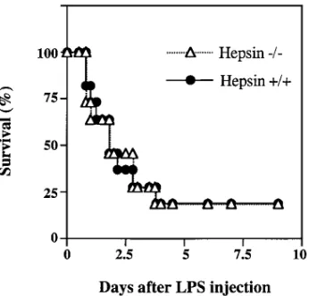
Thromboplastin-induced DIC and LPS-induced septic shock
models. To examine the potential function of hepsin under
pathological conditions, hepsin2/2 mice were challenged in
DIC and septic shock models. In the DIC model, thromboplas-tin was injected intravenously. The times at which breathing or heart beating stopped were similar in both hepsin2/2 mice and
wild-type controls (Table I), indicating that the tissue factor-dependent coagulation pathway was not altered in hepsin2/2
mice. This was supported by the results from a septic shock model, in which survival rates of hepsin2/2 and wild-type mice
after injection of a high dose of LPS were not significantly dif-ferent (Fig. 2).
Figure 1. Disruption of the mouse hepsin gene by homologous
[image:4.612.56.293.61.382.2]re-combination. (A) Structure of the hepsin gene and the targeting vec-tor. Exons (black boxes) and selected restriction sites in the hespin gene (wild-type allele) are indicated (top panel). The targeting vector was constructed by replacing of the KpnI/BamHI fragment with a neo gene cassette. A tk gene cassette was included at the 39 end of the vector. The predicted genomic structure after homologous recombi-nation (mutant allele) is shown. Locations of oligonucleotide primers for PCR analysis and a hepsin genomic probe for Southern blotting are indicated. (B) Genotyping by PCR and Southern analyses. Ge-nomic DNA was isolated from tail or ear biopsies. Genotypes were determined by PCR (left) and Southern (right) analyses. In the PCR reactions, oligonucleotide primers specific for the neo and wild-type hepsin genes were included. The PCR products were visualized in an agarose gel stained with ethdium bromide. DNA fragments from neo or hepsin genes represent the mutant or wild-type alleles, respec-tively. For Southern blotting, genomic DNA was digested with EcoRI and hybridized with the hepsin genomic probe (A). DNA fragments of the expected size from wild-type and mutant hepsin alleles are in-dicated. (C) Northern analysis of hepsin mRNA expression. Total RNA was isolated from liver samples from wild-type, heterozygous, and homozygous mice. Northern blot was probed with a mouse hep-sin cDNA. Hephep-sin mRNA was detected in wild-type and heterozy-gous mice but not in homozyheterozy-gous mice (top panel). As a control, the same blot was reprobed with a mouse prothrombin (PrT) cDNA. The expression of mouse prothrombin mRNA was not affected in hepsin2/2 mice (lower panel).
Table I. Functional Hemostasis Assays in Wild-type and Hepsin-deficient Mice
Hepsin1/1 Hepsin2/2
Tail bleeding time (s) 70.6619.7 77.3628.7 (n 5 5) (n 5 7) PT (s) 20.966.6 22.664.5 (n 5 6) (n 5 5) aPTT (s) 37.2611.2 38.469.1 (n 5 9) (n 5 6) Thromboplastin-induced DIC:
cessation of breathing (min) 1.3060.16 1.4260.42 (n 5 5) (n 5 6) cessation of heart beating (min) 1.8660.34 1.9760.45
(n 5 5) (n 5 6)
The tail bleeding time, plasma prothrombin time (PT) and activated partial thromboplastin time (aPTT) were performed with 9–12-wk-old mice, as described in Methods. In the thromboplastin-induced DIC model, acute death was ascertained as cessation of either breathing or
heart beating. Data are presented as mean6SD and the number of mice
in each experimental group is indicated in parentheses.
Figure 2. Survival of hepsin-deficient and wild-type mice after LPS
[image:4.612.315.556.83.238.2] [image:4.612.317.490.505.671.2]Blood chemistry analysis for liver function. In addition to blood coagulation, hepsin was reported to be required for growth of hepatocytes in vitro (13). To examine the effects of hepsin deficiency on liver function, blood samples from hep-sin-deficient mice were analyzed. As shown in Table II, serum concentrations of alanine aminotransferase (SGPT), g -glu-tamyltranspeptidase (GGT), bilirubin, albumin, globulin, and total protein were similar in hepsin2/2 and wild-type mice, and
values were within the normal ranges. Interestingly, serum al-kaline phosphatase levels were approximately twofold higher in hepsin2/2 mice when compared with wild-type litter mates
(Fig. 3). The elevation of serum alkaline phosphatase was found in both male and female hepsin2/2 mice at different ages
(Fig. 3). Alkaline phosphatase isoenzymes were analyzed, and results indicated that the elevated alkaline phosphatase in the serum from hepsin2/2 mice was mostly of bone origin (Fig. 4).
Morphologic and histologic analyses. Necropsies and his-tologic examinations were performed on hepsin2/2 mice at
var-ious ages. No gross abnormalities were indentified in major or-gans including liver, brain, lung, heart, kidney, pancreas, spleen, and bone. Livers from hepsin2/2 and wild-type mice
appeared similar and had comparable weights (Table II). In H & E–stained sections, tissues including liver and bone from hepsin2/2 mice were histologically indistinguishable from
those of wild-type mice (data not shown). X-ray examinations were also performed to detect potential defects in the skele-ton. No structural abnormalities were found in long bones, pel-vis, and vertebrae in hepsin2/2 mice at the age of 5 mo (data
not shown).
Discussion
Human recombinant hepsin expressed on the surface of BHK cells has been reported to specifically activate FVII (15), an es-sential enzyme that acts at the top of the tissue
factor–depen-dent coagulation pathway (21). This finding was confirmed in our own studies (data not shown), indicating a potential func-tion of hepsin in the initiafunc-tion of blood coagulafunc-tion. Genetic and site-directed mutagenesis studies show that the activation cleavage of the Age152-Ile peptide bond in FVII is required to convert the zymogen to an active enzyme (22, 23). The mecha-nism of FVII activation under physiological conditions is not completely understood. In addition to hepsin, factors IXa, Xa and XIIa, and thrombin have been reported to activate FVII in vitro (24–28). Recent studies with haemophilia patients sug-gest that FIXa is primarily responsible for the basal levels of FVIIa in vivo (29–31). However, the functional significance of the FVII activation mediated by other enzymes remains un-clear at this time.
To evaluate the physiological importance of hepsin, we generated hepsin-deficient mice by gene targeting technology. Hepsin2/2 mice were viable and did not bleed spontaneously.
[image:5.612.315.484.55.368.2]Furthermore, no functional defects were detected in the tail bleeding and blood clotting assays. These results suggest that hepsin deficiency did not significantly alter blood coagulation pathways. Due to lack of a suitable assay, we were unable to measure directly plasma FVIIa level in hepsin2/2 mice (32, 33). Table II. Liver Weight and Blood Chemistry Analysis for
Liver Function
Hepsin1/1 Hepsin2/2
Liver weight (grams) 0.9660.13 0.9260.08 (n 5 6) (n 5 5) SGPT (IU/liter) 25.7568.27 31.3867.86
(n 5 28) (n 5 29) GGT (IU/liter) 0.7160.75 1.060.58
(n 5 7) (n 5 7) Total protein (grams/dl) 5.4860.28 4.8760.43
(n 5 8) (n 5 13) Albumin (grams/dl) 2.8160.24 2.3260.15
(n 5 18) (n 5 13) Globulin (grams/dl) 2.6760.22 2.5560.31
(n 5 18) (n 5 13) Total bilirubin (mg/dl) 0.1760.07 0.1360.07
(n 5 14) (n 5 11)
Wild-type and hepsin2/2 mice (8–12-wk old) were killed under CO
2
nar-cosis. The liver was dissected out and weighed. Blood chemistry analy-sis was performed as described in Methods. Data are presented as
mean6SD and the number of mice in each experimental group is
[image:5.612.56.300.82.269.2]indi-cated in parentheses.
Figure 3. Serum alkaline phosphatase levels in hepsin-deficient and
control mice. Serum samples were prepared from male (top) and fe-male (bottom) hepsin2/2 mice and wild-type litter mates at 10, 12, and
14–16 wk of age. The number of mice in each group is four to five. Total serum alkaline phosphatase activity was measured by hydroly-sis of p-nitrophenylphosphate at pH 9.2 (39). Data are presented as mean6SD. Statistical differences between hepsin2/2 mice (open bars)
Since the prothrombin time and thromboplastin-induced DIC assays, which depend on FVIIa, were normal in these animals, it is very unlikely that the hepsin-mediated FVII activation is physiologically important.
In addition to FVII activation, hepsin was shown to play a role in hepatocyte growth (13). The observation is intriguing since serine proteases are known to have growth factor–like activities. For example, thrombin is a potent mitogen for vas-cular fibroblasts and smooth muscle cells (34). Furthermore, several growth factors, such as hepatocyte growth factor (HGF) and the product of growth arrest-specific gene 6 (Gas6), share striking sequence and structural similarities with blood coagulation proteases (35, 36), and HGF is critical for the development of several epithelial organs including liver and placenta (37, 38). The hepsin-dependent growth of hepa-tocytes in culture suggests that hepsin may play a role in pro-cesses such as cell differentiation and organogenesis. Surpris-ingly, hepsin2/2 mice developed normally. Liver weight and
serum levels of liver proteins in hepsin2/2 mice were similar to
those in wild-type controls (Table II). Histological examina-tions did not identify any structural abnormalities in major organs. These results indicate that the hepatocyte growth-stimulating activity of hepsin is not essential for embryonic de-velopment and maintenance of normal liver function.
An interesting observation in hepsin2/2 mice is that serum
alkaline phosphatase is increased in both male and female hepsin2/2 mice of various ages (Fig. 3). The significance of this
finding is not clear at this time. We consider several possibili-ties here. First, in humans, elevation of serum alkaline phos-phatase is most commonly associated with hepatobiliary disor-ders. In hepsin2/2 mice, however, hepatolibilary disease is
unlikely since the livers were histologically normal, and tests of liver function, such as bilirubin, SGPT, GGT (Table II), and liver-derived alkaline phosphatase (Fig. 4), were within normal ranges. Occasionally, high levels of alkaline phosphatases also are found in patients with malignant tumors that ectopically express the enzyme (39). However, this is not the case in
hepsin2/2 mice since they have not developed tumors. The
in-crease of serum alkaline phosphatase in hepsin2/2 mice could
be due to impaired catabolism of alkaline phosphatase, since the liver is proposed to be responsible for the clearance of al-kaline phosphatase from the blood (40). The mechanism of this process is not clear. If hepsin were responsible for the cleavage and removal of serum alkaline phosphatase, then hepsin deficiency could lead to the elevation of alkaline phos-phatase in serum. Finally, the selective increase in bone alka-line phosphatase in hepsin2/2 mice (Fig. 4) suggests the
possi-bility of certain skeletal disorders. In humans, for example, elevated alkaline phosphatase levels can be found in Paget’s disease, osteomalacia, and rickets (39). In our study, x-ray ex-aminations did not detect skeletal abnormalities in hepsin2/2
mice up to the age of 5 mo, but minor changes in bone density cannot be excluded completely. Further studies will determine whether skeletal abnormalities develop in older animals.
Acknowledgments
We thank L. Westfield (Howard Hughes Medical Institute, St. Louis, MO) for oligonucleotide synthesis, J. Xue (Washington University School of Medicine, St. Louis, MO) for stimulating discussions, J. Mc-Clary for DNA sequencing, M. Werner and P. Verhallen for assis-tance with mouse DIC models, V. Del Vecchio for assisassis-tance in histo-logical studies, and B. Lillis and staff members of Animal Facilities at Berlex (Richmond, CA) for animal care. We also thank W. Dole and G. Rubanyi for their encouragement and helpful discussions.
J.E. Sadler is a Howard Hughes Medical Institute Investigator.
References
1. Bond, J.S. 1991. Plasma membrane proteases: introductory remarks.
Biomed. Biochim. Acta. 50:775–780.
2. Chen, W.-T. 1993. Membrane proteases: role in tissue remodeling and tu-mour invastion. Curr. Opin. Cell Biol. 4:802–809.
3. Mignatti, P., and D.B. Rifkin. 1993. Biology and biochemistry of protein-ases in tumor invasion. Physiol. Rev. 73:161–195.
4. Leytus, S.P., K.R. Loeb, F.S. Hagen, K. Kurachi, and E.W. Davie. 1988. A novel trypsin-like serine protease (hepsin) with a putative transmembrane domain expressed by human liver and hepatoma cells. Biochemistry. 27:1067– 1074.
5. Kurachi, K., A. Torres-Rosado, and A. Tsuji. 1994. Hepsin. Methods
En-zymol. 244:100–114.
6. Kraut, J. 1977. Serine proteases: structure and mechanism of catalysis.
Annu. Rev. Biochem. 46:331–358.
7. Neurath, H. 1984. Evolution of proteolytic enzymes. Science. 224:350– 357.
8. Appel, L.F., M. Prout, R. Abu-Shumays, A. Hammonds, J.C. Garbe, D. Fristrom, and J. Fristrom. 1993. The Drosophila Stubble-stubbloid gene en-codes an apparent transmembrane serine protease required for epithelial mor-phogenesis. Proc. Natl. Acad. Sci. USA. 90:4937–4941.
9. Beaton, A., I. Kiss, D. Fristrom, and J.W. Fristrom. 1988. Interaction of the Stubble-stubbloid locus and the Broad-Complex of Drosophila
melano-gaster. Genetics. 120:453–464.
10. Tsuji, A., A. Torres-Rosado, T. Arai, S.-H. Chou, and K. Kurachi. 1991. Characterization of hepsin, a membrane bound protease. Biomed. Biochim.
Acta. 50:791–793.
11. Tsuji, A., A. Torres-Rosado, T. Arai, M.M. Le Beau, R.S. Lemons, S.-H. Chou, and K. Kurachi. 1991. Hepsin, a cell membrane-associated protease: characterization, tissue distribution, and gene localization. J. Biol. Chem. 266: 16948–16953.
12. Zhukov, A., U. Hellman, and M. Ingelman-Sundberg. 1997. Purification and characterization of hepsin from rat liver microsomes. Biochim. Biophys.
Acta. 1337:85–95.
13. Torres-Rosado, A., K.S. O’Shea, A. Tsuji, S.-H. Chou, and K. Kurachi. 1993. Hepsin, a putative cell-surface serine protease, is required for mammalian cell growth. Proc. Natl. Acad. Sci. USA. 90:7181–7185.
14. Tanimoto, H., Y. Yan, J. Clarke, S. Korourian, K. Shigemasa, T.H. Parmley, G.P. Parham, and T.J. O’Brien. 1997. Hepsin, a cell surface serine protease identified in hepatoma cells, is overexpressed in ovarian cancer.
[image:6.612.58.245.58.209.2]Can-cer Res. 57:2884–2887.
Figure 4. Analysis of alkaline phosphatase isoenzymes. Serum
sam-ples were collected from 14–16-wk-old female mice. Total serum al-kaline phosphatase activity was assayed by hydrolysis of p-nitrophe-nylphosphate at pH 9.2 (39). Determination of activity from isoenzymes was based on a quantitative assay described by Hoffmann et al. (20). Data are presented as mean6SD from 8 wild-type (filled
bars) and 14 hepsin2/2 (open bars) mice. Statistical differences are
15. Kazama, Y., T. Hamamoto, D.C. Foster, and W. Kisiel. 1995. Hepsin, a putative membrane-associated serine protease, activates human factor VII and initiates a pathway of blood coagulation on the cell surface leading to thrombin formation. J. Biol. Chem. 270:66–72.
16. Wetsel, R.A., D.T. Fleischer, and D.L. Haviland. 1990. Deficiency of the murine fifth complement component (C5): a 2-base pair gene deletion in a 59-exon.
J. Biol. Chem. 265:2435–2440.
17. Mortensen, R.M., D.A. Conner, S. Chao, A.A.T. Geisterfer-Lowrance, and J.G. Seidman. 1992. Production of homozygous mutant ES cells with a sin-gle targeting construct. Mol. Cell. Biol. 12:2391–2395.
18. Capecchi, M.R. 1989. Altering the genome by homologous recombina-tion. Science. 244:1288–1292.
19. Degen, S.J.F., L.A. Schaefer, C.S. Jamison, S.G. Grant, J.J. Fitzgibbon, J.-A. Pai, V.M. Chapman, and R.W. Elliott. 1990. Characterization of the cDNA coding for mouse prothrombin and localization of the gene on mouse chromosome 2. DNA Cell Biol. 9:487–498.
20. Hoffmann, W.E., N. Everds, M. Pignatello, and P.F. Solter. 1994. Auto-mated and semiautoAuto-mated analysis of rat alkaline phosphatase isoenzymes.
Toxicol. Pathol. 22:633–638.
21. Davie, E.W. 1995. Biochemical and molecular aspects of the coagula-tion cascade. Thromb. Haemostasis. 74:1–6.
22. Chaing, S., B. Clarke, S. Sridhara, K. Chu, P. Friedman, W. VanDusen, H.R. Roberts, M. Blajchman, D.M. Monroe, and K.A. High. 1994. Severe fac-tor VII deficiency caused by mutations abolishing the cleavage site for activa-tion and altering binding to tissue factor. Blood. 83:3524–3535.
23. Wildgoose, P., K.L. Berkner, and W. Kisiel. 1990. Synthesis, purifica-tion, and characterization of an Arg152→ Glu site-directed mutant of recombi-nant human blood clotting factor VII. Biochemistry. 29:3413–3420.
24. Broze, G.J., and P.W. Majerus. 1980. Purification and properties of hu-man coagulation factor VII. J. Biol. Chem. 255:1242–1247.
25. Wildgoose, P., and W. Kisiel. 1989. Activation of human factor VII by factors IXa and Xa on human bladder carcinoma cells. Blood. 73:1888–1895.
26. Kisiel, W., K. Fujikawa, and E.W. Davie. 1977. Activation of bovine fac-tor VII (proconvertin) by facfac-tor XIIa (activated Hageman facfac-tor).
Biochemis-try. 16:4189–4194.
27. Radcliffe, R., A. Bagdasarian, R. Colman, and Y. Nemerson. 1977. Acti-vation of bovine factor VII by Hageman factor fragments. Blood. 50:611–617.
28. Broze, G.J., J.E. Leykam, B.D. Schwartz, and J.P. Miletich. 1985.
Purifi-cation of human brain tissue factor. J. Biol. Chem. 260:10917–10920.
29. Wildgoose, P., Y. Nemerson, L.L. Hansen, F.E. Nielsen, S. Glazer, and U. Hedner. 1992. Measurement of basal levels of factor VIIa in hemophilia A and B patients. Blood. 80:25–28.
30. Eichinger, S., P.M. Mannucci, F. Tradati, A.A. Arbini, R.D. Rosenberg, and K.A. Bauer. 1995. Determinants of plasma factor VIIa levels in humans.
Blood. 86:3021–3025.
31. Miller, G., J.C. Martin, K.A. Mitropoulos, M.P. Esnouf, J.A. Cooper, J.H. Morrissey, D.J. Howarth, and E.G.D. Tuddenham. 1996. Activation of fac-tor VII during alimentary lipemia occurs in healthy adults and patients with congenital factor XII or factor XI deficiency, but not in patients with factor IX deficiency. Blood. 87:4187–4196.
32. Neuenschwander, P.F., and J.H. Morrissey. 1992. Deletion of the mem-brane anchoring region of tissue factor abolishes autoactivation of factor VII but not cofactor function. J. Biol. Chem. 267:14477–14482.
33. Morrissey, J.H., G. Macik, P.F. Neuenschwander, and P.C. Comp. 1993. Quantitation of activated factor VII levels in plasma using a tissue factor mu-tant selectively deficient in promoting factor VII activation. Blood. 81:734–744.
34. Fenton, J.W. 1986. Thrombin. Ann. NY Acad. Sci. 485:5–15.
35. Nakamura, T., T. Nishizawa, M. Hagiya, T. Seki, M. Shimonishi, A. Sug-imura, K. Tashiro, and S. Shimizu. 1989. Molecular cloning and expression of human hepatocyte growth factor. Nature. 342:440–443.
36. Manfioletti, G., C. Brancolini, G. Avanzi, and C. Schneider. 1993. The protein encoded by a growth arrest-specific gene (gas6) is a new member of the vitamin K-dependent proteins related to protein S, a negative coregulator in the blood coagulation cascade. Mol. Cell. Biol. 13:4976–4985.
37. Schimidt, C., F. Bladt, S. Goedecke, V. Brinkmann, W. Zschiesche, M. Sharpe, E. Gherardi, and C. Birchmeier. 1995. Scatter factor/hepatocyte growth factor is essential for liver development. Nature. 373:699–702.
38. Uehara, Y., O. Minowa, C. Mori, K. Shiota, J. Kuno, T. Noda, and N. Kitamura. 1995. Placental defect and embryonic lethality in mice lacking hepa-tocyte growth factor/scatter factor. Nature. 373:702–705.
39. McComb, R.B., G.N. Bowers, Jr., and S. Posen. 1979. Alkaline Phos-phatase. Plenum Press, New York. 986 pp.
40. Young, G.P., I.S. Rose, S. Cropper, S. Seetharam, and D.H. Alpers. 1984. Hepatic clearance of rat plasma intestinal alkaline phosphatase. Am. J.