0095-1137/96/$04.0010
Copyrightq1996, American Society for Microbiology
Rapid Identification of Candida Species by DNA
Fingerprinting with PCR
M. THANOS,
1G. SCHO
¨ NIAN,
2* W. MEYER,
3,4C. SCHWEYNOCH,
2Y. GRA
¨ SER,
2T. G. MITCHELL,
3W. PRESBER,
2AND
H.-J. TIETZ
1Department of Dermatology
1and Institute of Microbiology and Hygiene,
2Medical Faculty, Charite´ Hospital,
Humboldt University, Berlin, Germany; Department of Microbiology, Duke University Medical Center,
Durham, North Carolina
3; and Centre for Infectious Diseases and Microbiology,
Westmead Hospital, The University of Sydney, Sydney, Australia
4Received 7 August 1995/Returned for modification 10 October 1995/Accepted 7 December 1995
DNA polymorphisms in different species and strains of the genus
Candida
were assessed by amplifying
genomic DNA with single nonspecific primers. This PCR method employed an arbitrary primer (the 10-mer
AP3), a primer derived from the intergenic spacer regions (T3B), and the microsatellite primers (GTG)
5and
(AC)
10. Distinctive and reproducible sets of amplification products were observed for 26 different
Candida
and
8 other fungal species. The numbers and sizes of the amplification products were characteristic for each
species. All yeast species tested could be clearly distinguished by their amplification patterns. With all primers,
PCR fingerprints also displayed intraspecies variability. However, PCR profiles obtained from different strains
of the same species were far more similar than those derived from different
Candida
species. By comparing
species-specific PCR fingerprints of clinical isolates with those of reference strains, clinical isolates could be
identified to the species level even if they could not be identified by routine biochemical methods.
The anamorphic yeast genus Candida includes many
patho-genic species of yeasts that cause a variety of clinical
syn-dromes in humans, ranging from superficial infection to
inva-sive disease in immunocompromised patients. For compelling
prognostic, epidemiological, and therapeutic reasons, it is
es-sential to identify accurately the etiological species of a clinical
isolate of Candida. Routine procedures for species
identifica-tion involve the examinaidentifica-tion of colony and microscopic
mor-phologies and the assessment of various biochemical reactions
(50). Commercial carbohydrate assimilation systems are widely
used to identify species of Candida or other genera of
medi-cally important yeasts. However, some of the biochemical
re-actions ascribed to certain species vary among different strains
of the same species, and considerable strain variation may lead
to problems in identification (53).
DNA-based methods have been reported to recognize
dida species in culture or in clinical material. Isolates of
Can-dida have been identified by such methods as karyotyping by
pulsed-field gel electrophoresis, the analysis of restriction
frag-ment length polymorphisms, and Southern hybridization with
appropriate DNA probes (16, 22, 23, 25, 38, 39, 41, 42, 46, 48).
However, the characterization of Candida isolates by these
techniques is laborious and time-consuming. To detect
Can-dida species in clinical material, PCR assays have been based
on the amplification of genes that encode actin (17), heat
shock protein 90 (6), cytochrome P-450
lanosterol-a-demethy-lase (4, 5), and both nuclear (14, 15, 24, 35, 45) and
mitochon-drial (33, 34) ribosomal DNAs. Few of these assays are able to
detect Candida species other than C. albicans (5, 24, 33, 34).
In the absence of specific nucleotide sequence information,
species-specific DNA polymorphisms can be detected by PCR
with single arbitrary primers differing in length and nucleotide
composition. This technique of randomly amplified
polymor-phic DNA was originally described by Welsh and McClelland
(51) and Williams et al. (54). We have successfully applied a
similar method of PCR fingerprinting to biotype and analyze
the epidemiological patterns of clinical strains of C. albicans
(40) and other fungi (30–32). The PCR profiles of these
iso-lates consisted of various species-specific and
subspecies-spe-cific amplification products. Lehmann et al. (21) and Niesters
et al. (35) amplified species- and subspecies-specific
polymor-phic DNA fragments in an investigation of five and eight
cies of Candida, respectively. In this report, we describe
spe-cies-specific PCR fingerprints that were produced by using
different single primers to amplify genomic DNAs of 26 species
of Candida and 8 other fungal species. By comparing PCR
profiles derived from clinical isolates with those of reference
strains, different species of Candida could be identified, even if
they could not be identified with biochemical methods.
MATERIALS AND METHODS
Yeast isolates.The yeast strains used and their sources are listed in Tables 1 and 2. Yeasts were grown on Sabouraud glucose agar supplemented with chlor-amphenicol for 48 h at 378C. The identification of isolates of C. albicans was confirmed by the production of germ tubes and by the formation of chlamydos-pores (50). Other species of yeasts were identified by their carbohydrate assim-ilation patterns on ID 32C strips (bioMerieux SA, Marcy-l’Etoile, France). The test strips were incubated for up to 48 h and evaluated both manually with the analytical profile index and spectrophotometrically with the ATB reader and corresponding identification software. Yeasts were stored on slants of Sabouraud glucose agar at 48C or at2208C.
DNA extraction.Genomic DNA was extracted by three different methods. To obtain large amounts of DNA, the method of Gruber (12) was used as described previously (40). Because only small amounts of DNA are needed for PCR fingerprinting, two minipreparation methods were used to isolate DNA from yeasts. First, by the method of Lee (20), isolates were inoculated from agar plates or slants into 5 to 7 ml of YPD medium (20 g of glucose per liter, 10 g of yeast extract per liter, and 10 g of pancreatic peptone per liter), and the tubes were rotated at 200 rpm overnight at 378C. The cells were harvested by centrifugation (4,000 rpm for 5 min, Eppendorf centrifuge) at room temperature and resus-pended in 500ml of 1 M sorbitol solution containing 25mg Lyticase (Sigma Chemical Co., St. Louis, Mo.). The mixture was incubated for 30 min at 378C and centrifuged for 1 min, and the supernatant was discarded. The spheroblasts were resuspended in 0.5 ml of 33TE buffer (10 mM Tris-HCl [pH 7.4], 1 mM EDTA), * Corresponding author. Mailing address: Institut fu¨r Mikrobiologie
und Hygiene, Universita¨tsklinikum Charite´, Humboldt-Universita¨t zu Berlin, Dorotheenstr. 96, D-10098 Berlin, Germany. Phone: 49-30-2202411. Fax: 49-30-2292741. Electronic mail address: schoeni@rz. charite.hu-berlin.de.
615
on May 15, 2020 by guest
http://jcm.asm.org/
25ml of 20% sodium dodecyl sulfate solution was added, and the specimens were heated for 20 min at 658C. After the addition of 400ml of potassium acetate (5 M, pH 5.2), the lysates were stored for 30 min on ice and then centrifuged for 5 min. Seven hundred fifty microliters of the supernatant was mixed with the same volume of isopropanol for at least 30 min at room temperature. The pellet obtained by centrifugation for 5 min at room temperature was resuspended in 300ml of TE buffer. The DNA was then precipitated by adding 40ml of 3 M sodium acetate (pH 5.2) and 200ml of isopropanol. After a quick centrifugation, the pellet was washed with 70% ethanol and air dried.
The second method of DNA extraction was the cetyltrimethylammonium bromide minipreparation method of Gardes and Bruns (10). A few yeast colonies were taken from an agar plate or slant and resuspended in 300ml of extraction buffer (consisting of 100 mM Tris-HCl [pH 8.0], 1.4 M NaCl, 20 mM of EDTA, 2% cetyltrimethylammonium bromide, and 2%b-mercaptoethanol). The cells were weakened by three rounds of freezing and thawing and crushed with a micropestle. One volume of chloroform was added, and the mixture was vortexed and centrifuged at 13,0003g for 15 min. The upper phase was removed, and the DNA was precipitated with ca. 600ml of cold isopropanol. After centrifugation for 10 min, the pellet was washed with 70% ethanol and air dried. The DNA isolated by either method was dissolved in 13TE buffer and stored at 48C. The DNA concentration was estimated by measuring the optical densities at 260 and 280 nm.
PCR fingerprinting.The following oligonucleotides were used as single prim-ers in the PCR experiments: the 10-mer oligonucleotide AP3 (59-TCA CGA TGC A) (54), the simple repeat sequences (GTG)5(59-GTG GTG GTG GTG GTG) (1) and (AC)10(59-ACA CAC ACA CAC ACA CAC AC) (35), and T3B (59-AGG TCG CGG GTT CGA ATC C) (26), which was derived from tRNA intergenic spacers. Amplification reactions were performed in volumes of 50ml containing 1 to 25 ng of template DNA, reaction buffer (10 mM Tris-HCl [pH 8.0], 50 mM KCl, 1.5 mM MgCl2, 3 mM magnesium acetate), 200 mM each dATP, dCTP, dGTP, and dTTP (Pharmacia LKB Biotechnology Inc., Piscat-away, N.J.), and 1.5 U of Taq DNA polymerase (Perkin-Elmer Cetus, Norwalk, Conn.). Primer AP3, (AC)10, or T3B was added at a final concentration of 25 pmol/50-ml assay mixture, and (GTG)5was used at 5 pmol/50-ml assay mixture. Samples were overlaid with sterile, light mineral oil (Sigma), and amplified in a thermocycler (Perkin-Elmer 9600) as follows: initial denaturation for 5 min at 958C; denaturation for 15 s at 958C; annealing for 30 s at 368C for the AP3 primer, 508C for the (GTG)5primer, 528C for the T3B primer, or 548C for the (AC)10primer; and extension for 1.20 min at 728C. This was followed by a final extension cycle of 6 min at 728C. Reaction tubes were kept at 48C prior to analysis. The number of PCR cycles varied: 45 cycles were used with the AP3 primer, and 32 cycles were used with all other primers. Samples were concen-trated to a volume of approximately 20ml (Speed Vac; Savant, Hicksville, N.Y.) and electrophoresed in 1.2% agarose gels (5 mm by 25 cm by 20 cm) for 5 h at 3 V cm21in 0.53TBE buffer (0.045 M Tris-borate [pH 8.3], 1 mM EDTA). The gels were stained with ethidium bromide and photographed, and the DNA fragments were sized and compared by the use of scanner-associated computer hardware and software (RFLPscan, version 2.01; Scanalytics CSP Inc., Billerica, Mass.). Similarity indices, representing the ratio of shared bands to total bands within two lanes being compared during a matching operation, were estimated for the different yeast species and for isolates of the same species.
RESULTS
Generation of species-specific fingerprints by using single
nonspecific primers.
To determine whether the PCR
finger-print technique could be employed for species identification,
each sample of genomic DNA from 26 species of Candida and
8 other fungal species was amplified separately with four
prim-ers, the 10-mer AP3, the simple repeats (GTG)
5and (AC)
10,
and T3B, which was derived from the tRNA spacer. The PCR
protocols used with different primers were optimized with
re-gard to primer concentration, amount of template DNA,
an-nealing temperature, and cycling program, as previously
de-scribed (11). Each of the four primers generated different sets
of amplification products, which varied in band positions and
intensities. With all primers, different strains yielded up to 26
bands ranging from 0.2 to 4.0 kb in length. Figure 1 shows the
PCR profiles of 26 different Candida species and 8 other
spe-cies obtained with primers T3B and AP3. The number and
sizes of the amplification products were characteristic for each
species. Substantial interspecies variation was observed within
the genus Candida, and all species could be clearly
distin-guished by their PCR fingerprints. When different Candida
species were compared, similarity indices varied between 0.100
and 0.500.
To assess the stability of the PCR fingerprints, three strains,
C. albicans CBS 5983, C. albicans ATCC 14053, and C. famata
[image:2.612.58.296.79.556.2](Torulopsis candida) CBS 1795, were subcultured six times.
The PCR fingerprints revealed identical amplification patterns.
Figure 2 illustrates the PCR profiles of C. famata (T. candida)
CBS 1795 after the first, third, and sixth passages. In addition,
the method of DNA extraction had no influence on the PCR
TABLE 1. Strains used in this studyaSpecies Strainb
Comment
Candida albicans CBS 562NT Serotype A CBS 5983 Serotype B
CBS 1905NT C. stellatoidea (synonym) (identical to ATCC 11006)
ATCC 14053 ATCC 76615 Candida castelli CBS 4332T Candida ciferrii CBS 4856T Candida famata CBS 1795T
Torulopsis candida (alter-native)
Candida glabrata NCYC 350 Torulopsis glabrata (alter-native)
MB 1010
Candida guilliermondii ATCC 6260T Identical to CBS 566T Candida intermedia CBS 572T
Candida kefyr Y 601
ATCC 4135T C. pseudotropicalis (syn-onym) (identical to CBS 607T) Candida krusei CBS 573T
Candida lambica CBS 1876T Candida lipolytica CBS 599T Candida lusitaniae CBS 4413T Candida norvegica CBS 4238T Candida norvegensis CBS 1922T Candida parapsilosis CBS 604T Candida pulcherrima CBS 610T Candida rugosa CBS 613T Candida sake CBS 159T Candida sphaerica CBS 141T Candida tropicalis CBS 94T Candida utilis CBS 621T Candida valida CBS 638T Candida viswanathii CBS 4024T Candida zeylanoides CBS 619T Clavispora opuntiae CBS 7068T
Cryptococcus curvatus CBS 570T Formerly Candida curvata Cryptococcus humicolus CBS 2041T Formerly Candida
humi-cola Cryptococcus neoformans ATCC 3544
Issatchenkia orientalis MB 16 Pichia carsonii CBS 2285T Pichia etchellsii CBS 2011T Saccharomyces cerevisiae CBS 1171T Saccharomyces kluyveri CBS 3082T Saccharomycopsis capsularis CBS 2519NT Trichosporon cutaneum CBS 2466NT
Formerly Geotrichum cu-taneum, Mycoderma cutaneum
a
Strains were obtained from the Centraal bureau voor Schimmelcultures (CBS), Baarn, The Netherlands, the American Type Culture Collection (ATCC), Rockville, Md., or the National Collection of Yeast cultures, (NCYC), Norwich, United Kingdom, except C. (T.) glabrata MB 1010, C. kefyr Y 601, and I. orientalis MB16, which were supplied by Pfizer Laboratories, Illertissen, Germany.
bNT, neotype culture; T, type culture.
on May 15, 2020 by guest
http://jcm.asm.org/
profiles (Fig. 2). Since it was quicker and simpler than the two
other methods, the cetyltrimethylammonium bromide
mini-preparation was used to extract DNAs from subsequent yeast
isolates.
Variability of PCR fingerprints among strains of a given
species.
With all primers, the PCR fingerprints of different
isolates of the same species showed different degrees of
in-traspecies variation. Different strains of C. albicans (CBS 562,
CBS 5983, ATCC 14053, and CBS 1905) produced similar but
not identical amplification patterns (Fig. 1A and C, lanes 2 to
5, respectively). When the PCR fingerprints of these strains of
C. albicans were compared, the similarity indices varied
be-tween 0.884 and 0.955 with primer T3B, bebe-tween 0.800 and
0.880 with primer AP3 (with the exception of CBS 1905, as
shown below), and between 0.920 and 1.000 with primer
(GTG)
5. Similar results were observed with additional clinical
isolates of C. albicans, C. krusei (data not shown), and C.
tropicalis and Candida (Turolopsis) glabrata (Fig. 3). Only
mi-nor differences in the PCR profiles of isolates from different
geographical regions (Table 2) were found. With primer T3B,
comparisons of the PCR fingerprints of clinical isolates of
C. albicans, C. (T.) glabrata, and C. tropicalis yielded similarity
indices that ranged from 0.837 to 1.000, from 0.760 to 0.982,
and from 0.875 and 0.980, respectively. No intraspecies
varia-tion was found among the PCR fingerprints of seven strains of
C. parapsilosis, although they were isolated from patients in
different geographical areas.
PCR fingerprinting was helpful in establishing the
identifi-cation of isolates in several yeast taxa. Similar but not identical
PCR fingerprints were produced by all C. albicans strains,
including the CBS 1905 strain (formerly C. stellatoidea), with
primer T3B, which supports the classification of C. stellatoidea
as a subspecies of C. albicans (Fig. 1A) (19). However, primer
AP3 generated PCR fingerprints of C. albicans CBS 1905
(for-merly C. stellatoidea) that exhibited more variation (Fig. 1C),
and similarity indices of about 0.600 were calculated when the
fingerprints were compared with those of all other C. albicans
strains tested. This result with AP3 supports the observation
that the genotype of C. albicans subsp. stellatoidea is somewhat
different, as suggested by electrophoretic karyotyping (16, 19,
42, 53). The PCR fingerprints of C. kefyr Y 601 and ATCC
4135 (formerly C. pseudotropicalis) were highly similar (Fig. 4).
With all four primers, a similarity index of 0.990 was calculated
for these two isolates, which are considered to be synonymous
(9, 29). A high degree of uniformity was also observed between
the DNA of the anamorph C. krusei (CBS 573) and its
teleo-morph, Issatchenkia orientalis (MB 16) (Fig. 4). Among the
four primers, the similarity indices for these strains were 0.830
to 0.980.
Identification of problematical clinical isolates of
Candida
by PCR fingerprinting.
The production of species-specific
DNA polymorphisms within the genus Candida by PCR
fin-gerprinting prompted us to investigate whether this technique
could be used to identify species of unknown clinical yeast
isolates. Three Candida isolates (Table 2) were obtained from
a patient with AIDS who received antimycotic therapy for 8
months. These strains grew very slowly and were resistant to
fluconazole. They failed to produce chlamydospores on rice
agar but formed germ tubes. With the ID 32C system, all three
isolates generated a profile number (7367 1400) that did not
correspond to any species-specific carbohydrate assimilation
pattern. PCR fingerprints indicated that all three isolates were
a single strain of C. albicans (Fig. 5, left).
Another Candida isolate was derived from a patient with
superficial candidasis (Table 2). This strain was identified by
the ID 32C system either as C. famata (T. candida), which is
not known to cause cutaneous infections in humans, or C.
guilliermondii, with a low discrimination value. PCR
finger-printing identified this isolate as C. guilliermondii (Fig. 5, left),
which has been implicated as a cause of skin infections.
Another problematic strain of Candida was isolated from
the skin of a patient with psoriasis (Table 2). This isolate could
not be identified from its macroscopic and microscopic
appear-ance. The carbohydrate assimilation profile produced by the
ID 32C system suggested that the isolate was either C.
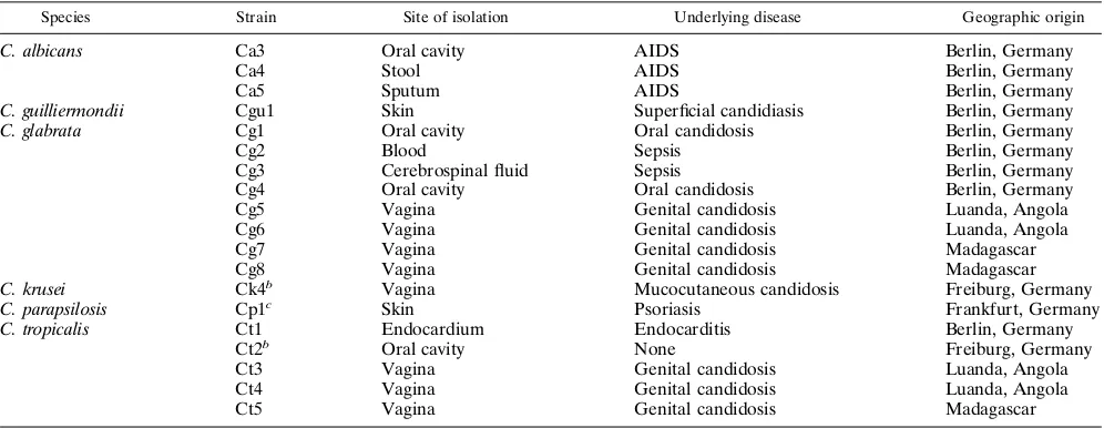
parap-TABLE 2. Clinical isolates used in this studyaSpecies Strain Site of isolation Underlying disease Geographic origin
C. albicans Ca3 Oral cavity AIDS Berlin, Germany
Ca4 Stool AIDS Berlin, Germany
Ca5 Sputum AIDS Berlin, Germany
C. guilliermondii Cgu1 Skin Superficial candidiasis Berlin, Germany C. glabrata Cg1 Oral cavity Oral candidosis Berlin, Germany
Cg2 Blood Sepsis Berlin, Germany
Cg3 Cerebrospinal fluid Sepsis Berlin, Germany
Cg4 Oral cavity Oral candidosis Berlin, Germany
Cg5 Vagina Genital candidosis Luanda, Angola
Cg6 Vagina Genital candidosis Luanda, Angola
Cg7 Vagina Genital candidosis Madagascar
Cg8 Vagina Genital candidosis Madagascar
C. krusei Ck4b Vagina Mucocutaneous candidosis Freiburg, Germany C. parapsilosis Cp1c Skin Psoriasis Frankfurt, Germany C. tropicalis Ct1 Endocardium Endocarditis Berlin, Germany
Ct2b Oral cavity None Freiburg, Germany
Ct3 Vagina Genital candidosis Luanda, Angola
Ct4 Vagina Genital candidosis Luanda, Angola
Ct5 Vagina Genital candidosis Madagascar
aExcept as indicated, all isolates were from the strain collection of the Department of Dermatology, Charite´ Hospital, Humboldt-University Berlin. b
Given to us by H. J. Grundmann, Institute of Environmental Medicine and Hospital Hygiene, University Freiburg/Br.
c
Obtained from I. Menzel, Department of Dermatology, Hospital of the Johann-Wolfgang-Goethe-University of Frankfurt/M.
on May 15, 2020 by guest
http://jcm.asm.org/
[image:3.612.58.556.83.276.2]silosis or C. sake. Comparison of the PCR fingerprints of this
unknown isolate with those of the reference strains of C.
parap-silosis and C. sake clearly indicated that the isolate was C.
parapsilosis (Fig. 5, right).
DISCUSSION
Species of Candida continue to be a frequent source of
hospital-acquired infections. Although C. albicans remains the
most common agent of fungal infections, an increasing number
of mycoses are caused by other species, including C. (T.)
gla-brata, C. tropicalis, C. parapsilosis, C. krusei, and C. lusitaniae
(3, 18, 27, 28, 44, 47). Several Candida species, such as C.
lusitaniae, C. tropicalis, C. krusei, C. guilliermondii, and C. (T.)
glabrata, have also been shown to develop resistance during
antifungal therapy or to possess intrinsic resistance to the
an-tifungal agents (7, 8, 13, 27, 35, 36, 49, 55). Since pathogenicity
and antifungal susceptibility often vary among species, a rapid
and accurate identification of the disease-causing species of
Candida is crucial for clinical treatment and epidemiological
studies.
This report describes the assessment of DNA
polymor-phisms in different species and strains within the genus
Can-dida by amplifying the genomic DNA with single nonspecific
primers. This PCR method employed an arbitrary primer (the
10-mer AP3) (54), a primer derived from intergenic spacers
(T3B) (26), and two microsatellite primers, (GTG)
5(1) and
(AC)
10(35). With these four primers, distinctive and
repro-ducible sets of amplification products were observed for all
Candida species tested. All 26 Candida species, as well as 8
other fungal species, could be easily distinguished by their PCR
fingerprint patterns. The discriminating powers of the four
primers used in this study were nearly the same. However,
more prominent and significant differences were observed
among the profiles generated by primers T3B and AP3, which
therefore might be better for the identification of Candida
species than (GTG)
5and (AC)
10. With all primers, the PCR
fingerprints revealed variability among isolates of a single
spe-FIG. 1. PCR fingerprints of different Candida species obtained with primers T3B (A and B) and AP3 (C and D). (A and C) Lanes 2 to 5, C. albicans (ATCC 14053, CBS 562, CBS 5983, and CBS 1905, respectively); lane 6, C. (T.) glabrata NCYC 350; lane 7, C. kefyr Y 601; lane 8, C. krusei CBS 573; lane 9, C. guilliermondii ATCC 6260; lane 10, C. parapsilosis CBS 604; lane 12, Cryptococcus humicolus CBS 2041; lane 13, C. ciferrii CBS 4856; lane 14, Cryptococcus curvatus CBS 570; lane 15, C. famata (T. candida) CBS 1795; lane 16, C. lipolytica CBS 599; lane 17, C. lusitaniae CBS 4413; lane 18, C. pulcherrima CBS 610; lane 19, C. rugosa CBS 613; lane 20, C. tropicalis CBS 94; lane 21, C. norvegensis CBS 1922; lanes 1, 11, and 22, molecular size markers in kilobases. (B and D) Lane 2, C. albicans ATCC 14053; lane 3, C. zeylanoides CBS 619; lane 4, C. intermedia CBS 572; lane 5, C. norvegica CBS 4238; lane 6, C. sake CBS 159; lane 7, C. sphaerica CBS 141; lane 8, C. utilis CBS 621; lane 9, C. valida CBS 638; lane 10, C. viswanathii CBS 4024; lane 12, C. castelli CBS 4332; lane 13, C. lambica CBS 1876; lane 14, Pichia etchellsii CBS 2011; lane 15, Pichia carsonii CBS 2285; lane 16, Trichosporon (Geotrichum, Mycoderma) cutaneum CBS 2466; lane 17, Saccharomyces cerevisiae CBS 1171; lane 18, Saccharomyces kluyveri CBS 3082; lane 19, Saccharomycopis capsularis CBS 2519; lane 20, Clavispora opuntiae CBS 7068; lane 21, Cryptococcus neoformans ATCC 3544; lanes 1, 11, and 22, molecular size markers in kilobases.on May 15, 2020 by guest
http://jcm.asm.org/
[image:4.612.61.556.76.421.2]cies. However, the variation among profiles obtained from
different strains of the same species was far less than the
variation observed among different species of Candida.
Comparison of PCR fingerprints of clinical isolates with
those of reference strains enabled the identification of different
Candida species, even if they could not be typed by
biochem-ical methods. A fluconazole-resistant Candida isolate, which
produced germ tubes but not chlamydospores and which could
not be identified by the carbohydrate assimilation system ID
32C, yielded a PCR fingerprint characteristic for C. albicans. In
two other cases, in which the ID 32C system was unable to
differentiate between C. famata (T. candida) and C.
guillier-mondii and between C. parapsilosis and C. sake, the correct
identification of C. guilliermondii and of C. parapsilosis,
respec-tively, was determined from the PCR fingerprints. Recently, we
described vaginal C. albicans isolates from Africa that were not
able to utilize the aminosugars glucosamine and
N-acetylglu-cosamine and that were therefore misidentified as C. sake by
the ID 32C technique. The correct species identification was
established only by PCR fingerprinting (43). A great advantage
of the PCR technique is that it permits the species
identifica-tion of clinical isolates with altered morphology, growth
char-acteristics, and biochemical properties.
Very similar amplification patterns were produced by the
anamorph yeast C. krusei and its teleomorph, I. orientalis.
Whether this PCR assay could be used for identification of
anamorph and teleomorph yeast pairs will require further
in-vestigation.
Our results agree with those of Lehmann et al. (21) and
Niesters et al. (35), who used similar PCR fingerprint
tech-niques to detect species-specific DNA polymorphisms within
the genus Candida. Using a different set of single nonspecific
primers [with the exception of primer (AC)
10, which was
[image:5.612.318.554.474.619.2]de-FIG. 2. To evaluate the stability of the PCR fingerprinting, DNA of C. famata (T. candida) CBS 1795 was passed through six generations in vitro. Lanes 2 to 4 (left), PCR profiles obtained after first, third, and sixth passages, respec-tively. The DNA of the same strain extracted by three different methods (see Material and Methods) yielded identical PCR patterns (lanes 2 to 4, right). Primer T3B was used in both experiments. Lanes 1, molecular size markers in kilobases.
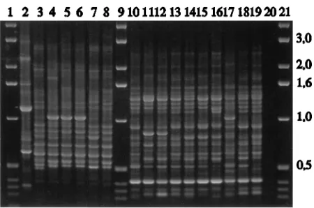
FIG. 3. PCR profiles of different C. tropicalis and C. (T.) glabrata strains amplified with primer T3B. Lane 2, C. albicans reference strain ATCC 14053; lane 3, C. tropicalis reference strain CBS 94; lanes 4 to 8, C. tropicalis wild-type strains (Ct1, Ct2, Ct3, Ct4, and Ct5, respectively), lanes 10 and 11, C. (T.) glabrata reference strains NCYC 350 and MB 1010, respectively; lanes 12 to 19, C. (T.) glabrata wild-type strains (Cg1, Cg2, Cg3, Cg4, Cg5, Cg6, Cg7, and Cg8, respectively); lane 20, control sample without DNA; lanes 1, 9, and 21, molecular size markers in kilobases.
FIG. 4. Comparison of T3B-primed PCR fingerprints of the synonymous isolates C. kefyr Y 601 and ATCC 4135 (formerly C. pseudotropicalis) as well of the anamorphic yeast C. krusei and its teleomorph I. orientalis. Lane 1, C. kefyr Y 601; lane 2, C. kefyr ATCC 4135; lane 3, C. krusei CBS 573; lane 4, C. krusei clinical isolate Ck4; lane 5, I. orientalis MB 16; lane 6, control sample without DNA; lane 7, molecular size markers in kilobases.
FIG. 5. Identification of clinical isolates by comparing their PCR profiles with those obtained from reference strains. (Left) Primer T3B. Lanes 2 to 6, C. albicans reference strains ATCC 14053, CBS 562, CBS 5983, CBS 1905, and ATCC 76615, respectively; lanes 7 to 9, fluconazole-resistant isolates derived from an AIDS patient (Ca3, Ca4, and Ca5, respectively); lane 11, C. guilliermon-dii reference strain ATCC 6260; lane 12, strain isolated from a patient with superficial candidiasis (Cgu1); lane 13, C. famata (T. candida) reference strain CBS 1795; lanes 1 and 10, molecular size markers in kilobases. (Right) Primer AP3, lanes 2, 5, and 8, isolate obtained from a patient suffering from psoriasis (Cp1); lane 1, C. albicans ATCC 14053; lane 3, C. (T.) glabrata NCYC 350; lane 4, C. parapsilosis CBS 604; lane 6, C. sake CBS 159; lane 7, Cryptococcus neo-formans ATCC 3544; lane 9, C. tropicalis CBS 94; lane 10, molecular size marker in kilobases.
on May 15, 2020 by guest
http://jcm.asm.org/
[image:5.612.67.292.511.660.2]scribed first by Niesters et al. (35)], these groups reported
species-specific amplification patterns for five and eight
differ-ent Candida species, respectively. Extending the investigation
to 26 different Candida species and 8 other fungal species, the
present study confirms their suggestion that PCR
fingerprint-ing with sfingerprint-ingle primers could be successfully applied to species
identification within the genus Candida. PCR fingerprinting is
faster and simpler to perform than most other methods of
genotypic analysis for species differentiation and has the
ad-vantage that a great variety of species can be identified by using
the same methodology.
It has been suggested that PCR fingerprints might be
accu-rate indicators of genetic distances because PCR
fingerprint-ing randomly samples sequence polymorphisms distributed
throughout the genome (52). Relatedness is deduced from the
number of amplified fragments that two strains or species have
in common. Importantly, these fragments are usually
nonal-lelic, being scored simply as present or absent (2). However, as
with restriction fragment length polymorphisms, the
probabil-ities of losing and regaining a band are unknown, and length
polymorphisms are usually indistinguishable from nucleotide
substitution polymorphisms, so that some of the characters are
not truly independent. We are currently comparing data
ob-tained by the PCR fingerprint method with those derived from
sequencing of the ribosomal DNA to determine whether PCR
fingerprints may be applied to phylogenetic studies within the
genus Candida.
ACKNOWLEDGMENTS
This work was supported by a grant from the Deutsche Forschungs-gemeinschaft to G. Scho¨nian and H. J. Tietz (Scho 448/3-1) and, in part, by grants from the Deutsche Forschungsgemeinschaft to W. Meyer (Me 1393/1-1) and from the National Institutes of Health to T. G. Mitchell (AI 28836).
REFERENCES
1. Ali, S., C. R. Mu¨ller, and J. T. Epplen.1986. DNA fingerprinting by oligo-nucleotide probes specific for simple repeats. Hum. Genet. 74:239–243. 2. Amos, B., and J. Pemberton. 1992. DNA fingerprinting in non-human
pop-ulations. Curr. Opin. Genet. Dev. 2:857–860.
3. Beck-Sague´, C. M., W. R. Jarvis, and the National Nosocomial Infections
Surveillance System.1993. Secular trends in the epidemiology of nosocomial fungal infections in the United States, 1980–1990. J. Infect. Dis. 167:1247– 1251.
4. Buchman, T. G., M. Rossier, W. G. Merz, and P. Charache. 1990. Detection of surgical pathogens by in vitro DNA amplification. I. Rapid identification of Candida albicans by in vitro amplification of a fungus-specific gene. Sur-gery 108:338–347.
5. Burgener-Kairuz, P., J. P. Zuber, P. Jaunin, T. G. Buchman, J. Bille, and M.
Rossier.1994. Rapid detection and identification of Candida albicans and Torulopsis (Candida) glabrata in clinical specimens by species-specific nested PCR amplification of a cytochrome P-450 lanosterol-a-demethylase (L1A1) gene fragment. J. Clin. Microbiol. 32:1902–1907.
6. Crampin, A. C., and R. C. Matthews. 1993. Application of the polymerase chain reaction to the diagnosis of candidosis by amplification of an HSP-90 gene fragment. J. Med. Microbiol. 39:233–238.
7. Dick, J., B. Rosengard, W. Merz, R. Stuart, G. Hutchins, and R. Saral. 1985. Fatal disseminated candidiasis due to amphotericin-B-resistant Candida guil-liermondii. Ann. Intern. Med. 102:67–68.
8. Drutz, D., and R. Lehrer. 1978. Development of amphotericin-B-resistant Candida tropicalis in a patient with defective leukocyte function. Am. J. Med. Sci. 276:77–92.
9. Fuson, G. B., H. J. Phaff, and H. L. Presley. 1987. Deoxyribonucleic acid base sequence relatedness among members of the yeast genus Kluyveromyces. Int. J. System. Bacteriol. 37:371–379.
10. Gardes, M., and T. D. Bruns. 1993. ITS primers with enhanced specificity for basidiomycetes—applications to the identification of mycorrhizae and rusts. Mol. Evol. 2:113–118.
11. Gra¨ser, Y., W. Meyer, W. Presber, and G. Scho¨nian.1993. Optimization of a PCR-based assay for fingerprinting microorganisms. Med. Microbiol. Lett.
2:379–385.
12. Gruber, F. 1990. Homologe und heterologe Transformation von Tri-choderma reesei mit den Orthidin-59-Phosphat-Decarboxylase-Genen als
Selektionsmarker. Ph.D. thesis, Technische Universita¨t Wien, Vienna. 13. Hadfield, T., M. Smith, R. Winn, M. Rinaldi, and C. Guerra. 1987. Mycoses
caused by Candida lusitaniae. Rev. Infect. Dis. 9:1006–1012.
14. Holmes, A. R., R. D. Cannon, M. G. Shepherd, and H. F. Jenkinson. 1994. Detection of Candida albicans and other yeasts in blood by PCR. J. Clin. Microbiol. 32:228–231.
15. Hopfer, R. L., P. Walden, S. Setterquist, and W. E. Highsmith. 1993. De-tection and differentiation of fungi in clinical specimens using polymerase chain reaction (PCR) amplification and restriction enzyme analysis. J. Med. Vet. Mycol. 31:65–75.
16. Iwaguchi, S.-I., M. Homma, and K. Tanaka. 1990. Variation in the electro-phoretic karyotype analysed by the assignment of DNA probes in Candida albicans. J. Gen. Microbiol. 136:2433–2442.
17. Kan, V. L. 1993. Polymerase chain reaction for the diagnosis of candidemia. J. Infect. Dis. 168:779–783.
18. Komshian, S. V., A. Uwaydah, J. D. Sobel, and L. Crane. 1989. Fungemia caused by Candida species and Torulopsis glabrata in hospitalized patients. Frequency, characteristics, and evaluation of factors influencing outcome. Rev. Infect. Dis. 11:379–390.
19. Kwon-Chung, K. J., W. S. Riggsby, R. A. Uphoff, J. B. Hicks, W. L. Whelan,
E. Reiss, B. B. Magee, and B. L. Wickes.1989. Genetic differences between type I and type II Candida stellatoidea. Infect. Immun. 57:527–532. 20. Lee, F. J. S. 1992. Modified protocol for yeast DNA mini-preparation.
BioTechniques 5:677.
21. Lehmann, P. F., D. Lin, and B. A. Lasker. 1993. Genotypic identification and characterization of species and strains within the genus Candida by using random amplified polymorphic DNA. J. Clin. Microbiol. 30:3249–3254. 22. Magee, B. B., T. M. D’Souza, and P. T. Magee. 1987. Strain and species
identification by restriction fragment length polymorphisms in the ribosomal DNA repeat of Candida species. J. Bacteriol. 169:1639–1643.
23. Magee, B. B., and P. T. Magee. 1987. Electrophoretic karyotypes and chro-mosome numbers in Candida species. J. Gen. Microbiol. 133:425–430. 24. Maiwald, M., R. Kappe, and H. G. Sonntag. 1994. Rapid presumptive
iden-tification of medically relevant yeasts to the species level by polymerase chain reaction and restriction enzyme analysis. J. Med. Vet. Mycol. 32:115–122. 25. Mason, M. M., B. A. Lasker, and W. S. Riggsby. 1987. Molecular probe for
identification of medically important Candida species and Torulopsis gla-brata. J. Clin. Microbiol. 25:563–566.
26. McClelland, M., C. Petersen, and J. Welsh. 1992. Length polymorphisms in tRNA intergenic spacer detected by using the polymerase chain reaction can distinguish streptococcal strains and species. J. Clin. Microbiol. 30:1499– 1504.
27. Merz, W. G. 1984. Candida lusitaniae: frequency of recovery, coloniza-tion, infeccoloniza-tion, and amphotericin B resistance. J. Clin. Microbiol. 20:1194– 1195.
28. Merz, W. G., J. E. Karp, D. Schron, and R. Saral. 1986. Increased incidence of fungemia caused by Candida krusei. J. Clin. Microbiol. 24:581–584. 29. Meyer, S. A., D. G. Ahearn, and D. Yarrow. 1984. Candida Berkhout, p.
585–844. In N. J. W. Kreger-van Rij (ed.), The yeasts: a taxonomic study, 3rd ed. Elsevier Science Publisher, Amsterdam.
30. Meyer, W., E. Lieckfeldt, K. Kuhls, E. Z. Freedman, T. Bo¨rner, and T. G. Mitchell.1993. DNA- and PCR-fingerprinting in fungi, p. 311–320. In S. D. J. Pena, R. Chakraborty, J. T. Epplen, and A. J. Jeffreys (ed.), DNA fingerprinting: state of the science. Birkha¨user Verlag, Basel.
31. Meyer, W., T. G. Mitchell, E. Z. Freedman, and R. J. Vilgalys. 1993. Hy-bridization probes for conventional DNA fingerprinting used as single prim-ers in the polymerase chain reaction to distinguish strains of Cryptococcus neoformans. J. Clin. Microbiol. 31:2274–2280.
32. Mitchell, T. G., R. L. Sandin, B. H. Bowman, W. Meyer, and W. G. Merz. 1994. Molecular mycology: DNA probes and application of PCR technology. J. Med. Vet. Mycol. 32(Suppl. 1):351–366.
33. Miyakawa, Y., T. Mabuchi, and Y. Fukazawa. 1993. New method for detec-tion of Candida albicans in human blood by polymerase chain reacdetec-tion. J. Clin. Microbiol. 31:3344–3347.
34. Miyakawa, Y., T. Mabuchi, K. Kagaya, and Y. Fukazawa. 1992. Isolation and characterization of a species-specific DNA for detection of Candida albicans by polymerase chain reaction. J. Clin. Microbiol. 30:894–900.
35. Niesters, H. G. M., W. H. F. Goessens, J. F. M. G. Meis, and W. G. V. Quint. 1993. Rapid, polymerase chain reaction-based identification assays for Can-dida species. J. Clin. Microbiol. 31:904–910.
36. Nobre, G., E. Mendes, M. Charrua, and O. Cruz. 1989. Ketoconazole resis-tance in Torulopsis glabrata. Mycopathologia 107:51–55.
37. Pappagianis, D., M. Collins, R. Hector, and J. Remmington. 1979. Devel-opment of resistance to amphotericin B in Candida lusitaniae infecting a human. Antimicrob. Agents Chemother. 16:123–126.
38. Sadhu, C., M. J. McEachern, E. P. Rustchenko-Bulgac, J. Schmid, D. R. Soll,
and J. B. Hicks.1991. Telomeric and dispersed repeat sequences in Candida yeasts and their use in strain identification. J. Bacteriol. 173:842–850. 39. Scherer, S., and D. A. Stevens. 1988. A Candida albicans dispersed, repeated
gene family and its epidemiologic applications. Proc. Natl. Acad. Sci. USA
85:1452–1456.
40. Scho¨nian, G., O. Meusel, H. J. Tietz, W. Meyer, Y. Gra¨ser, I. Tausch, W.
on May 15, 2020 by guest
http://jcm.asm.org/
Presber, and T. G. Mitchell.1993. Identification of clinical strains of Candida albicans by DNA fingerprinting with the polymerase chain reaction. Mycoses
36:171–179.
41. Smith, R. A., C. A. Hitchcock, E. G. V. Evans, C. J. N. Lacey, and D. J. Adams. 1989. The identification of Candida albicans strains by restriction fragment length polymorphism analysis of DNA. J. Med. Vet. Mycol. 27:431–434. 42. Suzuki, T., I. Kobayashi, I. Mizuguchi, I. Banno, and K. Tanaka. 1988.
Electrophoretic karyotypes in medically important Candida species. J. Gen. Appl. Microbiol. 34:409–416.
43. Tietz, H. J., A. Ku¨ssner, M. Thanos, M. Pinto De Andrade, W. Presber, and G. Scho¨nian.1995. Phenotypic and genotypic characterization of unusual vaginal isolates of Candida albicans from Africa. J. Clin. Microbiol. 33:2462–2465. 44. Tietz, H. J., G. Scho¨nian, and C. Schweynoch.1994. Candida glabrata: ein
Erreger im Aufwind? Hautnah Derm. 10:552–558.
45. Van Deventer, A. J. M., W. H. F. Goessens, A. Van Belkum, H. J. A. Van Vliet,
E. W. M. Van Etten, and H. A. Verbrugh.1995. Improved detection of Candida albicans by PCR in blood of neutropenic mice with systemic candidiasis. J. Clin. Microbiol. 33:625–628.
46. Vazquez, J. A., A. Beckley, S. Donabedian, J. D. Sobel, and M. J. Zervos. 1993. Comparison of restriction enzyme analysis versus pulsed-field gradient gel elec-trophoresis as a typing system for Torulopsis glabrata and Candida species other than C. albicans. J. Clin. Microbiol. 31:2021–2030.
47. Wade, J. C. 1993. Epidemiology of Candida infections, p. 85–107. In G. P. Bodey (ed.), Candidiasis: pathogenesis, diagnosis and treatment. Raven Press, New York.
48. Walmsley, R. M., B. M. Wilkinson, and T. H. Kong. 1989. Genetic finger-printing for yeasts. Bio/Technology 7:1168–1170.
49. Warnock, D. W., J. Burke, N. J. Cope, E. Johson, N. Von Fraunhofer, and E.
Williams.1988. Fluconazole resistance in Candida glabrata. Lancet ii:1310. 50. Warren, N. G., and K. C. Hazen. 1995. Candida, Cryptococcus, and other yeasts of medical importance, p. 723–737. In P. R. Murray, E. J. Baron, M. A. Pfaller, F. C. Tenover, and R. H. Yolken (ed.), Manual of clinical microbi-ology, 6th ed. American Society for Microbimicrobi-ology, Washington, D.C. 51. Welsh, J., and M. McClelland. 1990. Fingerprinting genomes using PCR
with arbitrary primers. Nucleic Acids Res. 18:7213–7218.
52. Welsh, J., C. Pretzman, D. Postic, I. Saint Girons, G. Baranton, and M.
McClelland.1992. Genomic fingerprinting by arbitrarily primed polymerase chain reaction resolves Borrelia burgdorferi into three distinct phyletic groups. Int. J. Syst. Bacteriol. 42:370–377.
53. Wickes, L. B., J. B. Hicks, W. G. Merz, and K. J. Kwon-Chung. 1992. The molecular analysis of synonymy among medical important yeasts within the genus Candida. J. Gen. Microbiol. 138:901–907.
54. Williams, J. K. G., A. R. Kubelik, K. J. Livak, J. A. Rafalski, and S. V.
Tingey.1990. DNA polymorphisms amplified by arbitrary primers are useful as genetic markers. Nucleic Acids Res. 18:6531–6535.
55. Woods, R., M. Bard, I. Jackson, and D. Drutz. 1974. Resistance to polyene antibiotics and correlated sterol changes in two isolates of Candida tropicalis from a patient with an amphotericin-B-resistant funguria. J. Infect. Dis. 129: 53–98.