Copyright © 2000, American Society for Microbiology. All Rights Reserved.
Molecular Typing and Exopolysaccharide Biosynthesis of
Burkholderia cepacia
Isolates from a Portuguese
Cystic Fibrosis Center
JOA˜ O A. RICHAU,1JORGE H. LEITA˜ O,1MANUELA CORREIA,1LUI´S LITO,2MARIA JOSE´ SALGADO,2
CELESTE BARRETO,3PAOLA CESCUTTI,4ANDISABEL SA´ -CORREIA1*
Centro de Engenharia Biolo´gica e Quı´mica, Instituto Superior Te´cnico, 1049-001 Lisbon,1and Laborato´rio de Bacteriologia2and Departamento de Fibrose Quı´stica,3Hospital de Santa Maria, 1500 Lisbon, Portugal, and Dipartimento di Biochimica,
Biofisica e Chimica delle Macromolecole, Universita` di Trieste, I-34127 Trieste, Italy4
Received 19 July 1999/Returned for modification 15 October 1999/Accepted 7 January 2000
This work describes the first epidemiological survey ofBurkholderia cepaciainvolved in pulmonary infections
among the Portuguese population with cystic fibrosis (CF) who attended the major CF treatment Center in Lisbon at Sta. Maria Hospital from 1995 to the end of 1997. The characterization of the genomic relatedness
of the isolates was based on the analysis of their ribopatterns (withEcoRI) followed by construction of a
ribotype-based phylogenetic tree. This study was complemented with macrorestriction fragment analysis by pulsed-field gel electrophoresis. After optimization of the solid growth medium, we found that
exopolysaccha-ride (EPS) production byB. cepaciaCF isolates is not as rare a phenomenon as was thought before; indeed,
70% of the isolates examined were EPS producers.
Burkholderia cepacia, originally described as a plant patho-gen (3), has become an important opportunistic pathopatho-gen in patients with cystic fibrosis (CF) (10, 11, 14, 26) and an infre-quent cause of nosocomial infection in patients without CF (13). There is increased evidence of transmission among pa-tients with CF by social contact (9) The environment is also regarded as a potential source of strains capable of infecting these patients (4, 9). However, one risk factor forB. cepacia
acquisition by patients with CF in the United States appeared to be hospitalization, and a recent hospital outbreak appar-ently involved patients both with and without CF (13). There are no clear data about the possible contribution of the poly-saccharide produced extracellularly by specificB. cepacia iso-lates (1, 5, 6, 20) to the colonization and persistence of the species in the infected host, as was ascribed to alginate in the respiratory infection of patients with CF by Pseudomonas aeruginosa(10). Nevertheless, the mucoid colonial morphotype is thought to be relatively rare amongB. cepaciaCF isolates (10). However, the exopolysaccharides (EPSs) produced by several gram-negative bacterial species infecting plants or an-imals have been considered important virulence factors due to their contributions to the colonization and persistence of the producing microorganism in the infected host (19).
In Portugal, the first clearly identified B. cepacia isolate recovered from the sputum of a patient with CF attending the CF treatment center at Sta. Maria Hospital in Lisbon was found in 1992. The CF Center is attended by approximately 85% of the population with CF residing in the Lisbon area and by patients with CF living in the south of Portugal and in the Madeira and Azores Islands. From 9 of a total of 140 patients with CF registered at the CF Center between 1995 and the end of 1997, 23 isolates capable of growing on the selective Burk-holderia Cepacia Selectatab medium (Mast Diagnostics, Mer-seyside, United Kingdom) were recovered at the Laboratory of
Bacteriology of Sta. Maria Hospital. The isolates (Table 1) were obtained on different dates of isolation from bronchial secretions of different patients with CF, each identified by a letter (Table 1), after 3 days of incubation at 35°C followed by another day of incubation at room temperature in the selective medium Burkholderia Cepacia Selectatab. The four putative
B. cepacia isolates obtained during 1996 were lost due to a prolonged storage period at the hospital and could not be examined in this study. To confirm that all 19 CF isolates obtained belonged to the speciesB. cepacia, the commercial systems API 20NE (Biomerieux, Marcy L’Etoile, France) and BIOLOG gram-negative (GN) (Biolog Inc., Hayward, Calif.) were used. The isolates identified as B. cepacia by the two systems were submitted to additional confirmation based on PCR amplification using the specific oligonucleotide primers CMG-16-1, C-16-21001, CMG-23-1, CM-16-2458, and G-23-2
proposed by Bauernfeind et al. (2). Further efforts to confirm the identification of the 19 isolates were undertaken at the Instituto Superior Te´cnico (IST) laboratory before comparing their genomic relatedness and their abilities to produce EPS, due to the increasing evidence of misidentification ofB. cepa-cia by standard laboratory procedures (2). This analysis re-vealed that only the 16 IST isolates listed in Table 1 could be confirmed to beB. cepacia. They were stored at⫺70°C in 40% (wt/vol) glycerol, and when in use, the cultures were routinely maintained on Pseudomonas isolation agar (Difco, Detroit, Mich.) plates.
B. cepaciaprevalence rate and clinical course.The value of the 3-year cumulative prevalence rate found during the period of surveillance (6.4%) did not suggest epidemic transmission of
B. cepacia. This value is close to the prevalence rate observed in other surveillance studies carried out in other countries (11, 21, 22, 25), while the colonization rates in one large United Kingdom regional center, experiencing the spread of an epi-demic strain, reached values close to the 40% prevalence ex-perienced in a major North American CF center (9). Consis-tent with previous reports, the clinical courses of the nine Portuguese patients with CF infected withB. cepaciawho were followed during this study were variable: patients A, D, and I * Corresponding author. Mailing address: Centro de Engenharia
Biolo´gica e Quı´mica, Instituto Superior Te´cnico, Av. Rovisco Pais, 1049-001 Lisbon, Portugal. Phone: 351-218417233. Fax: 351-218480072. E-mail: pcisc@alfa.ist.utl.pt.
1651
on May 15, 2020 by guest
http://jcm.asm.org/
exhibited stable pulmonary function after infection with B. cepacia; patient G died from the “cepacia syndrome”; patients B and H showed an increased deterioration of pulmonary function which was clearly associated with infection with the sameB. cepaciaisolate that continued to be isolated during 1998, and patients E and F died, although their deaths were considered unrelated toB. cepaciainfection. Patient C moved to another geographical area, which prevented his clinical ob-servation during the full duration of the present study.
Molecular typing. The genetic relatedness of the 16B.
ce-paciaisolates from Portuguese patients with CF was compared by ribotyping complemented with macrorestriction fragment analysis by pulsed-field gel electrophoresis (PFGE). The envi-ronmental type strain ATCC 25416 and the three highly trans-missible epidemic strains J2315 (a representative of the highly epidemic Edinburgh-Toronto lineage [11]) and C1394 and C1579 (epidemic representatives of outbreaks of B. cepacia
among patients with CF attending CF centers at Manchester [23] and Glasgow [27], United Kingdom, respectively) were used as reference strains. For ribotyping analysis, the isolation of total DNA, restriction withEcoRI (Gibco BRL Life Tech-nologies, Gaithersburg, Md.), DNA blotting, and hybridization with the acetylaminofluorene-labeled 16S⫹23S rRNA probe fromE. coli(Eurogentec, Seraing, Belgium) were carried out as previously described (16). The sizes of the hybridization restriction fragments were estimated with DNA Simdex soft-ware from GenetX withRaoulI (Eurogentec) as the reference molecular size marker. In order to transform the restriction fragment length polymorphism (RFLP) data obtained from ribotyping for numerical analysis, each band of an RFLP pro-file obtained for each isolate under study was treated as a unit character and scored as 1 when present or 0 when absent across all isolates. Variations in staining intensity were not taken into account for the construction of the matrix. From the resulting binary data, a triangular similarity matrix was gener-ated using the Dice similarity coefficient,SD (24). The
con-struction of the dendrogram from the similarity matrix was performed by the UPGMA method (unweighted-pair group
method using arithmetic means), which forms groups by suc-cessively pairing similar ribopatterns according to the magni-tudes of their observed distances (24). The software package used was the program NTSYSpc version 2.02 (Exeter Software, Inc.). The cophenetic correlation,r, between the similarity and the cophenetic matrices was 0.968. The use of the Jaccard similarity coefficient or the change of the order in which isolate data were input into the software did not lead to significant changes in the final dendrogram. The UPGMA method was chosen because it has been shown to give the most accurate representation of data (18).
Chromosomal analysis by PFGE was carried out as de-scribed previously (16). Genomic DNA was prepared using approximately 5⫻108cells (from an overnight culture of each
strain under analysis) per ml of agarose (0.5% [wt/vol]) plugs. Digestions with the rare-cutting endonucleases AseI or AflII (New England Biolabs, Beverly, Mass.), were carried out by using 20 U of enzyme per one-third of a plug, according to the manufacturer’s instructions. The plugs were loaded onto 1% (wt/vol) agarose gel prepared with the running buffer, 0.5⫻
Tris-borate-EDTA buffer, pH 8.0. The macrorestriction frag-ments generated were separated by PFGE using the Gene Navigator (Pharmacia, Uppsala, Sweden) apparatus under ap-propriate conditions (180 V for 22 h with pulse times in the interpolation mode ranging from 2 to 50 s). Bacteriophage lambda concatamers (New England Biolabs) were used as size standards. After electrophoretic separation, the gels were stained with ethidium bromide and photographed under UV illumination.
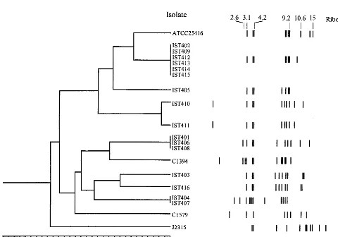
[image:2.612.57.551.85.309.2]Eight distinct ribopatterns were found with the 16 Portu-guese CF isolates examined. An example of the results ob-tained is given in Fig. 1, and the ribopatterns are schematically shown in Fig. 2. The ribopatterns obtained exhibited 7 to 12 bands, ranging from 1.25 to 27.0 kb. One band, approximately 4.2 kb, was common to all the isolates under study. Bands of approximately 4.0 and 3.1 kb were also consistently found for nearly all isolates, the exceptions being C1579 and J2315 (Fig. 1 and 2). The conserved 4.2-kbEcoRI band was also reported TABLE 1. Profiles of CF isolates used in the studya
Patient or
reference Data of isolation B. cepaciaisolate Ribopattern
EPS [g of total sugar䡠(g of cell protein)⫺1]b
A S
A August 95 IST401 2 0.24⫾0.09 4.30⫾0.11
A September 95 IST404 3 ND 0.60⫾0.06
B April 95 IST402 1 ND 0.80⫾0.08
B April 95 IST409 1 ND 1.1⫾0.5
C February 95 IST403 7 ND 0.10⫾0.02
D December 95 IST407 3 ND ND
E December 95 IST406 2 0.36⫾0.02 2.5⫾0.20
F January 95 IST408 2 0.39⫾0.01 2.20⫾0.15
G April 95 IST405 4 ND ND
G June 95 IST410 5 ND ND
G February 95 IST411 6 ND ND
H January 97 IST412 1 ND 0.50⫾0.04
H March 97 IST413 1 ND 0.80⫾0.02
H May 97 IST414 1 ND 0.80⫾0.06
H July 97 IST415 1 ND 0.80⫾0.09
I September 97 IST416 8 ND 0.40⫾0.02
Type strain ATCC 25416 12 ND 4.00⫾0.20
11 J2315 11 ND ND
23 C1394 9 ND ND
27 C1579 10 ND 0.50⫾0.01
aCF patient hosts are identified by letters. Ribopatterns are shown in Fig. 3.
bEPS produced (expressed as grams of total sugars present in the ethanol-precipitable material per gram of cell protein) after 5 days of incubation at 30°C on the
surface of A or S solid medium byB. cepaciaisolates. ND, not detectable.
on May 15, 2020 by guest
http://jcm.asm.org/
for all theB. cepaciaisolates from patients at six North Amer-ican CF centers (13). However, the other conserved EcoRI band in these North AmericanB. cepaciaisolates was reported to be 2.6 kb. The different size of the closer conserved band
that was calculated in the present study (3.1 kb) may result from the different molecular size reference marker used. The genomic diversity of the isolates examined should be consid-ered to be higher than the number that 8 ribopatterns for 16 isolates may suggest. Indeed, four of the isolates (IST412, IST413, IST414, and IST415), were serial isolates from the same patient, H (from January to July 1997), and exhibited the same ribopattern, 1; their RFLP-PFGE profiles withAseI and
[image:3.612.61.282.76.236.2]AflII were also indistinguishable (Fig. 3A and results not shown). Isolates IST404 and IST407, which gave rise to the same ribopattern (number 3), were obtained from patients A and D, but it was impossible to compare their RFLP-PFGE patterns because after repeated attempts these isolates pro-duced lanes with smeared DNA, probably due to the degrada-tion of the DNA by endogenous endonucleases. Addidegrada-tionally, three other isolates (IST401, IST406, and IST408) with the same ribopattern 2 were indistinguishable by RFLP-PFGE with both AseI (Fig. 3B) andAflII (results not shown); they were isolated from three different patients (A, E, and F). Because these patients were hospitalized in the Infectious Dis-eases Unit of Sta. Maria Hospital, although not simulta-neously, during the temporary closure of the CF hospitaliza-tion unit for repairs, these typing results strongly suggest that the three isolates examined are the same strain that was ac-quired during hospitalization. Interestingly, this strain is genet-ically related to the CF isolate C1394 (65% similarity), a rep-resentative of a Manchester, United Kingdom, outbreak (23).
FIG. 1. Example of the ribopatterns obtained with theB. cepaciaisolates indicated below after digestion of total DNA withEcoRI and hybridization with the acetylaminofluorene-labeled 16S⫹23S rRNA fromE. coli. Lane 1, molec-ular size standardRaoulI; lanes 2 to 12,B. cepaciaisolates IST403, IST406, IST407, IST401, IST408, IST410, IST404, IST411, IST409, IST402, and ATCC25416, respectively. Sizes in kilobases are given on the left.
FIG. 2. Dendrogram showing the results of clustering analysis using UPGMA for theB. cepaciastrains and isolates under study. The numbers in the horizontal axis indicate the percentage of similarity as determined with the Dice coefficient for the ribopatterns schematically represented on the left, with molecular sizes (in kilobases) arranged in a logarithmic scale.
on May 15, 2020 by guest
http://jcm.asm.org/
[image:3.612.62.542.362.698.2]It must be stressed thatB. cepacia-positive patients are rou-tinely segregated from B. cepacia-negative patients at Sta. Maria Hospital CF Center.
A cluster analysis was undertaken of the ribopatterns gen-erated during this study. Although we also had available the macrorestriction fragment profiles obtained by PFGE for all the Portuguese isolates examined in this study, due to the large number of bands generated, these profiles are difficult to an-alyze (Fig. 3A and B), and their cluster analysis was not con-sidered. The inspection of the ribotype-based phylogenetic tree that was constructed suggests the possible acquisition ofB. cepaciafrom environmental sources by one-third of the pa-tients with CF examined during the period of surveillance. Indeed, the ribopattern of the environmental strain used as a reference, B. cepacia ATCC 25416, was very closely related (80% similarity) to the ribopattern generated by six Portuguese isolates that were sequentially isolated from the same two patients, B (IST402 and IST409) and H (IST412, IST413, IST414, and IST415), who resided in distinct geographical ar-eas (on Madeira Island [B] and in the Lisbon area [H]) and who were never in contact or hospitalized. Isolates IST405, IST410, and IST411, with a similarity lower than 60%, were obtained (from February to June 1995) from patient G, who died of the cepacia syndrome. They gave rise to different, although related, ribotypes, particularly the isolates IST410 and IST411 (Fig. 3). Although it is possible that this patient harbored two or three different strains, we favor the hypothesis that the different ribotypes may result from genomic variations of the same colonizing strain. The results of this first epidemi-ological survey study ofB. cepaciainvolved in pulmonary in-fections among the Portuguese population with CF did not reveal genomic relatedness between the Portuguese isolates and the Edinburgh-Toronto epidemic strain J2315 (10) or the epidemic Glasgow isolate C1579 (27).
EPS biosynthesis. The mucoidy of B. cepaciaisolates was
assessed by comparing the morphologies of isolated colonies formed after incubation for 5 days at 30°C in the three different media examined, supplemented with 20 g of agar (Iberagar; Coina, Portugal) per liter. CDM and A media were described in the literature as leading to the production of EPS by a specificB. cepaciaCF isolate (1, 20), and S medium was suc-cessfully used in the IST laboratory to overproduce the EPS
gellan gum fromSphingomonas paucimobilisATCC 31461 (8, 15, 17). Moreover, the use of S agar plates allowed the differ-entiation of the mucoid colonial morphotypes ofS. paucimo-bilisvariants, which produced mutated gellan gum in different yields (15, 17). CDM contained, in grams per liter, NaCl (0.175), KCl (0.224), (NH4)2SO4(0.396), K2HPO4(0.205), and
glucose (10). Medium A contained, in grams per liter, only yeast extract (2) and glucose (10). S medium contained, in grams per liter, Na2HPO4(10), KH2PO4(3), K2SO4(1), NaCl
(1), MgSO4䡠7H2O (0.2), yeast extract (Difco) (1), Casamino
Acids (Difco) (1), CaCl2䡠2H2O (0.01), FeSO4䡠7H2O (0.001),
and glucose (20). Isolated colonies were obtained by spreading, after suitable dilution, 100l of liquid culture onto the surface of the solid growth medium. The liquid culture used to inoc-ulate the agar plates resulted from overnight cultivation at 30°C with orbital agitation (250 revolutions䡠min⫺1) in Luria
[image:4.612.134.469.73.255.2]broth (Sigma) medium. EPS production by the different iso-lates was quantified after 5 days of incubation at 30°C of confluent cell growth in either A or S solid medium. The plates were scraped, the material was resuspended in 0.9% (wt/vol) NaCl by vortexing, and the cells were separated by centrifuga-tion at 20,000⫻gfor 15 min. The EPS was precipitated from the cell-free supernatant by the addition of 2 volumes of cold ethanol, air dried, and redissolved in distilled water. The total sugar content was assessed by the phenol-sulfuric acid method (7) using the EPS produced by isolate IST408 as a standard. For this purpose, the EPS produced by isolate IST408 was further dialyzed against distilled water at 4°C for 24 h and recovered by freeze-drying. The cell pellets obtained from each plate were washed once with 0.9% NaCl, and the protein content was quantified by the biuret method (12) using bovine serum albumin fraction V (Merck) as a standard. The results of EPS production were expressed as grams of total sugars per gram of protein and are the means of at least three indepen-dent cultivations and of three determinations of total sugar and protein contents in each independent sample. After 5 days of incubation at 30°C on solid CDM medium, no mucoid colonies were detected by visual inspection. Confirming this observa-tion, we were unable to detect any ethanol-precipitable mate-rial from the cell-free supernatants. However, when grown on solid medium A, isolates IST401, IST406, and IST408 formed colonies that evidenced a very clear mucoid phenotype, while
FIG. 3. Comparison of theAseI macrorestriction fragment patterns of the genomic DNAs fromB. cepaciaisolates separated by PFGE. (A) Lanes: 1, size standard of concatamerized phageDNA; 2, IST409; 3, IST412; 4, IST413; 5, IST414; 6, IST415; 7, IST402. (B) Lanes: 1, IST401; 2, IST406; 3, IST408; 4, size standard of concatamerized phageDNA.
on May 15, 2020 by guest
http://jcm.asm.org/
the other isolates maintained the nonmucoid appearance (re-sults not shown). The indications of this visual observation were confirmed by quantitative analysis of EPS production (Table 1). The use of solid S medium to cultivate the whole collection of isolates allowed the identification of 12 Portu-guese isolates (out of 16) giving rise to mucoid colonies after 5 days of incubation at 30°C. The quantification of the EPS produced by the isolates during growth in this solid medium confirmed the colony morphotypes (Table 1). Only the isolates IST405, IST407, IST410, and IST411 (4 out of 16) were con-sistently unable to produce EPS in any of the three media tested. Among the three CF epidemic isolates used as refer-ence strains, only one was able to produce EPS in S medium while the environmental type strain ATCC 25416 produced high levels of EPS (Table 1). In general, the relative level of EPS produced on S plates by the different isolates was repro-duced in S liquid culture carried out in shake flasks (results not shown). The structural analysis of the EPS produced by IST408 in A agar plates indicated that the polymer is very similar, if not identical, to the EPS produced by a French clinical isolate (5; P. Cescutti, M. Bosco, F. Picotti, J. A. Richau, J. H. Leita˜o, and I. Sa´-Correia, Abstr. 10th Eur. Carbohydr. Symp., abstr. OB05, p. 85, 1999), and a detailed description will be published elsewhere. Moreover, the sugar composition of the EPS from
B. cepaciaIST408 (composed of glucose, mannose, rhamnose, galactose, and glucuronic acid in the molar ratio 1.0:1.0:1.0: 3.0:1.0 [Cescutti et al., Abstr. 10th Eur. Carbohydr. Symp.]) is similar to the compositions of the EPSs produced byB. cepacia
isolates from patients with CF in the United States (20) and in the United Kingdom (1).
Mucoid colonial morphotypes are considered rare in both environmental and clinical isolates ofB. cepacia (10). How-ever, although in solid CDM medium all the isolates were nonmucoid and in solid A medium only 3 of the 16 isolates examined were mucoid, 70% of the isolates were mucoid in solid S medium. It should be noted that the calculated per-centage of mucoid isolates among theB. cepaciaisolates from the Portuguese patients with CF (75% in S agar plates) could be overvalued. In fact, four of the mucoid isolates examined were serial isolates from the same patient, H, and exhibited the same ribopattern; three of them were indiscernible by RFLP-PFGE. Two other mucoid isolates are presumably the same strain obtained on different dates from patient B, and the strain acquired by three different patients, probably during their hospitalization, was also mucoid. Nevertheless, the results of the present study suggest that the concept that the mucoid phenotype is rarely found among B. cepaciaCF isolates may have resulted from the use of culture media unsuitable to the clear expression of EPS biosynthesis inB. cepacia. They also indicate that EPS production byB. cepaciamight not be as rare as initially thought and suggest that the B. cepacia EPS may indeed play a role in the colonization and persistence of B. cepaciain the lung in CF, as ascribed to alginate in CF infec-tion byP. aeruginosa.
This work was supported by Fundac¸a˜o para a Cieˆncia e a Tecnologia (FCT), by FEDER and the PRAXIS XXI program (grant PRAXIS/ PSAU/P/SAU/59/96), and Ph.D. and M.Sc. scholarships to J.R. and M.C., respectively.
We gratefully acknowledge the supply ofB. cepaciaJ2315, C1394, and C1579 by J. R. W. Govan (U. of Edinburgh Medical School, Edinburgh, United Kingdom).
REFERENCES
1.Allison, D. G., and M. J. Goldsbrough.1994. Polysaccharide production in
Pseudomonas cepacia. J. Basic Microbiol.34:3–10.
2.Bauernfeind, A., I. Schneider, R. Jungwirth, and C. Roller.1998.
Discrimi-nation ofBurkholderia gladiolifrom otherBurkholderiaspecies detectable in cystic fibrosis patients by PCR. J. Clin. Microbiol.36:2748–2751. 3.Burkholder, W. H.1950. Sour skin, a bacterial rot of onion bulbs.
Phytopa-thology50:115–117.
4.Butler, S. L., C. J. Doherty, J. E. Hughes, J. W. Nelson, and J. R. W. Govan.
1995. Burkholderia cepacia and cystic fibrosis: do natural environments present a potential hazard? J. Clin. Microbiol.33:1001–1004.
5.Ce´rantola, S., A. Lemassu-Jacquier, and H. Montrozier.1999. Structural elucidation of a novel exopolysaccharide produced by a mucoid clinical isolate ofBurkholderia cepacia. Characterization of a trisubstituted glucu-ronic acid in a heptasaccharide repeating unit. Eur. J. Biochem.260:373–383. 6.Ce´rantola, S., N. Marty, and H. Montrozier.1996. Structural studies of the acidic exopolysaccharide produced by a mucoid strain ofBurkholderia cepa-cia, isolated from cystic fibrosis. Carbohydr. Res.14:285:59–67.
7.Dubois, M., K. A. Gilles, J. K. Hamilton, P. A. Rebers, and F. Smith.1956. Colorimetric method for determination of sugars and related substances. Anal. Chem.28:350–356.
8.Fialho, A. M., L. O. Martins, M.-L. Donval, J. H. Leita˜o, M. J. Ridout, A. J. Jay, V. J. Morris, and I. Sa´-Correia.1999. Structures and properties of gellan polymers produced bySphingomonas paucimobilisATCC 31461 from lactose compared with those produced from glucose and from cheese whey. Appl. Environ. Microbiol.65:2485–2491.
9.Govan, J. R. W., P. H. Brown, J. Maddison, C. J. Doherty, J. W. Nelson, M. Dodd, A. P. Greening, and A. K. Webb.1993. Evidence for transmission of
Pseudomonas cepaciaby social contact in cystic fibrosis. Lancet342:15–19. 10. Govan, J. R. W., and V. Deretic.1996. Microbial pathogenesis in cystic
fibrosis: mucoidPseudomonas aeruginosaandBurkholderia cepacia. Micro-biol. Rev.60:539–574.
11. Govan, J. R. W., J. E. Hughes, and P. Vandamme.1996.Burkholderia cepa-cia: medical, taxonomic and ecological issues. J. Med. Microbiol.45:395–407. 12. Herbert, D., P. J. Phipps, and R. E. Strange.1971. Chemical analysis of microbial cells, p. 209–344.InJ. R. Norris and D. W. Ribbons (ed.), Methods in microbiology, vol. 5B. Academic Press, London, United Kingdom. 13. Holmes, A., R. Nolan, R. Taylor, R. Finley, M. Riley, R.-Z. Jiang, S.
Stein-bach, and R. Goldstein.1999. An epidemic ofBurkholderia cepaciatransmitted between patients with and without cystic fibrosis. J. Infect. Dis.179:1197–1205. 14. Isles, A., I. Maclusky, M. Corey, R. Gold, C. Prober, P. Fleming, and H. Levison.1984.Pseudomonas cepaciainfection in cystic fibrosis: an emerging problem. J. Pediatr.104:206–210.
15. Jay, A. J., I. J. Colquhoun, M. J. Ridout, G. J. Brownsey, V. J. Morris, A. M. Fialho, J. H. Leita˜o, and I. Sa´-Correia.1998. Analysis of structure and function of gellans with different substitution patterns. Carbohydr. Polym.
35:179–188.
16. Leita˜o, J. H., T. Alvim, and I. Sa´-Correia.1996. Ribotyping ofPseudomonas aeruginosaisolates from patients and water springs and genome fingerprinting of variants concerning mucoidy. FEMS Immunol. Med. Microbiol.13:287–292. 17. Martins, L. O., A. M. Fialho, P. L. Rodrigues, and I. Sa´-Correia.1996.
Gellan gum production and activity of biosynthetic enzymes in Sphingomo-nas paucimobilismucoid and non-mucoid variants. Biotechnol. Appl. Bio-chem.23:47–54.
18. Priest, F., and B. Austin.1993. Numerical taxonomy, p. 14–49.InF. Priest and B. Austin (ed.), Modern bacterial taxonomy, 2nd ed. Chapman & Hall, London, United Kingdom.
19. Roberts, I. S.1996. The biochemistry and genetics of capsular polysaccharide production in bacteria. Annu. Rev. Microbiol.50:285–315.
20. Sage, A., A. Linker, L. R. Evans, and T. G. Lessie.1990. Hexose phosphate metabolism and exopolysaccharide formation inPseudomonas cepacia. Curr. Microbiol.20:191–198.
21. Segonds, C., G. Chabanon, G. Couetdic, Y. Michel-Briand, and E. Bingen.
1996. Epidemiology of pulmonary colonization withBurkholderia cepaciain cystic fibrosis patients. Eur. J. Clin. Microbiol. Infect. Dis.15:841–842. 22. Shreve, M. R., S. Butler, H. J. Kaplowitz, H. R. Rabin, D. Stokes, M. Light,
and W. E. Regelmann.1999. Impact of microbiology practice on cumulative prevalence of respiratory tract bacteria in patients with cystic fibrosis. J. Clin. Microbiol.37:753–757.
23. Simpson, I. N., J. Finlay, D. J. Winstanley, N. Dewhurst, N. Nelson, S. Butler, and J. R. W. Govan.1994. Multi-resistance isolates possessing char-acteristics of bothBurkholderia(Pseudomonas)cepaciaandBurkholderia gladioli
from patients with cystic fibrosis. J. Antimicrob. Chemother.34:353–361. 24. Sneath, P. H. A., and R. R. Sokal.1973. Numerical taxonomy, p. 129–137.
W. H. Freeman and Co., San Francisco, Calif.
25. Taccetti, G., and S. Campana.1997. Microbiologic data overview of Italian cystic fibrosis patients. Eur. J. Epidemiol.13:323–327.
26. Vandamme, P., B. Holmes, M. Vancanneyt, T. Coenye, B. Hoste, R. Coop-man, H. Revets, S. Lauwers, M. Gillis, K. Kersters, and J. R. W. Govan.
1997. Occurrence of multiple genomovars ofBurkholderia cepaciain cystic fibrosis patients and proposal ofBurkholderia multivoranssp. nov. Int. J. Syst. Bacteriol.47:1188–1200.
27. Witheford, M. L., J. D. Wilkinson, J. H. McColl, F. M. Conlon, J. R. Michie, T. J. Evans, and J. Y. Paton.1995. Outcome ofBurkholderia(Pseudomonas)
cepaciacolonisation in children with cystic fibrosis following a hospital out-break. Thorax50:1194–1198.