Copyright © 2002, American Society for Microbiology. All Rights Reserved.
GUEST COMMENTARY
Bacterial Identification for Publication: When Is Enough Enough?
J. Michael Janda* and Sharon L. Abbott
Microbial Diseases Laboratory, Division of Communicable Disease Control, California Department of Health Services, Berkeley, California 94704-1011
The accurate and definitive identification of microorgan-isms, including bacteria, is one of the cornerstones forming the joint foundation of the fields of microbiology and infectious diseases. Identification is based upon the labeling of bacteria, parasites, and fungi with appropriate binomial names of Latin or Greek origin. Bacterial nomenclature thus provides the foundation from which host-parasite disease relationships are defined, therapeutic regimens are developed, and epidemio-logical investigations (e.g., comparative analysis of bacterial strains involved in outbreaks) are instigated.
A recent correspondence inClinical Infectious Diseases(W. Frederiksen and B. Tenning, Letter, Clin. Infect. Dis.32: 987-989, 2001) highlighted problems concerning the potential mis-identification of bacteria when commercial mis-identification sys-tems are used. However, this concern is only the tip of an iceberg of a potentially larger problem with more important ramifications. With the advent of the availability of commercial systems for rapid analysis and the use of molecular methods to provide genus and species identifications, the number of case reports linking old agents to new diseases and new or unusual (rare) agents to infectious processes has risen dramatically. However, equally important with regard to these published case reports describing new pathogenic taxa is the scientific accuracy of the identification of each species. Reliance on a single identification method when publishing can lead to mis-identification, which can translate into an inaccurate body of information in the medical literature concerning the clinical significance of many microbial species. A compounding prob-lem is the dramatic increase in the number of new bacterial species of medical or veterinary importance that are described on the basis of the sequence analysis of the 16S ribosomal DNA (rDNA) genes of one or two strains (3, 32). It is the goal of this article to briefly review pertinent aspects of this topic and to discuss the evolving fields of bacterial taxonomy and nomenclature as they relate to microbiology and infectious diseases.
BACTERIAL NOMENCLATURE
The first credible approaches to the systematic classification of bacteria began in the latter part of the 19th century. These early studies separated groups of bacteria primarily on the basis of morphology, size, and motility. A pioneer investigator
during this period was Ferdinand Cohn, who supported the concept of a diversity of microorganisms and argued that, within species, varieties emerged and transmitted their char-acteristics to the next generation (21). The subsequent devel-opment of agar-based media led to the in vitro isolation and propagation of pure cultures. This singular event fueled the first substantive biochemical investigations of bacterial species that occurred during the late 19th and early 20th centuries. However, because of the limited number of biochemical and phenotypic tests available, characterization of proposed spe-cies was inadequate and imprecise. This problem often led to confusion and resulted in the discovery and rediscovery of the same bacterial species by different investigators who gave the same taxa new names based upon slightly different morpho-logical, cultural, and phenotypic criteria. The gram-negative bacteriumKlebsiella pneumoniae provides an example of this taxonomic quagmire. This member of the family
Enterobacte-riaceaewas listed under six different genera, with seven
differ-ent species epithets, between 1885 and 1928. Between 1918 and 1960, a total of 84 descriptions of new motile species of bacteria or emendations of previously described motile species were published in the Journal of Bacteriology, yet of the 84 species, only 5 (6%) were properly described.
Over the first half of the 20th century, a number of ap-proaches to the identification and classification of bacteria were entertained. Among these were the use of physiologic tests to assess the diversity of bacteria, the first codification of biochemical test results, and the initial systematic approaches to classifying different groups of bacteria. In 1957, P. H. A. Sneath introduced the use of computers to analyze morpho-logical and biochemical characteristics as an approach to clas-sifying bacteria through numerical taxonomy methods. This groundbreaking work eventually led to the evolution of phen-etic analysis as a means to assess the relatedness of different bacteria or groups of bacteria to one another (29). Complex computer programs and large data matrices consisting of in-dependent covariant characters can be used to assess similarity or the likelihood that an unknown organism belongs to a given taxon. For instance, it is generally accepted that for an isolate to belong to a given species there must be at least 80 to 85% similarity based upon unweighted-pair-group method analysis. Many other different numerical methods have been proposed to identify taxa based upon probability and nonprobalistic data matrices and companion identification matrices. These meth-ods are reviewed in Wilcox et al. (29).
A major innovation in laboratory sciences occurred in the late 1960s, when manual miniaturized identification systems * Corresponding author. Mailing address: Microbial Diseases
Lab-oratory, 2151 Berkeley Way, Berkeley, CA 94704-1011. Phone: (510) 540-2242. Fax: (510) 540-2374. E-mail: jjanda@dhs.ca.gov.
1887
on May 15, 2020 by guest
http://jcm.asm.org/
were first introduced into the clinical microbiology laboratory. These early systems included the nine-test Enterotube (Roche, Basel, Switzerland) and two-tube R-B systems for identifying members ofEnterobacteriaceae. These systems offered several advantages over conventional testing, namely, all inocula for a set of tests performed sequentially were prepared from a single colony, preparation costs and the amount of storage space required for standard reagents and media were reduced, less incubation space was required, and in some instances identifi-cation times were reduced. However, biochemical reactions observed with commercial systems often did not correlate well with conventional test results, and because of the limited num-ber of tests employed in these systems, the percentage of strains correctly identified to the species level was less than satisfactory. These kits were rapidly followed by the landmark API 20E strip test (bioMérieux-Vitek, Hazelwood, Mo.), which was a micromethod employing 20 different biochemical tests. The 20E strip generated a septyl (7-digit) code in 18 to 21 h based upon biochemical reactions plus a screening oxidase test. The numeric code could then be located in a logbook that converted septyl codes into a final bacterial identification. The system identified species in the familyEnterobacteriaceaeand common nonfermenters, such asPseudomonas aeruginosa. Al-though a manual method, the API 20E strip test was so ad-vanced for its time that it is still commonly used in clinical microbiology labs throughout the world and is considered by many to be the “gold standard” commercial system against which all other such systems are measured (16).
In 1978, MicroScan (now Dade Behring MicroScan, West Sacramento, Calif.) developed the first combination panel pro-viding both organism identification and susceptibility profile determination (MIC determination) simultaneously. The 1980s saw continued improvements in diagnostic bacteriology with the introduction of automation into the laboratory (23). In 1983, MicroScan released the AS-3/touchSCAN system, the first widely accepted automated system for microbial identifi-cation and drug susceptibility testing in the clinical laboratory. Other systems, such as the Vitek AutoMicrobic system, quickly followed (23). These automated systems transformed pro-cesses for bacterial identification by reducing the time required to identify rapidly growing bacteria to as little as 2 to 4 h, in contrast to the 1 or more days that had been required previ-ously for a final identification by conventional test methodol-ogies.
The use of molecular biology and molecular techniques as an aid to bacterial taxonomy and identification was in its in-fancy in the 1960s. Early studies using DNA base composition could clearly distinguish between genomes that were unrelated based upon differences in G⫹C content (in moles percent) (21). However, the first major leap in molecular taxonomy and identification applicable to diagnostic microbiology occurred with the introduction of DNA-DNA hybridization studies pi-oneered by Don Brenner and his colleagues at the Walter Reed Army Institute of Research and later at the Centers for Disease Control and Prevention (3). The value of DNA hy-bridization was that it provided a quantitative definition of what constituted a species,⬃70% or greater DNA-DNA re-latedness with a‚Tmof 5°C or lower (26, 30). In cases where
new species were identified via DNA hybridization, it was also observed that in most instances the results of simple
biochem-ical tests would clearly separate newly recognized or redesig-nated genera or species from other established groups. This allowed for taxonomic advances made through DNA hybrid-ization studies to be easily adapted to the diagnostic laboratory through the use of new phenotypic identification schemes. However, not all newly recognized taxa that had been identi-fied by molecular techniques could be readily identiidenti-fied by biochemical tests in clinical laboratories.
The downside of DNA hybridization is that it is an expen-sive, technically complex, and labor-intensive procedure that, at its zenith, was restricted to a small number of research or public health centers around the world. Today, very few labo-ratories perform DNA hybridization by classic methods. The world of molecular taxonomy was revolutionized, however, in the mid-1980s with the advent of full sequence analysis of molecular chronometers such as rRNA (21). By the mid-1990s, sequencing of the small subunit (16S) rDNA genes had be-come commonplace, considered a standard tool of microbial taxonomists not only for elucidating phylogenetic relatedness but also as a means of bacterial identification (15, 21). The automation of 16S rDNA gene sequencing with such instru-ments as the ABI Prism 377 DNA sequencer (Applied Biosys-tems, Foster City, Calif.) allowed for a quick comparative anal-ysis of published sequences deposited in microbial genome databases (14). Today, bacterial strains that defy identification by conventional commercial methodologies are often sub-jected to 16S rDNA sequence analysis so that a useful label can be placed on the isolate in question (31).
IDENTIFICATION SYSTEM LIMITATIONS
All systems used to identify bacteria, whether phenotypic or genotypic, have limitations, because no single test methodol-ogy will provide results that are 100% accurate. Phenotypic systems, the most common approach used in clinical laborato-ries to identify bacteria, have several drawbacks. In contrast to the properties determined by molecular methods such as DNA hybridization, biochemical properties do not accurately reflect the entire extent of the genomic complexity of a given species. Furthermore, phenotypic properties can be unstable at times and expression can be dependent upon changes in environ-mental conditions, e.g., growth substrate, temperature, and pH levels (21). A further problem with commercial systems is the construction of databases, not to mention which biochemical tests are included on panels. Once biochemical tests are com-mercially produced, panel configurations are rarely changed, although reformulation of tests does occasionally occur. For instance, the tests included on the API 20E strip in 1975 were still the same tests on the strip in 2001. Yet the number of newly described taxa increased substantially between 1975 and 2001 (7). That means that when new taxa are added to existing commercial databases they must be identified on the basis of results of tests available on these preconfigured strips or pan-els. Often the best tests available to identify these newer spe-cies are not on the panels or cannot be formulated to fit a micromethod. This can lead to less reliable identification re-sults. The accuracy of commercial in-house databases used in the identification of bacterial species is dependent upon both the number of strains included in the database and the phe-notypic diversity of strains tested. For species encountered less
on May 15, 2020 by guest
http://jcm.asm.org/
frequently, even rarely, in the clinical laboratory, commercial databases may not have a sufficient number of bona fide strains to accurately provide a definitive identification for these iso-lates. Finally, most commercial systems do not consider clinical frequency or site of isolation in their bacterial identification matrices. One of the consequences of all of these limitations is that some commercial systems have great difficulty identifying certain groups of bacteria, such asPasteurellaspecies (10; Fre-deriksen and Tenning, letter) or Haemophilus species (11). Despite all of these shortcomings, commercial systems have served clinical microbiology laboratories well in the routine identification of infectious agents of medical importance. Given the number of isolates tested with these high-through-put systems, commercial systems are very accurate for the more common species and provide quick test results in a cost-effective manner.
16S rDNA gene sequencing is a powerful tool and by far the single most common molecular technique presently used for bacterial species identification (30). Although this technique relies on sequencing of the DNA that encodes the 16S rRNA subunit, like phenotypic tests, it surveys only a small portion of the microbial genome. Since ribosomal genes are highly con-served, sequence variation between strains belonging to differ-ent species from distinct genera is less eviddiffer-ent with rDNA gene sequencing than with DNA pairing studies, where DNA relat-edness values are used to compare strains. Another problem, one not faced in DNA hybridization studies (26), is that there are no consensus guidelines that define what constitutes a species based upon 16S rDNA gene sequence similarity or divergence. It is generally accepted that an unidentified isolate whose rDNA gene sequence is⬍97% similar to those of the isolate’s closest phylogenetic neighbors constitutes a new taxon. The general availability and ease with which ribosomal genes can be sequenced has fueled an explosion in the descrip-tion of new taxa. Over the past decade, approximately 40% of all newly described species have been described based upon the analysis of a single strain (4) (see, e.g., an article published in this journal) (32). This tendency is of immense concern when these species are of medical and/or veterinary impor-tance, since the published phenotype of the single (type) strain may or may not accurately reflect the typical biochemical pro-file of that species. Since clinical and veterinary laboratories continue to rely upon biochemical properties and not upon 16S rDNA gene sequencing to identify bacterial strains, reports of the biochemical properties of a new species that are based upon analysis of a single strain recovered from an infected site are essentially meaningless. Strains with 97% or greater simi-larity may or may not belong to the same species, and DNA pairing studies should be performed to resolve these issues (22). Because of the expense involved and the general lack of availability of DNA hybridization, various researchers or in-vestigators have attempted to establish cutoff values for strain relatedness based upon sequencing of 16S rDNA. One recent investigation (6) characterizing 177 unidentifiable environmen-tal, veterinary, and clinical bacterial isolates defined a species match as a strain exhibitingⱖ99% 16S rDNA gene sequence similarity (1% divergence) to strains previously deposited in GenBank. In another case report, 99.5% sequence relatedness was used to identify a strain asEnterobacter cloacae(31). How-ever, these arbitrary values may not always accurately reflect
taxonomic relatedness. Sequencing of the 16S subunit (⬃1539 bp) of bothEdwardsiella hoshinaeATCC 33379Tand
Edwards-iella ictaluriATCC 33202Tin our laboratory revealed only 0.19
to 0.65% divergence from the sequence of the type strain of
Edwardsiella tarda, ATCC 15947T. By 16S rDNA gene
se-quence analysis alone, many established breakpoints might recognize these as the same species, yet each of these three species is recognized as genetically distinct not only on the basis of DNA hybridization results but also because each group occupies a distinct ecologic niche.
A final issue regarding 16S identification concerns deposi-tion of sequences into established microbial genome databases. The accuracy of a partial or complete sequence of a 16S rDNA gene or of any other bacterial gene(s) deposited in GenBank or other databases is dependent upon how extensively the bacterial strain from which the sequences were derived has been characterized. Sequence heterogeneity in the literature can result from strain misidentification (5, 17). Since biochem-ical characteristics are still the touchstone for bacterial identi-fications, the choice in the use of commercial versus conven-tional methodologies for the identification of strains undergoing genetic characterization may largely influence how accurate the resulting label is. Thus, while the sequence data may be correct, if it is incorrectly associated with the wrong taxa, major errors in the literature can result.
ISSUES
Almost a decade ago, Weaver (27) raised the issue that case reports involving infrequently isolated bacteria should be ac-companied by at least a minimum of data regarding the bio-chemical characteristics of the organism and the methods used in identifying the species. A recent example of this possible problem can be seen in the case report of Varghese et al. (25) describing the recovery of an isolate identified asVibrio fluvia-lis from the wound of a 67-year-old woman subsequent to medicinal leech therapy. In this case report, reputed to be the first describing V. fluvialis as a wound pathogen after leech therapy, no information was provided on the methods used to identify this vibrio. In contrast to the authors’ findings, several things suggest this isolate was probably notV. fluvialis.
Aero-monas species live as symbionts in the gut of the medicinal
leech,Hirudo medicinalis(8), and there are numerous reports in the literature documenting aeromonad-associated wound infections following leech therapy. Furthermore, Aeromonas
species live in this environ apart from any other species (most often isolated in pure culture); rarely has any other microbial species ever been isolated from the gut of the leech. As indi-cated previously, there are problems associated with certain commercial systems in the identification of Pasteurella and
Haemophilus species (10, 11, 27; Frederiksen and Tenning,
letter), and the same situation exists withAeromonasspecies
and V. fluvialis. The results obtained with most commercial
systems for these species are nearly identical, and they often generate the same septyl or biotype codes. To distinguish these organisms, tests such as salt tolerance, susceptibility to 2,4-diamino-6,7-diisopropylpteridine, production of gas from D
-glucose, and the string test need to be performed. These tests are not available on any commercial bacterial identification system. Finally,V. fluvialisis a halophilic vibrio and requires
on May 15, 2020 by guest
http://jcm.asm.org/
media with salt supplementation. This species typically lives, in its natural environment, only in marine habitats, including es-tuaries. These cumulative facts strongly suggest that the isolate described by Varghese et al. (27) was notV. fluvialisbut rather
anAeromonasspecies.
There are many more examples in the literature of the mis-identification of bacteria by commercial systems. When these misidentifications are associated with the description of new infectious disease syndromes (either when a previously recog-nized disease agent is associated with a new syndrome or when a newly recognized disease agent is associated with a new disease), they take on added importance. A good example of this problem can be seen in the original descriptions by Albert and coinvestigators (1, 2) of several strains ofHafnia alveithat possessed someEscherichia coli-like virulence factors (attach-ing-effacing genes), produced diarrhea in animal models, and apparently caused gastroenteritis in young children. The re-ports by Albert et al. (1, 2) fueled a number of follow-up case studies concerning H. alvei-associated diarrhea (18, 28; J. Reina, J. Hervas, and N. Borrell, Letter, Clin. Infect. Dis.
16:443, 1993), and subsequent reports recognizedH. alveias a causative agent of intestinal disorders, including diarrhea (9, 20). However, in the original studies of Albert et al. (1, 2), the phenotypic properties of these strains were not reported and only a general reference to use of the API 20E strip and conventional test methodologies was made. Studies from sev-eral different laboratories have demonstrated that these strains
do not possess typical phenotypic or genotypic properties con-sistent with the genusHafnia (12, 13), and 16S rRNA gene sequencing and phoE gene probe assays indicate that these strains belong to the genusEscherichia(13, 19). Thus, there is no credible evidence at present thatH. alveiis an enteropatho-genic member of the familyEnterobacteriaceae.
PROPOSALS AND POSSIBLE SOLUTIONS
Concerns regarding appropriate methods to identify bacte-ria in published reports will become an increasingly difficult issue in the future unless standards are developed for the description of new taxa, reporting of DNA sequences into databases, and the submission of case reports involving un-usual (rare) bacterial species. Between 1980 and the end of 1996 there was a 238% increase in the total number of ap-proved names in the literature, with the number rising to 5,569 taxa (7). With the advent of the availability of automated molecular techniques to identify bacteria (e.g., 16S rDNA se-quencing), more species are being described based upon ex-aminations of five or fewer strains and a limited number of differential biochemical characteristics (21). Reliance on a sin-gle identification system, phenotypic or genotypic, to identify an organism provides more opportunity for misidentifying bac-terial species, and if the trend described above continues, the risk will become even greater in the future.
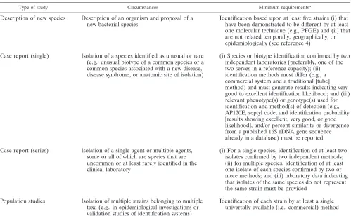
[image:4.587.43.542.81.389.2]Recently, Christensen and colleagues (4) made a formal TABLE 1. Proposed guidelines for identification of bacterial species for publication purposes
Type of study Circumstances Minimum requirementsa
Description of new species Description of an organism and proposal of a
new bacterial species Identification based upon at least five strains (i) thathave been demonstrated to be different by at least one molecular technique (e.g., PFGE) and (ii) that are not related temporally, geographically, or epidemiologically (see reference 4)
Case report (single) Isolation of a species identified as unusual or rare (e.g., unusual biotype of a common species or a common species associated with a new disease, disease syndrome, or anatomic site of isolation)
(i) Species or biotype identification confirmed by two independent laboratories (preferably, one of the two serves in a reference capacity); (ii) identification methods must differ (e.g., a commercial system and a traditional [tube] method) and must generate results indicating very good to excellent identification likelihood; and (iii) relevant phenotype(s) or genotype(s) used for identification and method(s) of detection (e.g., AP120E, septyl code, and identification probability [results showing excellent, very good, or good likelihood], and/or percent similarity or divergence from a published 16S rDNA gene sequence already in a database) must be reported
Case report (series) Isolation of a single agent or multiple agents, some or all of which are species that are uncommon or at least rarely identified in the clinical laboratory
(i) For a single species, identification of at least two isolates confirmed by two independent methods; (ii) for multiple species, identification of at least one isolate of each species confirmed by two or more methods; and (iii) laboratory data indicating that isolates of the same species do not represent the same strain must be provided
Population studies Isolation of multiple strains belonging to multiple taxa (e.g., in epidemiological investigations or validation studies of identification systems)
Identification of each strain by at least a single universally available (i.e., commercial) method
aPFGE, pulsed-field gel electrophoresis.
on May 15, 2020 by guest
http://jcm.asm.org/
proposal in this arena. They proposed that Recommendation 30b of theBacteriological Code(1990 Revision) be revised so that proposals to recognize new species are based not upon a single strain but rather upon a minimum of 5 to 10 strains from geographically and epidemiologically unrelated areas. We agree with this proposal and believe that similar (and perhaps expanded) guidelines should be applied to case reports or a limited series of case reports involving unusual (rare) bacterial species or infrequent biotypes (genotypes) of established (tra-ditional) pathogens. In addition, under these guidelines, pro-posals to recognize new species would require confirmation of the bacterial species or unusual phenotype or genotype by two independent laboratories (Table 1). In the case of species identification, secondary confirmation would be conducted by a recognized reference laboratory by a different methodology, preferably a nonautomated (commercial) one. The biochemi-cal results on any newly described strain need to be published in sufficient detail so that the reader can be confident of the accurate identification of the species and/or the phenotype or genotype.
Ideally, identification of any taxon is based upon a polypha-sic approach (24) that includes a combination of phenotypic testing methods (e.g., biochemical testing, cellular fatty acid analysis, and numerical analysis) and genotypic testing meth-ods (e.g., DNA-DNA hybridization, analysis of G⫹C content [in moles percent], and 16S rDNA gene sequencing). However, such methods are time consuming, expensive, and not easily adaptable to workflow in clinical microbiology laboratories. Table 1 lists an alternative proposal that should be both tech-nically and financially feasible and would help to reduce the number of publications with misidentifications. In all cases, as suggested by Weaver (27), critical information regarding the strain in question needs to be provided so that a assessment of the validity of the identification can be made. Only in this way can we attempt to ensure both the scientific and medical ac-curacy of the association of pathogenic bacteria with infectious processes in the medical literature.
REFERENCES
1.Albert, M. J., K. Alam, M. Islam, J. Montanaro, A. S. M. H. Rahman, K. Haider, M. A. Hossain, A. K. M. G. Kibriya, and S. Tzipori.1991.Hafnia alvei,a probable cause of diarrhea in humans. Infect. Immun.59:1507–1513. 2.Albert, M. J., S. M. Faruque, M. Ansaruzzaman, M. M. Islam, K. Haider, K. Alam, I. Kabir, and R. Robins-Browne.1992. Sharing of virulence-associated properties at the phenotypic and genetic levels between enteropathogenic Escherichia coliandHafnia alvei. J. Med. Microbiol.37:310–314. 3.Brenner, D. J.1977. Characterization and clinical identification of
Enter-obacteriaceaeby DNA hybridization. Prog. Clin. Pathol.7:71–117. 4.Christensen, H., M. Bisgaard, W. Federiksen, R. Mutters, P. Kuhnert, and
J. E. Olsen.2001. Is characterization of a single isolate sufficient for valid publication of a new genus or species? Proposal to modify Recommendation 30b of the Bacteriological Code (1990 Revision). Int. J. Syst. Evol. Microbiol. 51:2221–2225.
5.Clayton, R. A., G. Sutton, P. S. Hinkle, Jr., C. Bult, and C. Fields.1995. Intraspecific variation in small-subunit rRNA sequences in GenBank: why single sequences may not adequately represent prokaryotic taxa. Int. J. Syst. Bacteriol.45:595–599.
6.Drancourt, M., C. Bollet, A. Carlioz, R. Martelin, J.-P. Gayral, and D. Raoult.2000. 16S ribosomal DNA sequence analysis of a large collection of environmental and clinical unidentifiable bacterial isolates. J. Clin. Micro-biol.38:3623–3630.
7.Euzéby, J. P.1997. List of bacterial names with standing in nomenclature: a folder available on the internet. Int. J. Syst. Bacteriol.47:590–592. 8.Graf, J.2000. Symbiosis ofAeromonasandHirudo medicinalis, the medicinal
leech. ASM News66:147–153.
9.Günthard, H., and A. Pennekamp.1996. Clinical significance of extraintes-tinalHafnia alveiisolates from 61 patients and review of the literature. Clin. Infect. Dis.22:1040–1045.
10.Hamilton-Miller, J. M. T.1993. A possible pitfall in the identification of Pasteurellaspp. with the API system. J. Med. Microbiol.39:78–79. 11.Hamilton-Miller, J. M. T., and S. Shah.1996. Anomalous but helpful
find-ings from the BBL Crystal ID kit withHaemophilusspp. Lett. Appl. Micro-biol.23:47–48.
12.Ismaili, A., B. Bourke, J. C. S. de Azavedo, S. Ratnam, M. A. Karmali, and P. M. Sherman.1996. Heterogeneity in phenotypic and genotypic charac-teristics among strains ofHafnia alvei. J. Clin. Microbiol.34:2973–2979. 13.Janda, J. M., S. L. Abbott, and M. J. Albert.1999. Prototypal diarrheagenic
strains ofHafnia alveiare actually members of the genusEscherichia. J. Clin. Microbiol.37:2399–2401.
14.Jenks, P. J.1998. Sequencing microbial genomes—what will it do for mi-crobiology? J. Med. Microbiol.47:375–382.
15.Kolbert, C. P., and D. H. Persing.1999. Ribosomal DNA sequencing as a tool for identification of bacterial pathogens. Curr. Opin. Microbiol.2:299– 305.
16.O’Hara, C. M., D. L. Rhoden, and J. M. Miller.1992. Reevaluation of the API 20E identification system versus conventional biochemicals for identi-fication of members of the familyEnterobacteriaceae: a new look at an old product. J. Clin. Microbiol.30:123–125.
17.On, S. L. W.2001. Taxonomy ofCampylobacter,Arcobacter,Helicobacterand related bacteria: current status, future prospects and immediate concerns. J. Appl. Microbiol.90:1S-15S.
18.Ratnam, S.1991. Etiologic role ofHafnia alveiin human diarrheal illness. Infect. Immun.59:4744–4745.
19.Ridell, J., A. Siitonen, L. Paulin, O. Lindroos, H. Korkeala, and M. J. Albert. 1995. Characterization ofHafnia alveiby biochemical tests, random ampli-fied polymorphic DNA PCR, and partial sequencing of the 16S rRNA gene. J. Clin. Microbiol.33:2372–2376.
20.Rodriguez, L. A., J. Vivas, C. S. Gallardo, F. Acosta, L. Barbeyto, and F. Real.1999. Identification ofHafnia alveiwith the MicroScan WalkAway system. J. Clin. Microbiol.37:4186–4188.
21.Rosselló-Mora, R., and R. Amann. 2001. The species concept for pro-karyotes. FEMS Microbiol. Lett.25:39–67.
22.Stackebrandt, E., and B. M. Goebel. 1994. Taxonomic note: a place for DNA-DNA reassociation and 16S rRNA sequence analysis in the present species definition in bacteriology. Int. J. Syst. Bacteriol.44:846–849. 23.Stager, C. E., and J. R. Davis.1992. Automated systems for identification of
microorganisms. Clin. Microbiol. Rev.5:302–327.
24.Vandamme, P., B. Pot, M. Gillis, P. De Vos, K. Kersters, and J. Swings.1996. Polyphasic taxonomy, a consensus approach to bacterial systematics. Micro-biol. Rev.60:407–438.
25.Varghese, M. R., R. W. Farr, M. K. Wax, B. J. Chafin, and R. M. Owens. 1996.Vibrio fluvialiswound infection associated with medicinal leech ther-apy. Clin. Infect. Dis.22:709–710.
26.Wayne, L. G., D. J. Brenner, R. R. Colwell, P. A. D. Grimont, O. Kandler, M. I. Krichevsky, L. H. Moore, W. E. C. Moore, R. G. E. Murray, E. Stackebrandt, M. P. Starr, and H. G. Trüper.1987. Report of the ad hoc committee on the reconciliation of approaches to bacterial systematics. Int. J. Syst. Bacteriol.37:463–464.
27.Weaver, R. E.1992. In utero infection due toPasteurella multocida. Clin. Infect. Dis.15:881–882.
28.Westblom, T. U., and T. W. Milligan.1992. Acute bacterial gastroenteritis caused byHafnia alvei. Clin. Infect. Dis.14:1271–1272.
29.Wilcox, W. R., S. P. Lapage, and B. Holmes.1980. A review of numerical methods in bacterial identifications. Antonie Leeuwenhoek46:233–299. 30.Wilson, K. H.1995. Molecular biology as a tool for taxonomy. Clin. Infect.
Dis.20:S117-S121.
31.Woo, P. C. Y., E. Y. L. Cheung, K.-W. Leung, and K.-Y. Yuen.2001. Iden-tification by 16S ribosomal RNA gene sequencing of anEnterobacteriaceae species with ambiguous biochemical profile from a renal transplant recipient. Diagn. Microbiol. Infect. Dis.39:85–93.
32.Yuen, K.-Y., P. C. Y. Woo, J. L. L. Teng, K.-W. Leung, M. K. M. Wong, and S. K. P. Lau.2001.Laribacter hongkongensisgen. nov., a novel gram-negative bacterium isolated from a cirrhotic patient with bacteremia and empyema. J. Clin. Microbiol.39:4227–4232.