Copyright © 2001, American Society for Microbiology. All Rights Reserved.
Laribacter hongkongensis
gen. nov., sp. nov., a Novel Gram-Negative
Bacterium Isolated from a Cirrhotic Patient with
Bacteremia and Empyema
KWOK-YUNG YUEN,1,2* PATRICK C. Y. WOO,1JADE L. L. TENG,1KIT-WAH LEUNG,1 MICHELLE K. M. WONG,1ANDSUSANNA K. P. LAU1
Department of Microbiology, The University of Hong Kong, Queen Mary Hospital,1 and HKU-Pasteur Research Centre,2Hong Kong
Received 21 June 2001/Returned for modification 26 August 2001/Accepted 8 September 2001
A bacterium was isolated from the blood and empyema of a cirrhotic patient. The cells were facultatively anaerobic, nonsporulating, gram-negative, seagull shaped or spiral rods. The bacterium grows on sheep blood agar as nonhemolytic, gray colonies 1 mm in diameter after 24 h of incubation at 37°C in ambient air. Growth also occurs on MacConkey agar and at 25 and 42°C but not at 4, 44, and 50°C. The bacterium can grow in 1
or 2% but not 3, 4, or 5% NaCl. No enhancement of growth is observed with 5% CO2. The organism is
aflagellated and nonmotile at both 25 and 37°C. It is oxidase, catalase, urease, and arginine dihydrolase positive, and it reduces nitrate. It does not ferment, oxidize, or assimilate any sugar tested. 16S rRNA gene sequencing showed that there are 91 base differences (6.2%), 112 base differences (7.7%), and 116 base
differences (8.2%) between the bacterium and Microvirgula aerodenitrificans, Vogesella indigofera, and
Chro-mobacteriumspecies, respectively. The GⴙC content (mean and standard deviation) is 68.0%ⴞ2.43%, and the
genomic size is about 3 Mb. Based on phylogenetic affiliation, the bacterium belongs to theNeisseriaceaefamily
of the-subclass ofProteobacteria. For these reasons, a new genus and species,Laribacter hongkongensisgen.
nov., sp. nov., is proposed, for which HKU1 is the type strain. Further studies should be performed to ascertain the potential of this bacterium to become an emerging pathogen.
Since the discovery of PCR and DNA sequencing, compar-ison of the gene sequences of bacterial species has shown that the 16S rRNA gene is highly conserved within a species and among species of the same genus and hence can be used as the new gold standard for the identification of bacteria to the species level. Using this new standard, phylogenetic trees based on base differences between species can be constructed, and bacteria can be classified and reclassified into new genera (7, 8). Furthermore, noncultivable organisms and organisms with ambiguous biochemical profiles can be classified and tified (10, 11). Recently, this technique was used for the iden-tification of a strain ofMycobacterium neoaurumwith ambig-uous biochemical and whole-cell fatty acid profiles isolated from a patient with acute lymphoblastic leukemia (18), a strain ofEscherichia coliwith an ambiguous biochemical profile iso-lated from a bone marrow transplant recipient (16), a strain of Enterobacter cloacae with an ambiguous biochemical profile isolated from a renal transplant recipient (14), a strain of tube coagulase-negativeStaphylococcus aureusisolated from a pa-tient with refractory anemia with excessive blasts in transfor-mation (19), a strain ofArcobacter cryaerophilusisolated from a traffic accident victim (13), and a noncultivable strain of Pseudomonas veroniifrom a patient with a pseudotumor (3).
In this study, we report the isolation of a bacterial strain from the blood and empyema of a cirrhotic patient. The strain, named HKU1, exhibited phenotypic characteristics that do not
fit into the patterns of any known genus and species. 16S rRNA gene sequencing showed that there was only 93.8% base iden-tity between the 16S rRNA gene of HKU1 and that of the most closely related species. On the basis of these studies, we pro-pose a new genus and species,Laribacter hongkongensisgen. nov., sp. nov., to describe this bacterium.
MATERIALS AND METHODS
Patient and microbiological methods.All clinical data were collected
prospec-tively as described in a previous publication (5). The BACTEC 9240 blood culture system (Becton Dickinson, Cockeysville, Md.) was used. All isolates were identified by standard conventional biochemical methods (6). All tests were performed in triplicate with freshly prepared media on separate occasions. In addition, the Vitek system (bioMerieux Vitek, Hazelwood, Mo.) and the API system (bioMerieux Vitek) were used for the identification of the bacterial isolates in this study. Antimicrobial susceptibility was tested by the Kirby-Bauer disk diffusion method, and results were interpreted according to NCCLS criteria forE.coli(2).
SEM.The isolates were grown in brain heart infusion broth at 37°C. Bacterial cells were washed twice using Milli-Q water. A suspension of the bacterium was allowed to settle on a polycarbonate membrane (Nuclepore) with a pore size of 0.8m for 5 min. The membrane was fixed in 2.5% (wt/vol) glutaraldehyde for 30 min and washed once in 0.1 M sodium cacodylate buffer. Fixed material was dehydrated through a graded ethanol series from 20% to 80% in 20% steps, followed by two changes of absolute ethanol. Each of the stepwise changes was for 15 min. Dehydrated material in absolute ethanol was critical point dried in a BAL-TEC CPD O30 critical point drier using carbon dioxide as the drying agent. Critical-point-dried material was mounted on an aluminum stub and coated with palladium by use of a BAL-TEC SCD 005 scanning electron microscopy (SEM) coating system. Coated material was examined in a Leica Cambridge Stereoscan 440 scanning electron microscope operating at 12 kV and with the specimen stage tilted at 0°.
Extraction of bacterial DNA for 16S rRNA gene sequencing.Bacterial DNA
extraction was modified from a previously published protocol (17). Briefly, 80l of NaOH (0.05 M) was added to 20l of bacterial cells suspended in distilled water, and the mixture was incubated at 60°C for 45 min. Then, 6l of Tris-HCl
* Corresponding author. Mailing address: Department of Microbi-ology, The University of Hong Kong, University Pathology Building, Queen Mary Hospital, Hong Kong. Phone: (852) 28554892. Fax: (852) 28551241. E-mail: hkumicro@hkucc.hku.hk.
4227
on May 15, 2020 by guest
http://jcm.asm.org/
(pH 7.0) was added, achieving a final pH of 8.0. The resultant mixture was diluted 10-fold, and 5l of the diluted extract was used for PCR.
PCR, gel electrophoresis, and 16S rRNA gene sequencing.PCR amplification
and sequencing of the 16S rRNA gene were performed as described in previous publications (3, 13, 14, 15, 16, 18, 19). Briefly, DNase I-treated distilled water and PCR master mix (which contains deoxynucleoside triphosphates, PCR buffer, andTaqpolymerase [see below]) were used in all PCRs by adding 1 U of DNase I (Pharmacia, Uppsala, Sweden) to 40l of distilled water or PCR master mix and incubating the mixture at 25°C for 15 min and subsequently at 95°C for 10 min to inactivate the DNase I. The bacterial DNA extract and control were amplified with 0.5M primers (LPW264, 5⬘ -GAGTTTGATCMTGGCTCAG-3⬘, and LPW265, 5⬘-GNTACCTTGTTACGACTT-3⬘) (Gibco BRL, Rockville, Md.). The PCR mixture (50l) contained bacterial DNA, PCR buffer (10 mM Tris-HCl [pH 8.3], 50 mM KCl, 2 mM MgCl2, 0.01% gelatin), 200M each
deoxynucleoside triphosphate, and 1.0 U ofTaqpolymerase (Boehringer Mann-heim, MannMann-heim, Germany). The mixture was amplified for 40 cycles at 94°C for 1 min, 55°C for 1 min, and 72°C for 2 min and a final extension at 72°C for 10 min in an automated thermal cycler (Perkin-Elmer Cetus, Gouda, The Netherlands). DNase I-treated distilled water was used as the negative control. Ten microliters of each amplified product was electrophoresed in a 1.0% (wt/vol) agarose gel with a molecular size marker (lambda DNAAvaII digest; Boehringer) in parallel. Electrophoresis in Tris-borate-EDTA buffer was performed at 100 V for 1.5 h. The gel was stained with ethidium bromide (0.5g/ml) for 15 min, rinsed, and photographed under UV light illumination.
The PCR product was gel purified using a QIAquick PCR purification kit (QIAgen, Hilden, Germany). Both strands of the PCR product were sequenced twice using an ABI 377 automated sequencer according to the manufacturer’s instructions (Perkin-Elmer, Foster City, Calif.) with PCR primers LPW264 and LPW265 and additional sequencing primers (LPW266, 5⬘-TCCCAGTGTGGC AGATCAT-3⬘, and LPW267, 5⬘-GAAAGGGAGCGGTAACGCA-3⬘). The quence of the PCR product was compared with known 16S rRNA gene se-quences in the GenBank database by multiple sequence alignment using the CLUSTAL W program (12).
Determination of GⴙC content.Genomic DNA was prepared according to a
previously published protocol (1), and the G⫹C content was determined by thermal denaturation (4). Briefly, the temperature of the genomic DNA in SSC (0.15 M NaCl plus 0.015 M sodium citrate) buffer (25g/ml) was increased slowly (0.5°C/min) from 25°C, and the absorbance of the solution at 260 nm was monitored continuously against that of a blank containing SSC buffer only. The
Tmof the DNA was defined as the temperature at 50% hyperchromicity. The
G⫹C content of the genomic DNA was calculated with the following formula: percent G⫹C⫽2.44Tm⫺169.
Determination of genomic size.The genomic size of HKU1 was determined by
pulsed-field gel electrophoresis using a CHEF Mapper XA system (Bio-Rad Laboratories, Hercules, Calif.). The gel was subjected to electrophoresis for 72 h at 14°C, at 3 V/cm, with an included angle of 120°, and with pulse times of 3 to 15 min in 0.5⫻TBE (0.045 M Tris-borate, 0.001 M EDTA [pH 8.0]) buffer. After electrophoresis, the gel was stained with ethidium bromide, and the genomic DNA was visualized with a UV transilluminator. The size of the genomic DNA was determined by comparing the distance of migration with those of the CHEF DNA size markers,Hansenula wingelchromosomes andSaccharomyces cerevisiae
chromosomes (Bio-Rad).
Phylogenetic characterization.The phylogenetic relationships of strain HKU1
to other members of the-subclass ofProteobacteriawas determined using the CLUSTAL method with MegAlign 4.00 (DNAstar Inc., Madison, Wis.). A total of 1,398 nucleotide positions were included in the analysis.
Nucleotide sequence accession number.The 16S rRNA gene sequence of
HKU1 has been deposited in the GenBank sequence database under accession no. AF389085.
RESULTS
Patient. A 54-year-old Chinese man was hospitalized
be-cause of fever and shortness of breath for 4 days. He had alcoholic cirrhosis complicated by recurrent ascites. Examina-tion showed an oral temperature of 40.0°C, hepatosplenomeg-aly, ascites, and right pleural effusion. A chest radiograph and a contrast CT scan of the thorax showed right empyema and collapse-consolidation of the lower lobe of the right lung. The hemoglobin level was 10.8 g/dl; the total white cell count was 13.0⫻109/liter, with neutrophils at 11.7⫻109/liter,
lympho-cytes at 0.3⫻109/liter, and monocytes at 0.8⫻109/liter; and the platelet count was 75⫻109/liter. The serum bilirubin level was 50mol/liter, the albumin level was 20 g/liter, the alkaline phosphatase level was 95 U/liter, the alanine aminotransferase level was 13 U/liter, the aspartate aminotransferase level was 37 U/liter, and the␥-glutaryltransferase level was 521 U/liter. The serum urea and creatinine levels were within normal lim-its. The random serum glucose level was 15.9 mmol/level, the prothrombin time was 15.1 s, and the activated partial throm-boplastin time was 38.4 s. Blood culturing, thoracocentesis, and abdominal paracentesis were performed, and empirical intra-venous cefuroxime and netilmicin were administered. Pleural fluid examination revealed a white cell count of 18,540 ⫻
106/liter, with 93% neutrophils and 7% lymphocytes; a protein level of 50 g/liter; a glucose level of 4.9 mmol/liter; a lactate dehydrogenase level of 5,510 U/liter; and pH 7.0. Peritoneal fluid examination revealed a white cell count of 750⫻106/liter, with 35% neutrophils, 3% lymphocytes, and 62% monocytes; a protein level of 7.0 g/liter; and a glucose level of 16.1 mmol/ liter.
On day 3 postincubation, the blood culture turned positive with a gram-negative, seagull-shaped organism (strain HKU1). The same organism was also recovered from a pleural fluid culture (as demonstrated by the same biochemical profile, 16S rRNA gene sequence, and pulsed-field gel electrophoresis pat-tern afterXbaI digestion) (Fig. 1) but not in peritoneal fluid. The patient responded to cefuroxime and netilmicin and ade-quate drainage of the empyema and was discharged after 38 days.
Phenotypic characteristics. Strain HKU1 is a
seagull-shaped, gram-negative, non-spore-forming bacterium. It grows on sheep blood agar as nonhemolytic, gray colonies 1 mm in FIG. 1. Pulsed-field gel electrophoresis of genomic DNA of HKU1 recovered from blood (lane 2) and empyema (lane 3) after XbaI digestion andS. cerevisiaechromosomal DNA size marker (lane 1).
on May 15, 2020 by guest
http://jcm.asm.org/
diameter after 24 h of incubation at 37°C in ambient air. No enhancement of growth is observed with 5% CO2. It also grows on MacConkey agar, in a microaerophilic or anaerobic envi-ronment, and at 25 and 42°C but not at 4, 44, and 50°C. It can grow in 1 or 2% NaCl but not in 3, 4, or 5% NaCl. It is nonmotile at both 25 and 37°C. The biochemical profile of strain HKU1 is shown in Table 1. It produces catalase, cyto-chrome oxidase, urease, and arginine dihydrolase and reduces nitrate. It does not ferment, oxidize, or assimilate any sugar tested. It is sensitive to ampicillin, cephalothin, cefuroxime,
ceftazidime, ceftriaxone, imipenem, azetreonam, erythromy-cin, clarithromyerythromy-cin, gentamierythromy-cin, amikaerythromy-cin, ciprofloxaerythromy-cin, levo-floxacin, chloramphenicol, tetracycline, co-trimoxazole, and polymyxin B but resistant to vancomycin, clindamycin, metro-nidazole, and 0/129.
SEM.A scanning electron micrograph ofL.hongkongensisis shown in Fig. 2. Bacterial cells were aflagellated, spiral, slender rods which multiplied by longitudinal division.
Molecular characterization by 16S rRNA gene sequencing,
determination of GⴙC content and genomic size, and
phylo-TABLE 1. Biochemical profile determined for strain HKU1 by conventional biochemical tests and Vitek GNI⫹, API 20E, and API 20NE systems
Biochemical reaction or enzyme
Resultain the following test or system:
Conventional Vitek GNI⫹ API 20E API 20NE
Catalase ⫹
Cytochrome oxidase ⫹
Nitrate reduction ⫹ ⫹ ⫹
-Galactosidase ⫺ ⫺ ⫺ ⫺
Arginine dihydrolase ⫹ ⫹ ⫹ ⫹
Lysine decarboxylase ⫺ ⫺ ⫺
Ornithine decarboxylase ⫺ ⫺ ⫺
Citrate utilization ⫺ ⫺ ⫺ ⫺
Malonate utilization ⫺ ⫺
Mineral base medium acetate utilization ⫺
Acetamide utilization ⫺
H2S ⫺ ⫺ ⫺
Urease ⫹ ⫹ ⫹ ⫹
Tryptophan deaminase ⫺ ⫺ ⫺
Indole ⫺ ⫺ ⫺
Acetoin ⫺ ⫺
Gelatinase ⫺ ⫺ ⫺
Fermentation, oxidation, or assimilation of:
Glucose ⫺ ⫺ ⫺ ⫺
Mannitol ⫺ ⫺ ⫺ ⫺
Inositol ⫺ ⫺ ⫺
Sorbitol ⫺ ⫺ ⫺
Rhamnose ⫺ ⫺ ⫺
Sucrose ⫺ ⫺ ⫺
Melibiose ⫺ ⫺
Amygdalin ⫺ ⫺
Arabinose ⫺ ⫺ ⫺ ⫺
Lactose ⫺ ⫺
Maltose ⫺ ⫺ ⫺
Xylose ⫺ ⫺
Raffinose ⫺ ⫺
Mannose ⫺ ⫺
Adonitol ⫺ ⫺
Dulcitol ⫺
Salicin ⫺
N-Acetylglucosamine ⫺
Gluconate ⫺
Caprate ⫹
Adipate ⫹
Malate ⫹
Phenylacetate ⫺
Indoxyl--D-glucoside metabolism ⫺
Glucose fermentation in the
presence ofp-coumaric acid ⫺
Esculin hydrolysis ⫺ ⫺ ⫺
Polymyxin B resistance ⫺ ⫺
2,4,4⬘-Trichloro-2⬘
-hydroxydi-phenylether resistance ⫺
Identification (%b) Nonfermenting gram-negative Bacillussp. (asaccharolytic) (75);
Comamonas acidovorans(11)
Photobacterium damselae(81.7);
Bordetellasp.,Alcaligenessp., orMoraxellasp. (14.3)
Comamonas testosteronior Pseu-domonas alcaligenes(99.3);
Bordetella bronchiseptica(0.6) a⫹, positive;⫺, negative.
bPercent confidence of identification.
on May 15, 2020 by guest
http://jcm.asm.org/
[image:3.587.44.537.90.570.2]genetic characterization.PCR of the 16S rRNA gene of strain HKU1 showed a band at 1,495 bp. There were 91 base differ-ences (6.2%) between strain HKU1 andMicrovirgula aerodeni-trificans(GenBank accession no. U89333.1), 112 base differ-ences (7.7%) between strain HKU1 and Vogesella indigofera (GenBank accession no. AB021385.1), 119 base differences (8.2%) between strain HKU1 and the-subclass of Proteobac-teria(GenBank accession no. AB017489.1), and 116 base dif-ferences (8.2%) between strain HKU1 andChromobacterium species (GenBank accession no. AB017487.1). The G⫹C con-tent of strain HKU1 (mean and standard deviation) was 68.0%
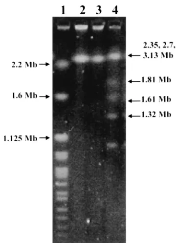
⫾2.43%. The genomic size of strain HKU1 was about 3 Mb (Fig. 3). Based on phylogenetic affiliation, HKU1 belongs to the Neisseriaceae family of the -subclass of Proteobacteria (Fig. 4).
DISCUSSION
We report the isolation of HKU1 from the blood and em-pyema of a Chinese cirrhotic patient. The clinical significance of the bacterium was evident by its isolation from sterile body fluids and the patient’s local and systemic responses (high fever, leukocytosis, and neutrophilia) to the bacterium. 16S rRNA gene sequence analysis unambiguously placed HKU1 in the-subclass of theProteobacteria. However, the 16S rRNA gene of HKU1 exhibited less than 94% nucleotide identity with the 16S rRNA gene of all previously described members of this subclass. The most closely related species isM. aerodenitrifi-cans, a new genus and species described in 1998; this organism was recovered from an upflow denitrifying filter inoculated with activated sludge and has never been implicated as a cause of human infections (9).
HKU1 exhibited phenotypic and genotypic characteristics that are very different from those of the other members of the
-subclass of theProteobacteria, as well as members of other
morphologically related pathogenic genera (Table 2). The G⫹C content of HKU1 is 68%, similar to those of M. aero-denitrificansandChromobacterium violaceum, two other mem-bers of the-subclass of theProteobacteria, but very different from those of representative species of other morphologically related pathogenic genera. The major characteristics for dis-FIG. 2. Scanning electron micrograph ofL. hongkongensis. Cells vary in length from 0.79 to 2.5m. The bacterium has a spiral curvature and is aflagellated. Bar, 1m.
FIG. 3. Pulsed-field gel electrophoresis of genomic DNA of HKU1 recovered from blood (lane 2) and empyema (lane 3),H. wingel chro-mosomal DNA size marker (lane 1), andS. cerevisiaechromosomal DNA size marker (lane 4), showing that the genome size of HKU1 is about 3 Mb.
on May 15, 2020 by guest
http://jcm.asm.org/
[image:4.587.329.509.430.678.2]tinguishing HKU1 fromM.aerodenitrificansare that HKU1 is nonmotile, assacharolytic, and urease and arginine dihydrolase positive. Phenotypically, HKU1 is most closely related to Pho-tobacterium damselae(identified by API 20E with 81.7% con-fidence) andComamonas testosteroniandPseudomonas alcali-genes(identified by API 20NE with 99.3% confidence). The major characteristics for distinguishing HKU1 fromP. dam-selaeare that HKU1 is assacharolytic and lysine decarboxylase negative butP.damselaeis glucose fermenting and lysine de-carboxylase positive; those for distinguishing HKU1 fromC. testosteroniorP. alcaligenesare that HKU1 is arginine dihy-drolase and urease positive butC.testosteroniorP.alcaligenes is arginine dihydrolase and urease negative. Since HKU1 is assacharolytic but urease and arginine dihydrolase positive, it probably utilizes proteins instead of carbohydrates as its energy sources. It is interesting that HKU1 has a seagull or spiral shape but is nonmotile, as all the other famous members of the Proteobacteriawith spiral or curved shapes (e.g.,M. aerodeni-trificansof the -subclass, Vibrio spp. of the ␥-subclass, and Campylobacterspp.,Helicobacter spp., andArcobacterspp. of theε-subclass) are all highly motile, and it is believed that the
curved or spiral shape actually assists in the movement of the organisms. Because of its interesting phenotypic characteris-tics, unique G⫹C content, and genome size and because it could not be assigned to any currently recognized genus, we propose a new genus and a new species for HKU1.
The reservoir of HKU1 and its route of transmission in our patient remain unknown. We speculate that the patient may have aspirated the bacteria into his lungs, thereby developing empyema and subsequent bacteremia. Endogenously, the most likely potential reservoir would be the oral cavity or the gas-trointestinal tract. However, screening of throat swab and stool samples from 30 volunteers did not show the presence of this bacterium (data not shown). The environment is another pos-sible source of the bacterium, and its tolerance to 2% NaCl makes aquatic environments possible reservoirs for HKU1. However, surveillance of water samples with different NaCl contents also failed to reveal the presence of this bacterium (data not shown).
Description ofLaribacter hongkongensisgen. nov., sp. nov.
Laribactermeans seagull-shaped rod;hongkongensis, in honor of Hong Kong, means the place where the bacterium was discovered.
Cells are facultatively anaerobic, nonsporulating, gram-neg-ative, seagull-shaped or spiral rods. The bacterium grows on sheep blood agar as nonhemolytic, gray colonies 1 mm in diameter after 24 h of incubation at 37°C in ambient air. Growth also occurs on MacConkey agar and at 25 and 42°C but not at 4, 44, and 50. It can grow in 1 or 2% NaCl but not in 3, 4, or 5% NaCl. No enhancement of growth is observed with 5% CO2. The organism is aflagellated and is nonmotile at both 25 and 37°C. It is oxidase, catalase, urease, and arginine dihy-drolase positive, and it reduces nitrate. It does not ferment, oxidize, or assimilate any sugar tested (Table 1). The moles percent G⫹C content of the DNA of the strain is 68.0% ⫾
2.43%. The genomic size of the strain is about 3 Mb. The organism was isolated from the blood and empyema of a cir-rhotic patient. The type strain of L. hongkongensis is strain HKU1. Its 16S rRNA gene sequence has been deposited within the GenBank sequence database under accession no. AF389085.
ACKNOWLEDGMENTS
[image:5.587.45.544.93.191.2]This work was partly supported by the University Research Grant Council and the Committee for Research and Conference Grants, The University of Hong Kong.
[image:5.587.52.278.481.661.2]FIG. 4. Phylogenetic tree showing the relationship ofL. hongkon-gensisgen. nov., sp. nov., to the other members of the-subclass of Proteobacteria. A total of 1,398 nucleotide positions in each 16S rRNA gene were included in the analysis. The scale bar indicates the esti-mated number of substitutions per 100 bases using the Jukes-Cantor correction; the length of the bar represents 1 substitution. Names and accession numbers are given as cited in the GenBank database.
TABLE 2. Comparison of phenotypic and genotypic characteristics ofL. hongkongensisand representative species of morphologically and/or phylogenetically related pathogenic generaa
Organism contentG⫹C (mol %)
Genome size
(Mb) Morphology
Oxygen
requirement Motility utilizationSugar Urease
2,4-Diamino- 6,7-diisopropyl-pteridine (0/129)
Campylobacter jejuni 30.6 1.64 Helical Microaerophilic Motile Asaccharolytic ⫺ NA
Arcobacter cryaerophilus 27–30 NA Curved Microaerophilic and aerobic Motile Asaccharolytic ⫺ NA
Vibrio cholerae 47.3 4.03 Curved Facultatively anaerobic Motile Saccharolytic ⫺ Sensitive
Chromobacterium violaceum 65–68 NA Straight Facultatively anaerobic Motile Saccharolytic Variable NA
Helicobacter pylori 39 1.64 Helical Microaerophilic Motile Asaccharolytic ⫹ NA
Microvirgula aerodenitrificans 65 NA Curved Aerobic Motile Asaccharolytic ⫺ NA
Laribacter hongleongensis 68 3 Seagull shaped Facultatively anaerobic Nonmotile Asaccharolytic ⫹ Resistant aNA, not available.⫺, negative;⫹, positive.
on May 15, 2020 by guest
http://jcm.asm.org/
REFERENCES
1.Ausubel, F. M., R. Brent, and R. E. Kingston (ed.).1998. Current protocols
in molecular biology, p. 2.4.1–2.4.2. John Wiley & Sons, Inc., New York, N.Y.
2.Bauer, A. W., W. M. M. Kirby, J. C. Sherris, and M. Turck.1966. Antibiotic
susceptibility testing by a single disc method. Am. J. Clin. Pathol.45:493.
3.Cheuk, W., P. C. Y. Woo, K. Y. Yuen, P. H. Yu, and J. K. C. Chan.2001.
Intestinal inflammatory pseudotumor with regional lymph node involvment: identification of a new bacterium as the etiologic agent. J. Pathol.192:289– 292.
4.Goodfellow, M.1985. Chemical methods in bacterial systematics, p. 67–93.
Academic Press Ltd., London, England.
5.Luk, W. K., S. S. Wong, K. Y. Yuen, P. L. Ho, P. C. Y. Woo, R. A. Lee, and
P. Y. Chau.1998. Inpatient emergencies encountered by an infectious
dis-ease consultative service. Clin. Infect. Dis.26:695–701.
6.Murray, P. R., E. J. Baro, M. A. Pfaller, F. C. Tenover, and R. H. Yolken
(ed.).1999. Manual of clinical microbiology, 7th ed. American Society for
Microbiology, Washington, D.C.
7.Olsen, G. J., R. Overbeek, N. Larsen, T. L. Marsh, M. J. McCaughey, M. A.
Maciukenas, W. M. Kuan, T. J. Macke, Y. Xing, and C. R. Woese.1992. The
ribosomal database project. Nucleic Acids Res. Suppl.20:2199–2200.
8.Olsen, G. J., and C. R. Woese.1993. Ribosomal RNA: a key to phylogeny.
FASEB J.7:113–123.
9.Patureau, D., J. J. Godon, P. Dabert, T. Bouchez, N. Bernet, J. P. Delgenes,
and R. Moletta.1998.Microvirgula aerodenitrificansgen. nov., sp. nov., a new
gram-negative bacterium exhibiting co-respiration of oxygen and nitrogen oxides up to oxygen-saturated conditions. Int. J. Syst. Bacteriol.48:775–782.
10.Relman, D. A., J. S. Loutit, T. M. Schmidt, S. Falkow, and L. S. Tompkins.
1990. The agent of bacillary angiomatosis. An approach to the identification of uncultured pathogens. N. Engl. J. Med.323:1573–1580.
11.Relman, D. A., T. M. Schmidt, R. P. MacDermott, and S. Falkow.1992.
Identification of the uncultured bacillus of Whipple’s disease. N. Engl. J. Med.327:293–301.
12.Thompson, J. D., D. G. Higgins, and T. J. Gibson.1994. CLUSTAL W:
improving the sensitivity of progressive multiple sequence alignment through sequence weighting, position-specific gap penalties and weight matrix choice. Nucleic Acids Res.22:4673–4680.
13.Woo, P. C. Y., K. T. K. Chong, K. W. Leung, T. L. Que, and K. Y. Yuen.2001.
Identification ofArcobacter cryaerophilusisolated from a traffic accident victim with bacteraemia by 16S ribosomal RNA gene sequencing. Diagn. Microbiol. Infect. Dis.40:125–127.
14.Woo, P. C. Y., E. Y. L. Cheung, K. W. Leung, and K. Y. Yuen.2001.
Identification by 16S ribosomal RNA gene sequencing of an Enterobacteri-aceaespecies with ambiguous biochemical profile from a renal transplant recipient. Diagn. Microbiol. Infect. Dis.39:85–93.
15.Woo, P. C. Y., A. M. Fung, S. S. Y. Wong, H. W. Tsoi, and K. Y. Yuen.2001.
Isolation and characterization of aSalmonella entericaserotype Typhi variant and its clinical and public health implications. J. Clin. Microbiol.39:1190– 1191.
16.Woo, P. C. Y., P. K. L. Leung, K. W. Leung, and K. Y. Yuen.2000.
Identi-fication by 16S ribosomal RNA gene sequencing of anEnterobacteriaceae
species from a bone marrow transplant recipient. Mol. Pathol.53:211–215.
17.Woo, P. C. Y., C. Y. Lo, S. K. Lo, H. Siau, J. S. M. Peiris, S. S. Y. Wong, W. K.
Luk, T. M. Chan, W. W. Lim, and K. Y. Yuen.1997. Distinct genotypic
distributions of cytomegalovirus (CMV) envelope glycoprotein in bone mar-row and renal transplant recipients with CMV disease. Clin. Diagn. Lab. Immunol.4:515–518.
18.Woo, P. C. Y., H. W. Tsoi, K. W. Leung, P. N. L. Lum, A. S. P. Leung, C. H.
Ma, K. M. Kam, and K. Y. Yuen.2000. Identification ofMycobacterium
neoaurumisolated from a neutropenic patient with catheter-related bacte-remia by 16S rRNA sequencing. J. Clin. Microbiol.38:3515–3517.
19.Woo, P. C. Y., A. S. P. Leung, K. W. Leung, and K. Y. Yuen.2001.
Identi-fication of slide-coagulase positive, tube-coagulase negativeStaphylococcus aureusby 16S ribosomal RNA gene sequencing. Mol. Pathol.54:244–247.