BIROn - Birkbeck Institutional Research Online
Dumontheil, Iroise and Hassan, B. and Gilbert, S.J. and Blakemore, S.J.
(2010) Development of the selection and manipulation of self-generated
thoughts in adolescence. Journal of Neuroscience 30 (22), pp. 7664-7671.
ISSN 0270-6474.
Downloaded from:
Usage Guidelines:
Please refer to usage guidelines at
or alternatively
Behavioral/Systems/Cognitive
Development of the Selection and Manipulation of
Self-Generated Thoughts in Adolescence
Iroise Dumontheil,
1Bano Hassan,
2Sam J. Gilbert,
1and Sarah-Jayne Blakemore
11Institute of Cognitive Neuroscience, University College London, London WC1N 3AR, United Kingdom, and2Great Ormond Street Children’s Hospital,
London WC1N 3JH, United Kingdom
The ability to select and manipulate self-generated (stimulus-independent, SI), as opposed to stimulus-oriented (SO), information, in a
controlled and flexible way has previously only been studied in adults. This ability is thought to rely in part on the rostrolateral prefrontal
cortex (RLPFC), which continues to mature anatomically during adolescence. We investigated (1) the development of this ability
behav-iorally, (2) the associated functional brain development, and (3) the link between functional and structural maturation. Participants
classified according to their shape letters either presented visually (SO phases) or that they generated in their head by continuing the
alphabet sequence (SI phases). SI phases were performed in the presence or absence of distracting letters. A total of 179 participants (7–27
years old) took part in a behavioral study. Resistance to visual distractors exhibited small improvements with age. SI thoughts
manipu-lation and switching between SI and SO thoughts showed steeper performance improvements extending into late adolescence.
Thirty-seven participants (11–30 years old) took part in a functional MRI (fMRI) study. SI thought manipulation and switching between SO and
SI thought were each associated with brain regions consistently recruited across age. A single frontal brain region in each contrast
exhibited decreased activity with age: left inferior frontal gyrus/anterior insula for SI thought manipulation, and right superior RLPFC for
switching between SO and SI thoughts. By integrating structural and functional data, we demonstrated that the observed functional changes with
age were not purely consequences of structural maturation and thus may reflect the maturation of neurocognitive strategies.
Introduction
Adolescence is a time of profound mental change, affecting many
aspects of behavior including planning, multitasking and the
ability to resist distractions (Blakemore and Choudhury, 2006).
These abilities recruit “executive functions,” which enable the
selection of actions that are consistent with one’s goal. While
most aspects of executive functions are well established by late
childhood, the ability to deploy them flexibly and consistently
continues to develop during adolescence (Luna et al., 2010). An
important aspect of executive function in adults is the ability to
modulate the balance between cognition that is provoked by
per-ceptual experience (stimulus-oriented, SO) and that which
oc-curs in the absence of sensory input (stimulus-independent, SI)
(Burgess et al., 2007). No previous study has directly investigated
the development of this ability. In children, manipulation of SI
thoughts has been studied in the context of fluid intelligence
(Wright et al., 2008; Crone et al., 2009) and working memory
(WM) tasks (Crone et al., 2006), while the ability to resist
dis-tracting SO information has been studied in perceptual (Bunge et
al., 2002; Booth et al., 2003) and WM tasks (Olesen et al., 2007).
Here, we used a single task that could be performed on the basis of
either SO or SI information, without high WM requirements
(Gilbert et al., 2005, 2007, 2008). Distracting SO information was
present or absent during SI phases of the task.
The studies mentioned above report observations during
ad-olescence of improvements in WM and fluid reasoning
perfor-mance, and to a lesser extent of the resistance to perceptual
distractors, as well as changes in neural activity, in particular in
the prefrontal cortex (PFC). Decreases are frequently interpreted
as being due to developmental reductions in gray matter volume,
presumably related to synaptic pruning, while increases are
thought to relate to improved and more localized task-specific
processing, potentially facilitated by faster long-range
connec-tions due to increased axonal myelination and size (Luna et al.,
2010). Understanding the link between structural and functional
changes is critical in understanding the mechanisms of
neurocog-nitive development, yet very few studies have directly compared
structural and functional data within the same individuals
(Olesen et al., 2003; Lu et al., 2009).
An a priori region of interest was the rostrolateral prefrontal
cortex (RLPFC), which has been suggested to support the
selec-tion and manipulaselec-tion of SI thoughts, as opposed to SO
informa-tion, in a controlled and flexible way (Christoff and Gabrieli,
2000; Burgess et al., 2005; Gilbert et al., 2005; Dumontheil et al.,
2008). In a behavioral study and a functional magnetic resonance
imaging (fMRI) study, we investigated: (1) how performance on
a task requiring selection and manipulation of SI thoughts
devel-ops between late childhood and adulthood, differentiating
be-tween three aspects of the task: selection and manipulation of SI
versus SO thoughts; switching versus maintaining the selection of
SI or SO thoughts; and resistance to distractors; (2) how neural
Received March 17, 2010; accepted April 19, 2010.
This research was supported by grants from the Royal Society to S.J.B. and S.J.G. and a Fellowship from the European Commission to I.D. We are grateful to A. Ranpura for his help with the VBM analyses.
Correspondence should be addressed to Dr. Iroise Dumontheil, UCL Institute of Cognitive Neuroscience, 17 Queen Square, London WC1N 3AR, UK. E-mail: [email protected].
DOI:10.1523/JNEUROSCI.1375-10.2010
activity underlying these processes develops between early
ado-lescence and adulthood; and (3) whether age-related changes
could be accounted for by changes in gray and white matter
volumes.
Materials and Methods
Both studies were restricted to female participants so as to reduce vari-ance attributable to sex differences in brain maturation and cognitive development (Giedd et al., 1999, 2006). Written informed consent was obtained before both studies from all participants, or from the parent of participants aged under 18. The studies were approved by the University College London (UCL) National Hospital for Neurology and Neurosur-gery Ethics Committee. Stimulus presentation was programmed in Co-gent (www.vislab.ucl.ac.uk/CoCo-gent/index.html) running in Matlab 6.5 (MathWorks), which recorded participants’ responses.
Behavioral study
Participants.A total of 179 female volunteers between the ages of 7.3 and 27.5 years were recruited for this study. Children and adolescents (n⫽143) were recruited from two London schools for girls, while adults (n⫽36) were recruited from the UCL Psychology Department volunteer database. All participants spoke English as their first language. Verbal ability was mea-sured in children and adolescents using the BPVS II scores (Dunn et al., 1997), and in adults using the Vocabulary subtest of the WASI (Wechsler, 1999). Data from one adolescent who had a verbal intelligence quotient (IQ) score of⬍75 were excluded from the analysis. There was no signif-icant correlation between verbal IQ and age (Pearsonr⫽0.008,p⬎0.8). Design.The behavioral task had three within-subjects factors (Condi-tion: Distractor or No-Distractor; Block type: SO or SI; and Trial type: Switch or Stay), and one between-subjects factor (Age). We used a computer-based task adapted from Gilbert et al. (2005), which tests the control of the allocation of attention between SO and SI information (Fig. 1). In SO blocks a single upper case letter was presented in the center of the screen in red. Participants were required to classify the letter ac-cording to its shape by pressing one of two “Ctrl” buttons on a laptop keyboard, using their left and right index fingers. Participants were asked to press the left key if the letter was composed entirely of straight lines (e.g., the letter A) or the right key if the letter shape contained a curve
(e.g., the letter B). After each button press a new letter was presented, following the sequence of the alphabet. During SI blocks, participants were asked to continue to go through the alphabet sequence and to continually classify as curved/straight-lined each of the lettersin their headand press the corresponding keys. In the No-Distractor condition, the screen during SI blocks was blank. In the Distractor condition, a blue letter, pseudo-randomly picked from the alphabet with the constraint that it was different from the current and previous letters the participant had in her head, was shown in the middle of the screen, with a new letter after each button press. Participants were instructed to ignore this letter. Eventually a new SO phase began and the red letters reappeared continu-ing the alphabet sequence. The Distractor condition enabled us to explore the proposal that selection between SI and SO thoughts is partic-ularly important when it does not occur naturally or automatically (as in the absence of competing SI or SO information) but is instead imposed in a controlled way (when both SI and SO information are present) (Bur-gess et al., 2005). Analyses differentiated between: (1) performance on the first trial of SO and SI blocks, i.e., “switch” trials, which indicates participants’ ability to switch between selecting SO and SI information; and (2) performance on the subsequent trials of SO and SI blocks, i.e., “stay” trials, which indicates participants’ ability to maintain the selec-tion and processing of SO or SI informaselec-tion.
SO and SI phases alternated and lasted on average 6.5 trials (range 5– 8 trials). Switches between SO and SI phases were cued by the appearance of a letter (red letter: SO; blue letter or blank: SI). Participants performed one run of the No-Distractor condition and one run of the Distractor condition; order was counterbalanced between participants. Each run comprised six SO and six SI phases. All conditions were self-paced with no interval between the displays of stimuli.
Instructions for the task were displayed on the screen and read out loud by the experimenter. Participants first performed for practice the SO task for two repetitions of the alphabet sequence (52 trials). Relevant additional instructions regarding SI phases and a run of 26 practice trials with alternating SO and SI phases preceded the runs of the Distractor and No-Distractor condition.
Participants were tested individually in a quiet room at their school or at home (for the children and adolescents) or in the laboratory (for the adults).
Data analysis. Mean accuracy and median response times (RT) in correctly responded trials were calculated for each participant in each condition. Main effects were investigated using pairedttests. Changes in performance with age for accuracy and RT overall and for the effects of each factor separately were investigated using regression analyses. Linear, quadratic and cubic age regressors were added in turn to the model until further improvement to the model was not significant. Bayesian infor-mation criterion (BIC), or Schwartz criterion, was calculated and in all cases confirmed the final model selection was not driven by increased model complexity only (Schwartz, 1978; Liddle, 2007). The models were subsequently used to calculate for each factor the amplitude of accuracy or RT changes between the youngest age and the age at the first inflection of the fit curve (or the oldest age for linear fits).
Neuroimaging study
Participants.Thirty-seven right-handed, female participants aged be-tween 11.0 and 30.4 years, with no history of psychiatric or neurological disorder, took part in the study. Adolescents (n⫽24) were recruited through advertisements at their schools or following their parents’ re-sponse to an article in a newspaper. Adults (n⫽13) were recruited following their participation via an advert in a local community elec-tronic newsletter or in the department. General ability of the participants was measured using the Vocabulary and Matrix Reasoning subtests of the WASI (Wechsler, 1999). There was no significant correlation between IQ and age (Pearson correlationr⫽0.028,p⬎0.8).
Experimental design.The fMRI task was a further adaptation of the alphabet task (Gilbert et al., 2005). The task had two factors: Block type (SO, SI) and Trial type (Stay, Color switch, SOSI switch). In all trials a single upper case letter was presented on the screen, in one of four pos-sible colors (Fig. 1). Two colors were associated with SO blocks (e.g., red and green) and two colors with SI blocks (e.g., blue and yellow). Color Figure 1. Examples of sequences of trials from the alphabet task used in the behavioral and
neuroimaging studies.A, In the behavioral task, participants were required to classify letters as consisting of straight lines only or containing a curve. In SO phases, participants classified the red letter presented on the screen. In SI phases, participants classified the letter in their head, generated by continuing the alphabet sequence. During SI phases either a random blue letter was presented on the screen (Distractor condition) or the screen was blank (No-Distractor con-dition). Switch trials were the first trial after a change between SI/SO phases; stay trials were defined as all subsequent trials.B, The neuroimaging task included the Distractor condition only. Letters were red or green in SO blocks, blue or yellow in SI blocks. Participants were trained to learn this association. A change in color of the letters within SO or SI blocks was coded as a Color switch, while a change in color of the letters between SO and SI blocks was coded as an SOSI switch.
switch trials were included as a control condition in which the stimulus color changed (as in SOSI switch trials) but the Block type (SO, SI) did not. Instructions for the SO and SI conditions were the same as in the Distractor condition of the behavioral study (see above). Participants used their right index and middle fingers to respond.
The task was self-paced with no interval between the presentations of successive letters. SO and SI phases alternated: SO phases lasted 10.5 s on average (average range 3.4 –15.5 s); SI phases lasted 9.3 s on average (average range 2.8 –16.5 s). Color switches occurred on average 5.3 s after the start of an SO (average range 2.1–7.5 s) or SI block (average range 2.6 – 8.1 s). Task blocks and fixation blocks alternated, and lasted on average 40.5 s (average range 40.0 – 45.3 s) and 14.7 s (average range 13.6 –15.0 s), respectively. There were two scanning sessions, each com-prising eight task blocks and seven fixation blocks.
Before the scanning participants performed supervised practice of the tasks, which was continued until participants performed at least 20 cor-rect responses in a row, after a minimum of three blocks of the task and an initial practice of two sets of 26 SO trials.
MRI data acquisition.A 1.5 Tesla Siemens Avanto MRI scanner was used to acquire both 3D T1-weighted fast-field echo structural images and multislice T2*-weighted echo-planar volumes with blood oxygen level-dependent (BOLD) contrast (TR⫽3 s; TE⫽50 ms; TA⫽2.9143 s). For each subject, functional data were acquired in two scanning ses-sions of the alphabet task of 7.45 min duration. This was preceded by two 7 min scanning sessions of a separate task and acquisition of the struc-tural images. The first 2 volumes of each session were discarded to allow for T1equilibrium effects. Each functional brain volume comprised 35 axial slices with a resolution of 3⫻3⫻3 mm, positioned to cover the whole brain.
Statistical analysis.Mean of the participants’ median RTs and percent-age accuracy were calculated. Main effects were investigated using paired ttests. Changes in performance with age in overall accuracy, overall RT, and in the effects of interest (SI stay vs SO stay; SOSI switch vs Color switch) were investigated using regression analyses. Linear, quadratic and cubic age regressors were added in turn to the model until further improvement to the model was not significant.
fMRI data were analyzed using statistical parametric mapping imple-mented in SPM5 (http://www.fil.ion.ucl.ac.uk/spm). Volumes were re-aligned (using fourth-degree-B-spline interpolation), corrected for differences in slice acquisition times, normalized into 3 mm cubic voxels using the subjects’ structural (n⫽35) or mean functional scans (n⫽2, subjects with lower quality structural scans) to the standard T1 or EPI template based on the Montreal Neurological Institute (MNI) reference brain, respectively, and spatially smoothed with an isotropic 8 mm, full-width, half-maximum Gaussian kernel.
Analysis of the movement parameters obtained during realignment showed that translation within each session was⬍3 mm in all partici-pants except in the second session of one participant (translation inz plane⬎8 mm). This session was excluded from all analyses. Correlation analyses performed on the participants’ means across sessions showed a significant linear decrease in rotation in they-axis with age (Pearsonr⫽
⫺0.365,p⫽0.026). The five other movement parameters did not show significant correlations with age (allp⬎0.1).
The volumes acquired during the two sessions were treated as separate time series. For each series, the variance in the BOLD signal was decom-posed with a set of regressors in a general linear model (Friston et al., 1995). The regressors were four boxcar regressors representing the instructions, fixation, SO and SI blocks, and three event-related re-gressors representing color switches, SOSI switches, and error trials. All regressors were convolved with a canonical hemodynamic re-sponse function and, together with regressors representing residual movement-related artifacts and the mean over scans, comprised the full model for each session. The data and model were high-pass fil-tered to a cutoff of 1/128 Hz.
Parameter estimates calculated from the least mean squares fit of the model to the data were used in a one samplettest (color switch, SOSI switch) or pairwise contrasts (SO⬎fixation, SI⬎fixation) at the indi-vidual subject level. The participants were divided into three groups of similarn(age 11–14,n⫽12; age 14 –18,n⫽12; age 22–30,n⫽13). First
level contrasts were entered into two separate 2 (Condition)⫻3 (age group) factorial design second level analyses, where ‘subject’ was treated as a random effect. The within-subjects factors were block type (SO vs SI) and switch type (color switch vs SOSI switch) respectively, and age group was the between-subjects factor. These analyses had two aims: first, to study any effect of age group as a categorical variable; second, to obtain a main effect of condition unbiased by age to define regions of interests in which to test for effects of age as a continuous variable on neural activity and on local gray and white matter volumes. Main effects of block type and switch type and their interaction with age were analyzed using weighted linear contrasts and determined using thet-statistic on a voxel by voxel basis. Tests were corrected for family-wise errors (FWE) across the whole brain, and in our region of interest (ROI) RLPFC, using an anatomically defined Brodmann area 10 (BA10) mask from WFU Pick-Atlas Tool Version 2.4 (Maldjian et al., 2003, 2004). Parameter estimates of the peak voxel of clusters showing an effect of experimental condition were extracted and analyzed for effects of age using Pearson tests of correlations. It should be noted that this is an unbiased method as the voxels of interest were defined from a fully balanced contrast orthogonal to the effect of age (Kriegeskorte et al., 2009). In a second step, multiple regressions were performed on the peak voxels showing correlation be-tween BOLD signal and age to test whether age still accounted for changes in BOLD signal when individual structural and performance differences were taken into account.
Voxel-based morphometry.Voxel-based morphometry (VBM) (Ashburner and Friston, 2000) investigating gray and white matter tissue volumes was performed on all participants using SPM5 VBM5 toolbox (v1.15 http://dbm.neuro.uni-jena.de/vbm/). Each participant’s structural T1 image was normalized to the standard T1 MNI template. Segmentation was performed without using prior tissue probability maps, which is the option favored when testing children and adolescents. Structural scans were segmented into CSF, gray and white matter; modulation for non-linear warping only was performed using the Jacobian determinants. This method approximates a proportional adjustment of volumes for overall head size (O’Brien et al., 2006). Images were resampled into 1.5⫻ 1.5⫻1.5 voxels and smoothed with an isotropic 12 mm, full-width, half-maximum Gaussian kernel. The MarsBaR toolbox for SPM5 (http:// marsbar.sourceforge.net/) was used to calculate mean gray and white matter adjusted volumes within spheres of 5 mm in radius, centered around the peak voxels of the main contrasts that exhibited significant age effects. Gray and white matter adjusted volumes were investigated for effects of age using Pearson tests of correlation.
Results
Behavioral study
We instructed 178 participants aged 7.3–27.5 years to classify
according to their shape letters that were either presented on the
screen (SO) or not (SI), and in the presence or absence of
distrac-tors (Fig. 1). Age-related changes in overall performance and in
the effect of each factor (Distractor–No-Distractor, SI–SO, and
Switch–Stay) were investigated separately for accuracy and
re-sponse time (RT) using regression analyses. Linear, quadratic and
cubic age regressors were added in turn to the model until further
improvement to the model was not significant.
Participants were slower and made more errors in the
Distrac-tor condition [mean (SD) accuracy and RT: DistracDistrac-tor: 93.8%
(7.3), 1106 ms (372); No-Distractor: 95.0% (5.0), 986 ms (324)
兴
,
SI blocks
关
(SI 93.9% (6.8), 1142 ms (430); SO: 94.9% (5.2), 951
ms (273)], and Switch trials [Switch: 94.1% (6.4), 1270 ms (466);
Stay: 94.8% (4.6), 822 ms (228)] than in their respective control
conditions (accuracy: all
t
177⬎
1.99,
p
⬍
0.05; RT: all
t
177⬎
8.38,
p
⬍
0.001). Note that the presence of distractors in SI phases
affected both SO and SI RTs (all
t
177⬎
5.78,
p
⬍
0.001) and SO
accuracy in the Distractor condition (SO:
t
177⫽
2.69,
p
⫽
0.008;
Dis-tractor SO: 94.1% (7.8), 991 ms (304);
No-Distractor SI: 94.3% (7.8), 1062 ms
(424); Distractor SI: 93.5% (9.8), 1221 ms
(480)].
Average accuracy was
⬎
90% in all
conditions. Regression analyses indicated
that overall accuracy improved linearly
with age (
F
(1,176)⫽
4.44,
p
⫽
0.037,

age⫽
0.16,
R
2⫽
0.02) (Fig. 2
A
). Of the three
main effects, only Condition (Distractor
vs No-Distractor) was associated with age,
with the difference in accuracy between
Distractor and No-Distractor blocks
fol-lowing a quadratic function (
F
(2,175)⫽
5.72,
p
⫽
0.004,

age⫽ ⫺
1.39,

age2⫽
1.24,
R
2⫽
0.06) (Fig. 2
B
). This reflected an
improvement with age in accuracy in the
Distractor (
p
⫽
0.010) but not the
No-Distractor condition (
p
⬎
0.29). The
in-flection of the quadratic curve occurred at
age 18.8 years, suggesting that the
accu-racy cost of the presence of Distractors
reached adult levels by this age.
Overall RT decreased with age following
a cubic function (
F
(3,174)⫽
48.49,
p
⬍
0.001,

age⫽ ⫺
6.52,

age2⫽
9.95,

age3⫽ ⫺
4.03,
R
2⫽
0.46) (Fig. 2
C
). The first inflection of
this cubic curve occurred at age 18.9 years.
All main effects showed changes with age,
with decreases in the RT difference between
Distractor and No-Distractor (linear model:
F
(1,176)⫽
5.16,
p
⫽
0.024,

age⫽ ⫺
0.17,
R
2⫽
0.03), SI and SO (cubic model:
F
(3,174)⫽
27.96,
p
⬍
0.001,

age⫽ ⫺
9.17,

age2⫽
16.48,

age3⫽ ⫺
7.86,
R
2⫽
0.33) and Switch
and Stay trials (quadratic model:
F
(2,175)⫽
40.25,
p
⬍
0.001,

age⫽⫺
2.40,

age2⫽
1.94,
R
2⫽
0.32) (Fig. 2
D
). Inflections of the
esti-mated functions occurred at 16.2 years for
the SI–SO difference, and at 20.7 years for
the Switch–Stay difference. The difference
between [Switch–Stay] and [SI–SO] did not
show any significant change with age (
p
⬎
0.15), however, which suggests that the time
courses of the two effects did not differ
significantly.
Neuroimaging study
Behavioral results
In the fMRI study, 37 participants aged
11.0 –30.4 years were scanned while
carry-ing out an adapted version of the alphabet
task in which there was no No-Distractor
condition (Fig. 1). Changes in
perfor-mance with age in overall accuracy and
RT, and in the differences in accuracy and
RT between SI and SO stay trials and
be-tween SOSI and Color switch trials, were
investigated using regression analyses.
Average accuracy was
⬎
90% in all
conditions. Regression analyses indicated
that overall accuracy increased linearly
with age (
F
(1,35)⫽
6.92,
p
⫽
0.013,

age⫽
Figure 2. Accuracy and RT data from the behavioral study and fMRI study. Regression analyses were performed to test for linear, quadratic or cubic changes in performance with age. Significant effects observed in the behavioral study (A–D) or in the fMRI study (E,F) are shown in this figure.A, Scatterplot of the overall accuracy (percentage) against age. The line represents the significant linear fit of the data.B, Scatterplot of the difference in accuracy between No-Distractor and Distractor conditions against age. The curve represents the significant quadratic fit of the data.C, Scatterplot of the overall RTs (in milliseconds) against age. The curve represents the significant cubic fit of the data.D, Scatterplot of the three main effects of the task on RT (in milliseconds) against age: in light gray, difference between Distractor and No-Distractor, with a significant linear fit; in dark gray, difference between SI and SO blocks, with a significant cubic fit; in black, switch cost (SOSI Switch–Stay), with a significant quadratic fit.E, Scatterplot of the overall accuracy (percentage) against age. The line represents the significant linear fit of the data.F, Scatterplot of the RT switch cost (SOSI switch–Color switch) against age. The line represents the significant linear fit of the data.
Figure 3. Neuroimaging results of the main effect SI–SO.A, Results of the SI–SO contrast averaged across the three age groups are rendered on the brain surface (outer panels) and shown on the sagittal slice (x⫽3) (middle panel) (FWE,p⬍0.05). The white circles indicate the approximate location of the regions in which the parameter estimates were extracted in the peak voxel and gray and white matter volumes were calculated in 5 mm spheres around the peak.B, Changes in brain structure and function with age are presented for the only region showing an effect of age on the SI–SO contrast, the left IFG. Left, Bar chart representing IFG activation in SI and SO blocks vs fixation blocks in the three age groups (mean⫾SE). Middle, Scatterplot of the BOLD response difference between SI and SO blocks as a function of age. Right, Scatterplot of gray (gray circles) and white (black circles) matter volume as a function of age. The full lines represent significant correlations between functional or structural measures and age.
0.41,
R
2⫽
0.17) (Fig. 2
E
). Participants made more errors in SI vs
SO stay blocks (
t
36⫽
2.38,
p
⬍
0.05), but not in SOSI switch vs
Color switch trials (
p
⬎
0.6). The differences in accuracy between
SI and SO stay trials, and between SOSI switch and Color switch
trials, did not show significant changes with age.
Participants were slower in SI vs SO stay blocks, and SOSI
switch vs Color switch trials (both
t
36⬎
4.99,
p
⬍
0.001). Overall
RT, and the RT difference between SI and SO stay trials, did not
change with age. The RT switch cost (SOSI switch–Color switch)
decreased linearly with age (
F
(1,35)⫽
6.06,
p
⫽
0.019,

age⫽
⫺
0.38,
R
2⫽
0.15) (Fig. 2
F
).
These fMRI behavioral results suggest later performance
im-provement for switching between SI and SO thoughts than for
maintaining the selection of SI thoughts, while the
developmen-tal time course of these two factors did not differ in the behavioral
study. The differences observed may arise from the narrower and
older age range used in the fMRI study, and more extensive task
practice in the fMRI study.
fMRI results
Main effects of condition.
The effects of the two experimental
fac-tors were assessed using two 2 (condition)
⫻
3 (age group)
fac-torial analyses (see Materials and Methods). [SI–SO] and [SOSI
switch–Color switch] main effect contrasts were performed
aver-aging across participants divided into three age groups [early
adolescence: 11–14 years old (
n
⫽
12); mid adolescence: 14 –17
years old (
n
⫽
12); adults (
n
⫽
13)]. Resulting activations
surviv-ing whole brain or within BA10 FWE correction at
p
⬍
0.05 are
reported.
The SI–SO block comparison revealed bilateral activation of
the inferior frontal gyri (BA46/44/45), extending into the insula
(Fig. 3
B
, see bottom left plot), and of the premotor cortex,
pari-etal cortex, medial frontal cortex, and the precuneus (Table 1, Fig.
3). Activations within our BA10 ROI were observed in left ([
⫺
36
45 21],
Z
⫽
4.72; [
⫺
39 48 12],
Z
⫽
4.69; [
⫺
30 51 9],
Z
⫽
4.32)
and right RLPFC ([36 42 30],
Z
⫽
5.92; [36 48 18],
Z
⫽
4.84; [30
51 3],
Z
⫽
4.06).
The SOSI switch–Color switch comparison revealed greater
activations in right superior RLPFC, cingulate gyrus (BA32),
pre-cuneus (BA7/18), pre-cuneus (BA17) and superior temporal gyrus
(BA22) (Table 1, Fig. 4). Activations within our BA10 ROI were
observed in left ([
⫺
33 51 24],
Z
⫽
4.09) and right superior
RLPFC ([36 45 30],
Z
⫽
6.48) (Fig. 4
B
, see bottom left plot).
Note there were no significant interactions between condition
and age group or main effects of age group in these whole-brain
analyses (FWE
p
⬍
0.05), indicating that age group as a
categor-ical variable did not affect neural activations in the SI–SO and
SOSI switch–Color switch contrasts.
Effects of age on brain structure and function.
Parameter
esti-mates were extracted from the peak voxels of the main effect
analyses and evaluated using orthogonal contrasts testing for
sig-nificant changes in brain activation with age as a continuous
variable. Defining voxels of interest based on functional data can
be biased (Kriegeskorte et al., 2009). Here, the contrasts defining
our voxels of interest were orthogonal to age. It should be noted
that the results are reported without correction for multiples
comparisons and therefore should be interpreted with caution
and require further replication. VBM was used to extract
av-erage volumes of gray and white matter adjusted for head size
in 5 mm spheres around the peak voxels exhibiting age-related
effects to test for structural changes with age in these regions.
Finally, multiple regression analyses were performed to test
whether the observed age differences in BOLD signal reflect
differences in brain structure and/or performance (accuracy
and RT).
SI–SO.
No change in BOLD signal with age was observed in
the RLPFC in the SI–SO contrast. However, the left IFG [
⫺
30 24
0] (extending into the insula in the whole-brain analysis)
exhib-ited a significant linear decrease in activation with age (Pearson
r
⫽ ⫺
0.417,
p
⫽
0.010). VBM analyses revealed that gray matter
volume in this region exhibited a significant decrease with age
(
r
⫽ ⫺
0.695,
p
⬍
0.001), while white matter volume in this region
exhibited a significant increase with age (
r
⫽ ⫺
0.511,
p
⫽
0.001)
(Fig. 3
B
).
To investigate whether the correlation between age and BOLD
signal was maintained when taking into account individual
dif-ferences in structural development and in performance, a
regres-sion analysis was performed with gray and white matter adjusted
volumes and differences in accuracy and RT between SI and SO
stay trials as regressors. Results indicated that age still showed a
trend toward accounting for BOLD response variance (

⫽
⫺
0.378,
p
⫽
0.099), while none of the structural or performance
regressors showed significant effects (all
p
⬎
0.26) (Table 2).
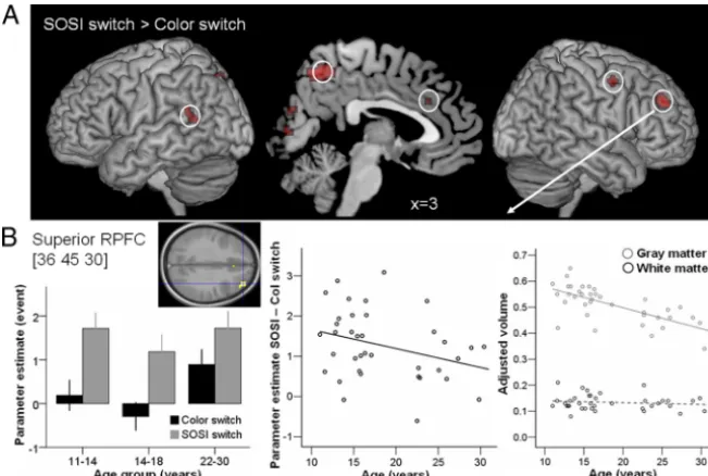
[image:6.594.299.546.92.453.2]SOSI switch– color switch.
Again, only one region observed in
the whole-brain analysis of the main effect of SOSI switch–Color
switch exhibited changes in BOLD signal with age. This was the
right superior RLPFC region [36 45 30], where the difference in
BOLD signal between SOSI switch and Color switch marginally
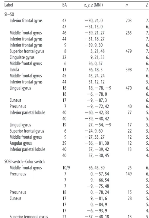
Table 1. Whole-brain analyses (FWE,p<0.05, 10 voxels or more) of the main effects of interest, SI–SO and SOSI switch–Color switch, averaged across the three age groups
Label BA x,y,z(MNI) n Z
SI–SO
Inferior frontal gyrus 47 ⫺30, 24, 0 203 7.51 47 ⫺51, 15, 0 6.15 Middle frontal gyrus 46 ⫺39, 21, 27 265 7.49 Inferior frontal gyrus 44 ⫺51, 18, 27 7.16 Inferior frontal gyrus 9 ⫺39, 9, 30 6.56 Superior frontal gyrus 8 3, 21, 48 479 7.38 Cingulate gyrus 32 9, 21, 33 6.55 Middle frontal gyrus 6 36, 0, 57 6.47 Insula 13 36, 18, 3 398 7.36 Middle frontal gyrus 45 45, 24, 24 6.19 Inferior frontal gyrus 44 51, 12, 12 5.95 Lingual gyrus 18 18,⫺78,⫺9 470 6.66 18 ⫺6,⫺78, 0 6.58 Cuneus 17 ⫺9,⫺87, 3 6.27 Precuneus 7 ⫺9,⫺72, 42 40 6.26 Inferior parietal lobule 40 ⫺60,⫺42, 33 77 5.93 40 ⫺39,⫺48, 42 5.89 Lingual gyrus 19 27,⫺54,⫺9 17 5.61 Superior frontal gyrus 6 ⫺24, 9, 60 22 5.54 Middle frontal gyrus 9 ⫺27, 33, 27 12 5.33 Angular gyrus 39 ⫺36,⫺81, 30 12 5.33 Inferior parietal lobule 40 57,⫺39, 42 13 5.26 40 57,⫺30, 45 4.98 SOSI switch–Color switch
Middle frontal gyrus 10/9 36, 45, 30 25 6.48 Precuneus 7 0,⫺57, 54 149 6.19 7 9,⫺66, 54 5.79 7 ⫺9,⫺75, 48 5.61 Precuneus 18 0,⫺78, 24 15 5.43 Cuneus 17 9,⫺81, 6 28 5.23 17 0,⫺84, 9 5.17 17 ⫺6,⫺93, 9 4.83 Superior temporal gyrus 22 ⫺57,⫺48, 18 13 5.1
significantly decreased with age (
r
⫽ ⫺
0.318,
p
⫽
0.055). VBM
analyses revealed that gray matter volume in the right superior
RLPFC decreased with age (
r
⫽ ⫺
0.682,
p
⬍
0.001). White
matter volume did not show significant changes with age (
p
⬎
0.7) (Fig. 4
B
).
A regression analysis was performed with gray and white
matter adjusted volumes and differences in accuracy and RT
between SOSI and Color switch trials as regressors. Results
indi-cated that age still marginally significantly accounted for BOLD
response variance (

⫽ ⫺
0.466,
p
⫽
0.055), while none of the
[image:7.594.47.373.62.281.2]structural or performance regressors
showed significant effects (all
p
⬎
0.41)
(Table 2).
Discussion
This is the first study to combine a large
scale behavioral study, and structural and
functional imaging, to investigate the
de-velopment of the selection and
manipula-tion of self-generated thoughts, abilities
that rely on RLPFC. The first aim of the
study was to investigate the time course of
performance changes from late childhood
to adulthood on the alphabet task, which
requires the flexible selection between SO
and SI thoughts. Behavioral results
re-vealed differences in the developmental
trajectories of the different components of
the task. Our second aim was to
investi-gate the neural basis of the late-maturing
components of the task. fMRI results
re-vealed a decrease in activity with age as a
continuous variable in the left IFG during
the selection and manipulation of SI
thoughts, and in the right superior RLPFC
when switching between SO and SI
thoughts. Our third aim was to evaluate
the relationship between brain structure
and function. The results demonstrated
that changes in brain activation in this
task do not entirely reflect local structural
changes in gray and white matter volumes
with age and may reflect the maturation of neurocognitive
strat-egies. Previous developmental fMRI studies of executive function
within the age range investigated here have mostly used WM and
inhibition tasks. The alphabet task differs from WM tasks in two
ways: (1) the representation that is manipulated has been
self-generated; and (2) only one item of information has to be
manipulated on each trial. This is the first time neurocognitive
development has been investigated using a task that requires
flexible selection between stimulus-oriented and self-generated
thoughts.
In the behavioral study, participants aged 7–27 years
per-formed a task requiring the flexible selection of SO or SI
infor-mation, in the presence or absence of visual distractors. The
different components of the alphabet task developed at different
rates (Fig. 2). The ability to resist visual distractors exhibited the
smallest changes with age, showing a small linear decrease in RTs
(
⬃
130 ms) and a small but prolonged quadratic decrease in
ac-curacy (
⬃
7%), with adult performance reached by age 18.8 years
(Fig. 2
B
,
D
). Studies of the interference of distracting
percep-tually derived information on the processing of task-relevant
perceptually derived information also suggest limited
devel-opmental changes in the age range studied here (Adleman et
al., 2002; Bunge et al., 2002; Booth et al., 2003; Schroeter et al.,
2004), although WM load can amplify age differences (Olesen
et al., 2007).
The ability to attend toward and manipulate SI (relative to
SO) thoughts exhibited a larger improvement in RT with age
(
⬃
620 ms). There was no change in accuracy, which indicates
that participants of all ages were able to keep the letter sequence in
mind appropriately (Fig. 2
D
). By age 16, participants were able to
perform the additional task demands in SI trials, i.e., attend
to-Figure 4. Neuroimaging results of the main effect SOSI switch–Color switch.A, Results of the SOSI switch–Color switch contrast averaged across the three age groups are rendered on the brain surface (outer panels) and shown on the sagittal slice (x⫽3) (middle panel) (FWE,p⬍0.05). The white circles indicate the approximate location of the regions in which the parameter estimates were extracted in the peak voxel and gray and white matter adjusted volumes were calculated in 5 mm spheres around the peak.B, Changes in brain structure and function with age are presented for the only region showing an effect of age on the SOSI–Color switch contrast, the right superior RLPFC. Left, Bar chart representing event-related RLFPC activation in SOSI switch and Color switch trials in the three age groups (mean⫾SE). Middle, Scatterplot of the BOLD response difference between SOSI switch and Color switch trials as a function of age. Right, Scatterplot of gray (gray circles) and white (black circles) matter adjusted volume as a function of age. The full lines represent significant correlations between functional or structural measures and age. The dashed line represents nonsignificant correlation with age.
Table 2. Results of multiple regression analyses performed to test whether the BOLD signal changes with age were a consequence of structural and performance differences
 t p
关⫺30, 24, 0兴IFG SI–SO parameter estimate
R2⫽0.220
Gray matter adjusted volume IFG 0.233 0.87 0.390 White matter adjusted volume IFG 0.253 1.13 0.267 SI stay–SO stay RT ⫺0.047 ⫺0.29 0.774 SI stay–SO stay accuracy 0.106 0.66 0.512 Age ⫺0.378 ⫺1.70 0.099
关36, 45, 30兴Superior RLPFC SOSI–Color switch parameter estimate
R2⫽0.135
Gray matter adjusted volume RLPFC ⫺0.206 ⫺0.83 0.412 White matter adjusted volume RLPFC 0.038 0.21 0.832 SOSI switch–Color switch RT ⫺0.030 ⫺0.15 0.880 SOSI switch–Color switch accuracy 0.075 0.42 0.681 Age ⫺0.466 ⫺1.99 0.055
Measures of performance, adjusted volumes of gray and white matter, and age were entered together. The results showed that the association between BOLD and age remained marginally significant when taking into account structural and performance parameters.
ward and manipulate self-generated thoughts, as quickly as were
adults. This development in the speed of manipulating
self-generated thoughts may underlie improvements during
adoles-cence in planning, reasoning and abstract thinking, abilities that
rely on the manipulation of thoughts that are not directly derived
from the environment (Anderson, 2001; De Luca et al., 2003;
Rosso et al., 2004; Huizinga et al., 2006).
There was a large decrease in the RT cost of switching between
selecting SI and SO thoughts (switch vs stay trials) with age
(
⬃
610 ms), which followed a quadratic function flattening
dur-ing late adolescence, with an inflection at 20 years. In adults,
switching between tasks, in particular switching between two sets
of stimulus-response mappings, is associated with slower
re-sponses and more errors immediately after a task switch (for
review, see Monsell, 2003). Previous studies have shown
im-provement in task switching until mid-adolescence, with adult
performance reached between 14 and 16 years (Reimers and
Maylor, 2005; Crone et al., 2006; Davidson et al., 2006). Our
results show a similar prolonged developmental change in
per-formance when, rather than switching between
stimulus-response mappings, participants switch between using SO and
SI information. Note that although switch trials could also be
described as switches from the alphabet sequence (SO) to
ran-dom letters (SI), or from no-distractor to distractor trials, it
was precisely the ability to focus on self-generated thoughts
despite the presence of SO information that was our focus of
interest.
To investigate the recruitment of brain regions associated with
the behavioral developmental changes observed, we performed a
neuroimaging study using an adapted version of the alphabet
task. The results revealed a core neural network in which activity
in the majority of regions was not modulated by age. The
com-parison of SI vs SO blocks revealed activations in bilateral RLPFC,
lateral PFC, ACC/preSMA, and parietal cortex. Fewer loci were
activated in the SOSI switch vs Color switch contrast, with
acti-vations mostly located in the superior right RLPFC and the
pre-cuneus. The recruitment of RLPFC when switching between
selecting SI and SO thoughts replicates earlier findings (Gilbert et
al., 2005), while using a stricter comparison (Color switch). Our
results suggest that by early adolescence the neural network
in-volved in selecting and manipulating self-generated thoughts is
recruited in a similar way as in adults (Gilbert et al., 2005, 2007,
2008). This is also the case for brain networks supporting
re-sponse inhibition and WM (Luna et al., 2010).
Changes in BOLD response with age were investigated in the
activation peaks from the main effects. Only two of these regions
exhibited changes in activation with age. In the SI–SO contrast,
the left IFG extending into the anterior insula showed a linear
decrease in activation with age. This brain area is part of a
net-work consistently recruited in functional imaging studies
com-paring two conditions differing in their need for cognitive control
(Duncan, 2006), and shows a condition-specific decrease in
acti-vation with age in the flanker task (Bunge et al., 2002) and WM
tasks (Scherf et al., 2006). In the SOSI switch–Color switch
con-trast, the right superior RLPFC exhibited a linear decrease in
activation with age. Previous studies that have observed
develop-mental changes in RLPFC have obtained inconsistent results.
De-creases in RLPFC activation in adults compared with children
(aged
⬃
8 –12) have been observed in response inhibition and
response competition paradigms (Casey et al., 1997; Booth et al.,
2003; Konrad et al., 2005), and during voluntary suppression of
emotion (Le´vesque et al., 2004). Increases in RLPFC activation
have been observed in children compared with adults in a
rela-tional reasoning task (Crone et al., 2009), and during childhood
and adolescence in spatial WM tasks (Kwon et al., 2002;
Schweinsburg et al., 2005; Olesen et al., 2007). These latter four
studies involved the integration of visuospatial aspects of the
stimuli, a requirement that was absent from the alphabet task or
from other tasks that showed decreased RLPFC activation with
age. Note that age differences in RLPFC activation time course
have also been observed in fluid reasoning tasks, with delayed
activations in children compared with adults (Wright et al., 2008;
Crone et al., 2009). In addition, RLPFC activation has been found
to be specific to the more difficult problems in adults but not in
children (Crone et al., 2009). Resistance to distractors has been
associated with a decrease in activation in the superior frontal
sulcus between childhood and adulthood (Olesen et al., 2007);
this result was not replicated here, possibly because of the low
WM demands of the alphabet task.
A critical question this study aimed to address was whether
age-related changes in brain activation could be accounted for by
structural changes (Rivera et al., 2005). Gray and white matter
volumes were evaluated in 5 mm spheres around the IFG and
superior RLPFC peaks. Structural changes with age were
ob-served in both regions. Gray matter volume, like brain activity,
decreased with age, while white matter volume increased with age
in IFG only. These results are consistent with the findings of
increases in white matter volumes and decreases in gray matter
volumes with age in the frontal cortex during adolescence (Giedd
et al., 1999; Sowell et al., 1999, 2004; Barnea-Goraly et al., 2005;
Shaw et al., 2008; Tamnes et al., 2010). Multiple regressions
indi-cated that, for both the IFG and superior RLPFC, individual
dif-ferences in gray and white matter volumes did not account for the
observed decrease in activation with age. This suggests that the
age-related functional changes are not purely consequences of
structural maturation and may reflect the maturation of
neuro-cognitive strategies. Consistent with these results, a recent study
of visuospatial training in adolescent girls revealed that cortical
thickness increases and brain activation changes after training
did not occur in the same brain regions (Haier et al., 2009).
By combining behavioral, structural and functional data, this
study shows that adolescence is associated with parallel changes
in neural structure and activation, as well as performance, during
the selection and manipulation of self-generated thoughts in the
presence of perceptual distractors. These changes show
indepen-dent associations with age, suggesting that adolescent cognitive
development is characterized by a complex combination of
be-havioral and neural strategy changes and anatomical maturation.
References
Adleman NE, Menon V, Blasey CM, White CD, Warsofsky IS, Glover GH, Reiss AL (2002) A developmental fMRI study of the Stroop color-word task. Neuroimage 16:61–75.
Anderson V (2001) Assessing executive functions in children: biological, psychological, and developmental considerations. Pediatr Rehabil 4:119 –136.
Ashburner J, Friston KJ (2000) Voxel-based morphometry—the methods. Neuroimage 11:805– 821.
Barnea-Goraly N, Menon V, Eckert M, Tamm L, Bammer R, Karchemskiy A, Dant CC, Reiss AL (2005) White matter development during childhood and adolescence: a cross-sectional diffusion tensor imaging study. Cereb Cortex 15:1848 –1854.
Blakemore SJ, Choudhury S (2006) Development of the adolescent brain: implications for executive function and social cognition. J Child Psychol Psychiatry 47:296 –312.
Bunge SA, Dudukovic NM, Thomason ME, Vaidya CJ, Gabrieli JD (2002) Immature frontal lobe contributions to cognitive control in children: evidence from fMRI. Neuron 33:301–311.
Burgess PW, Simons JS, Dumontheil I, Gilbert SJ (2005) The gateway hy-pothesis of rostral prefrontal cortex (area 10) function. In: Measuring the mind: speed, control, and age (Duncan J, Phillips L, McLeod P, eds). Oxford: Oxford UP.
Burgess PW, Dumontheil I, Gilbert SJ (2007) The gateway hypothesis of rostral prefrontal cortex (area 10) function. Trends Cogn Sci 11:290 –298. Casey BJ, Trainor RJ, Orendi JL, Schubert AB, Nystrom LE, Giedd JN, Cas-tellanos FX, Haxby JV, Noll DC, Cohen JD, Forman SD, Dahl RE, Rap-oport JL (1997) A developmental functional MRI study of prefrontal activation during performance of a Go-No-Go task. J Cogn Neurosci 9:835– 847.
Christoff K, Gabrieli JDE (2000) The frontopolar cortex and human cogni-tion: evidence for a rostrocaudal hierarchical organization within the hu-man prefrontal cortex. Psychobiology 28:168 –186.
Crone EA, Wendelken C, Donohue S, van Leijenhorst L, Bunge SA (2006) Neurocognitive development of the ability to manipulate information in working memory. Proc Natl Acad Sci U S A 103:9315–9320.
Crone EA, Wendelken C, van Leijenhorst L, Honomichl RD, Christoff K, Bunge SA (2009) Neurocognitive development of relational reasoning. Dev Sci 12:55– 66.
Davidson MC, Amso D, Anderson LC, Diamond A (2006) Development of cognitive control and executive functions from 4 –13 years: evidence from manipulations of memory, inhibition, and task switching. Neuropsycho-logia 44:2037–2078.
De Luca CR, Wood SJ, Anderson V, Buchanan JA, Proffitt TM, Mahony K, Pantelis C (2003) Normative data from the CANTAB I: development of executive function over the lifespan. J Clin Exp Neuropsychol 25: 242–254.
Dumontheil I, Burgess PW, Blakemore SJ (2008) Development of rostral prefrontal cortex and cognitive and behavioural disorders. Dev Med Child Neurol 50:168 –181.
Duncan J (2006) EPS Mid-Career Award 2004: brain mechanisms of atten-tion. Q J Exp Psychol 59:2–27.
Dunn LM, Dunn LM, Whetton C, Burley J (1997) The British picture vo-cabulary scale, Ed 2. Windsor: nfer-Nelson.
Friston KJ, Holmes AP, Poline JB, Grasby PJ, Williams SC, Frackowiak RS, Turner R (1995) Analysis of fMRI time-series revisited. Neuroimage 2:45–53.
Giedd JN, Blumenthal J, Jeffries NO, Castellanos FX, Liu H, Zijdenbos A, Paus T, Evans AC, Rapoport JL (1999) Brain development during childhood and adolescence: a longitudinal MRI study. Nat Neurosci 2:861– 863. Giedd JN, Clasen LS, Lenroot R, Greenstein D, Wallace GL, Ordaz S, Molloy
EA, Blumenthal JD, Tossell JW, Stayer C, Samango-Sprouse CA, Shen D, Davatzikos C, Merke D, Chrousos GP (2006) Puberty-related influences on brain development. Mol Cell Endocrinol 254 –255:154 –162. Gilbert SJ, Frith CD, Burgess PW (2005) Involvement of rostral prefrontal
cortex in selection between stimulus-oriented and stimulus-independent thought. Eur J Neurosci 21:1423–1431.
Gilbert SJ, Williamson ID, Dumontheil I, Simons JS, Frith CD, Burgess PW (2007) Distinct regions of medial rostral prefrontal cortex supporting social and nonsocial functions. Soc Cogn Affect Neurosci 2:217–226. Gilbert SJ, Bird G, Brindley R, Frith CD, Burgess PW (2008) Atypical
re-cruitment of medial prefrontal cortex in autism spectrum disorders: an fMRI study of two executive function tasks. Neuropsychologia 46:2281– 2291.
Haier RJ, Karama S, Leyba L, Jung RE (2009) MRI assessment of cortical thickness and functional activity changes in adolescent girls following three months of practice on a visual-spatial task. BMC Res Notes 2:174. Huizinga M, Dolan CV, van der Molen MW (2006) Age-related change in
executive function: developmental trends and a latent variable analysis. Neuropsychologia 44:2017–2036.
Konrad K, Neufang S, Thiel CM, Specht K, Hanisch C, Fan J, Herpertz-Dahlmann B, Fink GR (2005) Development of attentional networks: an fMRI study with children and adults. Neuroimage 28:429 – 439. Kriegeskorte N, Simmons WK, Bellgowan PS, Baker CI (2009) Circular
analysis in systems neuroscience: the dangers of double dipping. Nat Neu-rosci 12:535–540.
Kwon H, Reiss AL, Menon V (2002) Neural basis of protracted develop-mental changes in visuo-spatial working memory. Proc Natl Acad Sci U S A 99:13336 –13341.
Le´vesque J, Joanette Y, Mensour B, Beaudoin G, Leroux JM, Bourgouin P, Beauregard M (2004) Neural basis of emotional self-regulation in child-hood. Neuroscience 129:361–369.
Liddle AR (2007) Information criteria for astrophysical model selection. MNRAS 377:L74 –L78.
Lu LH, Dapretto M, O’Hare ED, Kan E, McCourt ST, Thompson PM, Toga AW, Bookheimer SY, Sowell ER (2009) Relationships between brain ac-tivation and brain structure in normally developing children. Cereb Cor-tex 19:2595–2604.
Luna B, Padmanabhan A, O’Hearn K (2010) What has fMRI told us about the development of cognitive control through adolescence? Brain Cogn 72:101–113.
Maldjian JA, Laurienti PJ, Kraft RA, Burdette JH (2003) An automated method for neuroanatomic and cytoarchitectonic atlas-based interroga-tion of fMRI data sets. Neuroimage 19:1233–1239.
Maldjian JA, Laurienti PJ, Burdette JH (2004) Precentral gyrus discrepancy in electronic versions of the Talairach Atlas. Neuroimage 21:450 – 455. Monsell S (2003) Task switching. Trends Cogn Sci 7:134 –140.
O’Brien LM, Ziegler DA, Deutsch CK, Kennedy DN, Goldstein JM, Seidman LJ, Hodge S, Makris N, Caviness V, Frazier JA, Herbert MR (2006) Ad-justment for whole brain and cranial size in volumetric brain studies: a review of common adjustment factors and statistical methods. Harv Rev Psychiatry 14:141–151.
Olesen PJ, Nagy Z, Westerberg H, Klingberg T (2003) Combined analysis of DTI and fMRI data reveals a joint maturation of white and grey matter in a fronto-parietal network. Brain Res Cogn Brain Res 18:48 –57. Olesen PJ, Macoveanu J, Tegne´r J, Klingberg T (2007) Brain activity related
to working memory and distraction in children and adults. Cereb Cortex 17:1047–1054.
Reimers S, Maylor EA (2005) Task switching across the life span: effects of age on general and specific switch costs. Dev Psychol 41:661– 671. Rivera SM, Reiss AL, Eckert MA, Menon V (2005) Developmental changes
in mental arithmetic: evidence for increased functional specialization in the left inferior parietal cortex. Cereb Cortex 15:1779 –1790.
Rosso IM, Young AD, Femia LA, Yurgelun-Todd DA (2004) Cognitive and emotional components of frontal lobe functioning in childhood and ad-olescence. Ann N Y Acad Sci 1021:355–362.
Scherf KS, Sweeney JA, Luna B (2006) Brain basis of developmental change in visuospatial working memory. J Cogn Neurosci 18:1045–1058. Schroeter ML, Zysset S, Wahl M, von Cramon DY (2004) Prefrontal
activa-tion due to Stroop interference increases during development-an event-related fNIRS study. Neuroimage 23:1317–1325.
Schwartz G (1978) Estimating the dimension of a model. Ann Statist 6:461– 464.
Schweinsburg AD, Nagel BJ, Tapert SF (2005) FMRI reveals alteration of spatial working memory networks across adolescence. J Int Neuropsychol Soc 11:631– 644.
Shaw P, Kabani NJ, Lerch JP, Eckstrand K, Lenroot R, Gogtay N, Greenstein D, Clasen L, Evans A, Rapoport JL, Giedd JN, Wise SP (2008) Neurode-velopmental trajectories of the human cerebral cortex. J Neurosci 28: 3586 –3594.
Sowell ER, Thompson PM, Holmes CJ, Jernigan TL, Toga AW (1999) In vivo evidence for post-adolescent brain maturation in frontal and striatal regions. Nat Neurosci 2:859 – 861.
Sowell ER, Thompson PM, Leonard CM, Welcome SE, Kan E, Toga AW (2004) Longitudinal mapping of cortical thickness and brain growth in normal children. J Neurosci 24:8223– 8231.
Tamnes CK, Ostby Y, Fjell AM, Westlye LT, Due-Tønnessen P, Walhovd KB (2010) Brain maturation in adolescence and young adulthood: regional age-related changes in cortical thickness and white matter volume and microstructure. Cereb Cortex 20:534 –548.
Wechsler D (1999) Wechsler abbreviated scale of intelligence (WASI). San Antonio, TX: Harcourt Assessment.
Wright SB, Matlen BJ, Baym CL, Ferrer E, Bunge SA (2008) Neural corre-lates of fluid reasoning in children and adults. Front Hum Neurosci 1:8.