R E S E A R C H
Open Access
Impact of poly(A)-tail G-content on
Arabidopsis
PAB binding and their role in
enhancing translational efficiency
Taolan Zhao
1,2,3†, Qing Huan
1,2†, Jing Sun
1†, Chunyan Liu
1†, Xiuli Hou
3,4, Xiang Yu
5, Ian M. Silverman
5, Yi Zhang
6,
Brian D. Gregory
5, Chun-Ming Liu
3,4, Wenfeng Qian
1,2,3*and Xiaofeng Cao
1,3,7*Abstract
Background:Polyadenylation plays a key role in producing mature mRNAs in eukaryotes. It is widely believed that the poly(A)-binding proteins (PABs) uniformly bind to poly(A)-tailed mRNAs, regulating their stability and
translational efficiency.
Results:We observe that the homozygous triple mutant of broadly expressedArabidopsis thalianaPABs, AtPAB2, AtPAB4, and AtPAB8, is embryonic lethal. To understand the molecular basis, we characterize the RNA-binding landscape of these PABs. The AtPAB-binding efficiency varies over one order of magnitude among genes. To identify the sequences accounting for the variation, we perform poly(A)-seq that directly sequences the full-length poly(A) tails. More than 10% of poly(A) tails contain at least one guanosine (G); among them, the G-content varies from 0.8 to 28%. These guanosines frequently divide poly(A) tails into interspersed A-tracts and therefore cause the variation in the AtPAB-binding efficiency among genes. Ribo-seq and genome-wide RNA stability assays show that AtPAB-binding efficiency of a gene is positively correlated with translational efficiency rather than mRNA stability. Consistently, genes with stronger AtPAB binding exhibit a greater reduction in translational efficiency when AtPAB is depleted.
Conclusions:Our study provides a new mechanism that translational efficiency of a gene can be regulated through the G-content-dependent PAB binding, paving the way for a better understanding of poly(A) tail-associated regulation of gene expression.
Keywords:Poly(A) tails, Poly(A)-binding proteins, PAB binding efficiency, Poly(A)-tail G-content, mRNA stability, Translational efficiency,Arabidopsis
Background
Various RNA-binding proteins regulate almost every step of an mRNA’s life, from birth (transcription) to
death (degradation) [1, 2]. A conserved family of
RNA-binding proteins, the cytoplasmic poly(A)-binding
proteins (PABs) [3, 4], was first purified from
mamma-lian cells as proteins covering the poly(A) tails of
mRNAs [5,6]. PABs are encoded by a single gene in the
budding yeast (Saccharomyces cerevisiae) and fruit flies
(Drosophila melanogaster) and by duplicate genes in Caenorhabditis elegans,Xenopus, mice, and humans [7].
EightArabidopsis thaliana PABgenes have been
identi-fied based on sequence similarity [8]. Three of them, AtPAB2,AtPAB4, andAtPAB8, are highly expressed in a wide range of tissues and developmental stages [8]. The
double mutants atpab2 atpab4 and atpab2 atpab8
exhibited pleiotropic developmental abnormalities in leaf shape, silique growth, plant height, and flowering time [9, 10]. They also displayed reduced ethylene sensitivity
[11], enhanced Turnip mosaic virus resistance [9], and
defects in pattern-triggered immunity [12]. These observations indicate a key role of AtPABs in basic cellular functions.
© The Author(s). 2019Open Access This article is distributed under the terms of the Creative Commons Attribution 4.0 International License (http://creativecommons.org/licenses/by/4.0/), which permits unrestricted use, distribution, and reproduction in any medium, provided you give appropriate credit to the original author(s) and the source, provide a link to the Creative Commons license, and indicate if changes were made. The Creative Commons Public Domain Dedication waiver (http://creativecommons.org/publicdomain/zero/1.0/) applies to the data made available in this article, unless otherwise stated.
* Correspondence:wfqian@genetics.ac.cn;xfcao@genetics.ac.cn
†Taolan Zhao, Qing Huan, Jing Sun and Chunyan Liu contributed equally to
this work.
1State Key Laboratory of Plant Genomics and National Center for Plant Gene
Research, Institute of Genetics and Developmental Biology, Chinese Academy of Sciences, Beijing 100101, China
It is generally assumed that PABs uniformly bind
poly(A)-tailed mRNAs [6, 13]. Based on this idea, a
method was developed to isolate tissue-specific mRNA; the epitope-tagged PAB was expressed from a tissue-specific promoter and was used to indiscriminately
im-munoprecipitate mRNA in the tissue [14, 15].
Intri-guingly, recent studies discovered the integration of non-A nucleotides (C, G, or U) into the poly(A) tail [16–19]. In HeLa cells, guanosine was identified as the most abundant non-A nucleotide in poly(A) tails and
exhibited a variable frequency among genes [16, 19].
These non-A nucleotides potentially affect PAB binding since the RNA recognition motifs of PABs mainly bind to A-tracts [20–22]. For example, a stretch of 11 or 12 consecutive A’s is required for human PAB or yeast Pab1p binding, respectively [20,23].
In spite of the importance of PABs and their ubiqui-tous binding to mRNA poly(A) tails, the molecular func-tion of PAB binding remains unclear. Although the regulatory roles of PABs in mRNA stability and transla-tional efficiency have been reported, most evidence was
from cell-free systems and reporter assays [3, 24, 25].
For example, depletion of PABs in cell extracts
pro-moted the degradation of poly-adenylated β-globin
mRNAs, while refilling PABs to the system re-stabilized those reporter mRNAs [26]. In addition, PABs facilitated the recruitment of the 40S ribosomal subunit [27] as well as the assembly of the 80S ribosome initiation com-plex in an in vitro translation system [28,29]. Moreover, PABs interacted with eukaryotic translation initiation factor 4G (eIF4G) to synergistically promote the transla-tion of luciferase reporters [30–32]. Collectively, the regulatory roles of PABs on the expression of endogen-ous genes remain largely unknown.
Results
AtPABs are essential in plants
AtPAB2, AtPAB4, and AtPAB8 are highly and consist-ently expressed (Additional file 1: Figure S1). To study the biological functions of them, we constructed their mutants (Fig.1a; Additional file 1: Figure S2a, b). Triple
mutants where one gene was heterozygous (atpab2+/−
atpab4 atpab8, atpab2 atpab4+/− atpab8, and atpab2 atpab4 atpab8+/−) were obtained and exhibited various levels of phenotypic abnormality (Fig. 1a), although the single mutants did not [9], implying functional
redun-dancy among AtPAB2, AtPAB4, and AtPAB8. The
atpab2+/− atpab4 atpab8 mutant (refer to as mut hereafter) was used for further analysis since the other two heterozygous mutants barely flowered.
The homozygous triple mutant atpab2 atpab4 atpab8
was embryonic lethal, as supported by the following four lines of evidence. First, we failed to generate the homozy-gous triple mutant from the self-cross of themut. Second, a self-cross of themutplants producedatpab2+/+atpab4 atpab8andmutprogeny in a 1:2 ratio (P= 0.56, the bino-mial test, Additional file 2: Table S1). Third, ~ 25% of seeds were aborted in the mutsiliques (P= 0.33, Fig. 1b; Additional file2: Table S2). Fourth, we observed the
asyn-chronous development of embryos within the same mut
silique (Additional file 2: Tables S3 and Table S4); at the time normal embryos reached the walking stick stage, ~ 25% of embryos in mutsiliques were arrested at the tor-pedo stage and later degenerated (Fig.1c; Additional file1: Figure S2c; Additional file2: Table S4).
To understand why homozygous triple mutant was embryonic lethal, we detected the expression of AtPAB2, AtPAB4, and AtPAB8 during embryo development. We constructed reporters by fusing each AtPAB with the
Fig. 1AtPABs play essential roles in plants.aHeterozygousatpabtriple mutants exhibited various phenotypic abnormalities, such as dwarf, multi-branches, premature senility, serrated leaves, and sterility.bAbout 25% aborted ovules (indicated by red triangles) were observed in the siliques of themut(atpab2+/−atpab4 atpab8).cEmbryos ofatpab2 atpab4 atpab8degenerated when normal embryos reached the cotyledon stage.
[image:2.595.61.537.513.683.2]green fluorescent protein (GFP) and hemagglutinin (HA) and expressed the fusion proteins from their respective native promoters (Additional file 1: Figure S3a, b). The fusion proteins (AtPAB2-GFP-HA, AtPAB4-GFP-HA,
and AtPAB8-GFP-HA) rescued the phenotypes ofatpab
double mutants (Additional file1: Figure S3c, d), indicat-ing that they retained AtPAB function. Usindicat-ing these complementation lines, we observed universally and constitutively cytoplasmic expression patterns of AtPABs
during seed generation (Additional file 1: Figure S4).
These observations indicate that AtPABs play ubiquitous roles in basic cellular functions.
mRNAs bind AtPABs with various efficiencies
To understand how AtPABs function in a cell, we investi-gated the RNA-binding profiles of AtPABs. We performed electrophoretic mobility shift assays (EMSA), in which un-labeled RNA fragments competed with the radio-un-labeled ones for protein binding so that the direct interaction be-tween protein and RNA was detected. We observed a strong binding of these three AtPABs to radio-labeled oli-go(A)30(Additional file1: Figure S5a), indicating AtPABs as functional poly(A)-binding proteins.
We further characterized the RNA-binding landscape of AtPABs in vivo. We performed protein-RNA cross-linking and immunoprecipitation (CLIP)-seq [33] to identify the direct RNA-binding targets of AtPABs (Fig. 2a; Additional file 1: Figure S5b-e). More than 15 million raw reads were obtained in each CLIP library (Additional file 2: Table S5). Sequencing reads contain-ing consecutive A’s dominated the AtPAB-CLIP libraries
(Fig. 2b), suggesting that AtPAB-binding RNAs were
successfully captured. To identify transcripts binding to AtPABs, we removed consecutive A’s from the 3′-end of each read and mapped the remaining sequence to the Arabidopsis genome (Fig. 2a). Mapped reads were enriched at the 3′-terminus of mRNAs (Fig.2c, d), indi-cating that the non-poly(A) sequences in the AtPAB-CLIP reads likely hitchhiked on poly(A) tails during AtPAB-CLIP. Therefore, mRNA poly(A) tails are the major targets of AtPABs in plant cells.
To identify transcripts that are bound by AtPABs, we clustered uniquely mapped reads into binding sites using Pyicoclip [34]. 8857, 7311, and 6300 binding clusters were identified in 8322, 6292, and 5356 genes, for AtPAB2,
AtPAB4, and AtPAB8, respectively (Additional file 2:
Table S5). To validate the binding genes detected by CLIP-seq, we randomly chose ten genes and successfully confirmed their binding with AtPABs by RNA immuno-precipitation coupled reverse transcription and
polymer-ase chain reaction (RIP-RT-PCR; one example in Fig. 2e;
others in Additional file1: Figure S6). The global nature of the RNA-binding targets of AtPABs echoes the essen-tial role of AtPABs in basic cellular functions (Fig.1).
PABs were generally considered to uniformly bind poly(A)-tailed mRNAs [6, 13, 15]. To determine if this is true, we plotted the read abundance of the genes in the AtPAB-CLIP against mRNA levels (Fig.2f; Additional file1: Figure S7). If all mRNAs are bound uniformly, we would expect them on a diagonal line; however, we observed a
substantial deviation from the diagonal (Fig. 2f;
Additional file1: Figure S7), indicating that mRNAs vary in their propensity for AtPAB binding. To assess the variation quantitatively, we defined the AtPAB-binding efficiency for each mRNA as the abundance of reads mapped to the gene in the AtPAB-CLIP normalized to its
mRNA level (Fig. 2f). A large variation was observed
among genes (Fig.2f; Additional file1: Figure S7) and not-ably, a > 10-fold difference in binding efficiency detected by CLIP-seq was validated with RIP-RT-coupled quantita-tive PCR (RIP-RT-qPCR; Fig.2g).
The mRNA-binding landscape was highly correlated
among AtPAB2, AtPAB4, and AtPAB8 (r> 0.9 in all
three pairwise comparisons, Pearson’s correlation,
Fig.2h), indicating their largely undifferentiated roles in binding various mRNAs. This observation, echoing the absence of growth defects in theatpabsingle mutants in spite of the lethal phenotype of the triple mutant (Fig.1), implies the functional redundancy among AtPAB2, AtPAB4, and AtPAB8.
G-content in the poly(A) tail contributes to the variance in AtPAB-binding efficiency among genes
What causes the variation in the AtPAB-binding efficiency among genes? Considering the non-A nucleosides (espe-cially guanosine) discovered in human poly(A) tails [19] and the A-tracts (or AU-tracts) binding preference of
PABs [20, 22, 23], we propose a hypothesis that AtPAB
binding is inhibited by G-residues in the poly(A) tail. To test this hypothesis, we developed an approach that directly sequenced the full-length poly(A) tails (poly(A)-seq) inArabidopsis, by ligating RNA adaptors to the 3′-ends of poly(A) tails (Fig.3a; Additional file2: Table S6). To avoid the low-quality reads caused by polymerase slippage on the mononucleotide A-runs, we estimated the G-content (G%) using the reads of which the 3′-adaptor was recognizable at the 3′-terminus (Fig. 3a). In these reads, 15.8% of poly(A)-tails contained at least one non-A residue, among which guanosine had the highest proportion; 11.5% of poly(A)-tails and 34.2% of genes contained at least one guanosine, with G% varying from 0.8 to 28% among transcripts (Fig. 3b; Additional file 1: Figure S8a). These guanosines frequently cut the poly(A) tails into fragments with < 12 consecutive A’s (Fig. 3b). Consistently, the average G% in the poly(A) tails of an mRNA was negatively correlated with its AtPAB-binding efficiency among Arabidopsis genes (ρ=−0.18, P= 3 ×
AtPAB4 and AtPAB8 in Additional file 1: Figure S8b). Collectively, these observations suggest that the in vivo poly(A)-binding landscape of AtPABs depends on G% in the poly(A) tail.
Three lines of evidence indicate that this observation cannot result from sequencing errors. First, the filtered G-containing poly(A) tails had an average Phred quality score ~ 35, meaning ~ 3 sequencing errors per 10,000 sites. This error rate is much lower than the observed G%. Second, among these reads, the quality score of G-sites was significantly greater than that of 3′-adaptors (P< 10−100, Mann-Whitney U test); the validity of the latter can be confirmed by the pre-knowledge of the adaptor sequence. Third, we removed the reads where any G has a quality score smaller than 20 and still ob-served the G%-dependent poly(A)-binding landscape (Additional file1: Figure S8c).
To determine whether a change in G% during evolu-tion can cause a difference in the AtPAB-binding efficiency, we identified 3587 paralogous gene pairs in Arabidopsiswith different G% in their poly(A) tails. The
AtPAB-binding efficiency increased as G% decreased
(P= 0.001, the paired Mann-Whitney Utest, AtPAB2 in
Fig.3d; AtPAB4 and AtPAB8 in Additional file1: Figure S8d), indicating the coevolution between G% and the AtPAB-binding efficiency.
AtPAB binding enhances translational efficiency
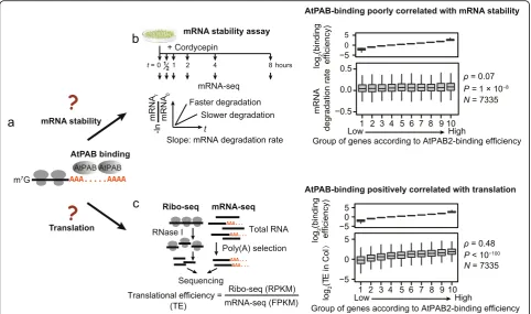
Does AtPAB binding affect mRNA stability and transla-tional efficiency [7]? To address these questions (Fig. 4a), we estimated mRNA stability and translational efficiency in the 2-week-old seedlings of the wild-type Col and calcu-lated their respective correlations with the AtPAB-binding efficiency among genes. We estimated the stability of an mRNA from the rate of change in abundance throughout the time course after the transcriptional inhibitor cordyce-pin was added (Fig.4b; Additional file2: Tables S7 and S8) [35, 36]. We estimated translational efficiency using ribo-some profiling (ribo-seq) that sequenced riboribo-some- ribosome-protected fragments [37,38]. Reads from ribo-seq showed a clear 3-nucleotide periodicity (Additional file 1: Figure S9a), indicating that the ribosome-protected fragments Fig. 3G-content contributes to the binding variance of AtPABs.aSchematic of the poly(A)-seq experiment that directly sequences the poly(A) tails of mRNAs. High-quality reads with recognizable Illumina 3′-adaptor sequences were used for further analysis.bExample reads from the poly(A)-seq show different propensities for AtPAB binding among genes. Yeast Pab1p and human PABP require at least 12 and 11 consecutive A’s for binding, respectively. Guanosines can inhibit AtPAB binding by cutting the poly(A) tail into fragments with < 12 consecutive A’s. Ref represents the referenceArabidopsisgenome.cThe G% was negatively correlated with the AtPAB-binding efficiency of a gene. Genes were divided into ten equal-size groups according to the average G% in the poly(A) tails.Pvalues were given by the Spearman’s correlation.d
[image:5.595.59.542.89.381.2]were successfully captured. After normalized by its mRNA level, the abundance of ribosome-protected fragments reflected the translational efficiency of a gene (Fig.4c; Add-itional file2: Tables S8, S9, and S10). We found that the AtPAB-binding efficiency was marginally correlated with mRNA degradation rate (ρ= 0.07,P= 1 × 10⁻⁸, AtPAB2 in Fig. 4b; AtPAB4 and AtPAB8 in Additional file 1: Figure S9b, c) but was positively correlated with translational effi-ciency (ρ= 0.48, P< 10−100, Fig. 4c; AtPAB4 and AtPAB8 in Additional file 1: Figure S9d, e), suggesting the major consequence of PAB binding in translational enhancement. Considering that G% is a determinant of
AtPAB-binding efficiency (Fig. 3c, d), we ask if G% in the
poly(A) tail contributes to the variation in translational efficiency among genes. To this end, we built linear models and used Akaike information criterion (AIC) to estimate the relative quality of models; a lower AIC value represents a better model. We found that integrat-ing G% into the linear model predictintegrat-ing translational
efficiency significantly reduced AIC (Table 1, model 1
vs. model 2). Genome-wide associations are sometimes confounded by other factors; therefore, we built a set of linear models that include the mRNA level and/or the poly(A) length as covariants. Including G% into the
models reduced AIC in all scenarios (Table1, models 3–
8), indicating the inhibitory role of guanosines in the
regulation of translation efficiency, likely through AtPAB binding.
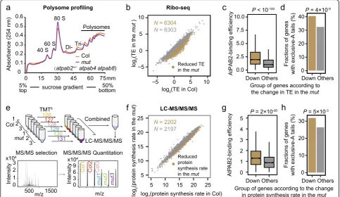
To further test the role of AtPABs in enhancing trans-lation, we compared polysome profiles between the
wild-type Col and the mut seedlings at the 2-week-old
[image:6.595.57.540.88.373.2]stage. The latter exhibited a reduced abundance of poly-somes (Fig. 5a; Additional file 1: Figure S10a). We also performed polysome profiling using the 6-week-old plants ofatpab2 atpab4(Additional file 1: Figure S10b), which displayed more severe developmental defects; consistently, a greater decrease in polysome abundance Fig. 4AtPAB-binding efficiency positively correlates with translational efficiency.aThe possible consequences of AtPAB binding to mRNAs.bThe AtPAB-binding efficiency was poorly correlated with the mRNA degradation rate in Col.cThe AtPAB-binding efficiency was positively correlated with the translational efficiency (TE) in Col
Table 1Models on features that predict translational efficiency (TE)
Model AIC
1 Null model 15,773
2 TE ~ G% 15,770
3 TE ~ mRNA level1 15,031
4 TE ~ mRNA level + G% 14,968
5 TE ~ poly(A)-tail length2 15,773
6 TE ~ poly(A)-tail length + G% 15,769
7 TE ~ mRNA level + poly(A)-tail length 15,033
8 TE ~ mRNA level + poly(A)-tail length + G% 14,968
1
The mRNA level of each gene in Col
2
[image:6.595.304.540.589.715.2]was observed (Additional file 1: Figure S10b). Both observations suggest a global reduction in translational efficiency when AtPABs are in short supply.
Further-more, we performed ribo-seq in the mut and identified
6304 genes with a reduction in translational efficiency (Fig.5b). These genes exhibited significantly higher AtPAB-binding efficiencies (P< 10−100, Fig. 5c; Additional file 1: Figure S10c) and higher fractions of exclusive-A tails (Fig. 5d,P= 4 × 10⁻⁹, Fisher’s exact test), indicating that AtPABs likely promote translation through the binding to the poly(A) tails of the respective genes.
To determine if the difference in AtPAB-binding ef-ficiency affects protein levels, we performed the Tan-dem Mass Tag (TMT)-based quantitative proteomics
on the 2-week-old seedlings of Col and the mut,
re-spectively (Fig. 5e). Genes with reduced protein
syn-thesis rate per mRNA in the mut showed higher
AtPAB-binding efficiencies (Fig. 5f, g; Additional file 1: Figure S10d) and higher fractions of exclusive-A tails
(Fig. 5h, P= 0.005), again indicating the role of the
“purity” of poly(A) tails and AtPAB binding in enhan-cing protein synthesis.
Discussion
The poly(A) tail is a hallmark of the eukaryotic messen-ger RNA [1,2]. It has been widely accepted that poly(A) tails are exclusively adenosines and regulate mRNA stability through the number of adenosines in the tail and probably the number of PABs bound to them. Re-cent studies reported that non-A nucleosides (especially guanosine) existed in the poly(A) tails of human cells [16–19]. In this study, we echoed in plants that guano-sine was prevalent in poly(A) tails and furthermore demonstrated the role of G-content in regulating trans-lational efficiency through AtPAB binding. Our study showcases the power of the next-generation genome-biology tools in understanding the basic principles underlying the central dogma.
We did not observe a significant effect of AtPAB-binding efficiency on mRNA stability, likely because the regulation of mRNA stability by its tail is a complex and dynamic process. On the one hand, PABs can protect mRNA from degradation by preventing deadenylase complex from poly(A) tails [39–41]. On the other hand, guanosines in poly(A) tails may slow down its trimming Fig. 5AtPABs enhance translational efficiency.aPolysome profiling shows the global reduction in TE in themut. Thex-axis indicates the
[image:7.595.56.540.89.369.2]process mediated by deadenylase and consequently in-hibit mRNA degradation [19]. Since guanosines nega-tively regulate PAB binding to the poly(A) tail, these two opposing effects could cancel out, leading to a net negotiable effect of PAB binding on the stability
of mRNA (Fig. 4b).
We identified the role of AtPABs in translational en-hancement. It could be related to the protein complex that PAB forms with translational initiation factors eIF4G and eIF4E. Such complex leads to a head-to-tail
looping structure of mRNA [3, 42], which may
promote translation through facilitating ribosome
re-cycling [27, 29, 42]. More experiments are required to
fully understand the mechanism.
The poly(A) tails are not static; rather, they are dynamically regulated during development and cell cycle [43–45]. For example, during the oocyte-to-embryo
transformation in Drosophila, changes in the length of
poly(A) tails were reported [43, 44]. Dramatic changes
were also observed in the length of poly(A) tails during human somatic cell cycles [45]. It remains unclear whether the composition of the poly(A) tail, especially G%, is also variable in different cell states and contrib-utes to translational regulation. It will be interesting to analyze the composition of poly(A) tail in various condi-tions, such as tissues, developmental stages, or environ-ments. Such work will lead to a future direction for the ultimate understanding of poly(A)-tail-mediated regula-tion of gene expression [46–48].
Conclusions
The Arabidopsis thaliana pab mutants exhibit various levels of phenotypic abnormality. Analysis of the RNA-binding landscape revealed a wide variation in AtPAB-binding efficiency among genes, which can be partly explained by the G% in the poly(A) tail. AtPAB binding enhances translation; genes with stronger binding to AtPABs exhibit more reduction in translational
effi-ciency in theatpab mutant. These observations indicate
that AtPABs can precisely enhance the translational efficiency of the genes with more uniform poly(A) tails.
Methods
Plasmid construction
The reporters for AtPABs were constructed in the back-ground of a vector containing the GFP-HA tag (pCAM-BIA1300 backbone). The promoters (2.0-kb regions upstream of the transcription start site) and genomic re-gions between the transcription start site and the site right before the stop codon were amplified with primers
(cx8366 and cx8367 forAtPAB2; cx8370 and cx8371 for
AtPAB4; cx8374 and cx8375 for AtPAB8) and were inserted before the GFP tag. The 3′-untranslated regions (UTRs) and terminator fragments (0.5 kb downstream to
the transcription end site) were obtained by PCR
ampli-fication with primers (cx8368 and cx8369 for AtPAB2;
cx8372 and cx8373 forAtPAB4; cx8376 and cx8377 for
AtPAB8) and were inserted after the HA tag. Primers are listed in Additional file2: Table S11.
Plant materials
All Arabidopsis thaliana materials used in this study
were in the Columbia (Col-0) background. Theatpab2-1
(SALK_026293),atpab4-2(SAIL_740_D08), and
atpab8-1 (SALK_022160) mutants were obtained from the
Arabidopsis Biological Resources Center at Ohio State (https://abrc.osu.edu/). The primers used for genotyping are listed in Additional file2: Table S11.
Seeds were sterilized in 75% ethanol for 1 min and treated with 10% bleach for 15 min. Seeds were sown on Murashige and Skoog (MS) media (0.43 g/L MS salts, 3 g/L sucrose, 0.8% agar, pH 5.8) and were treated at 4 °C for 3 days. Seeds were further grown at 23 °C under long-day conditions (cool-white fluorescent light on for 16 h and off for 8 h). Whole seedlings were harvested at 2 weeks for CLIP-seq, ribo-seq, poly(A)-seq, RNA stability assay, and mass spectrometry.
The transgenic plants AtPAB2-GFP-HA atpab2
atpab8, AtPAB8-GFP-HA atpab2 atpab8, and AtPAB4-GFP-HA atpab2 atpab4were generated by transforming AtPAB reporters into the double mutants with the floral
dipping method [49]. MS plates containing 25μg/mL
hygromycin were used for the selection of transgenic
plants. For each AtPAB, multiple single-copy insertion
lines were obtained (Additional file1: Figure S3c). Plants were planted on soil mix (2/3 vermiculite and 1/3 nutrient soil) under long-day conditions for pheno-typing. The total leaf number (including both rosette and cauline leaves) was used for the quantification of flowering time.
Microscopy
To identify phenotypic abnormality in embryos, seeds were cleared in Herr’s solution (lactic acid:chloral hy-drate:phenol:clove oil:xylene, 2:2:2:2:1, w/w) for 5–30 min until the embryonic morphology was clear under the microscope (Olympus BX51) in the differential inter-ference contrast mode.
EMSA
We followed the protocol of EMSA/Gel-Shift Kit (Beyo-time, GS002) with modifications. The oligo-RNAs were synthesized chemically by Genscript (Nanjing, China).
Oligo(A)-RNA was labeled with γ-32P-ATP by T4
poly-nucleotide kinase (T4 PNK, NEB, M0201) in the
reac-tion mix (1000 Ci γ-32P-ATP, 3.5 pmol oligo(A)-RNA,
probe (~ 0.035 pmol) was incubated with ~ 100 ng
AtPAB protein at 25 °C for 10 min in a 10-μL reaction
system. The reaction mix contains 4% glycerol, 1 mM MgCl2, 0.5 mM EDTA, 0.5 mM DTT, 50 mM NaCl, 10 mM Tris-HCl (pH 7.5), and 0.05 mg/mL poly
(dI-dC)•poly (dI-dC). The 32
P-labeled oligo(A)-RNA was added together with the unlabeled competing oligo-RNA. The reaction mix was separated by native poly-acrylamide gel (1 × Tris-glycine buffer, 5% glycerol, 6% polyacrylamide) electrophoresis (PAGE) in the 2 × Tris-glycine buffer at 80 V for 1 h. 1 × Tris-Tris-glycine buffer con-tains 25 mM Tris (pH 8.5), 190 mM glycine, and 1 mM EDTA. The gel was wrapped in plastic film and exposed to a storage phosphor screen (GE Healthcare) overnight, and signals were read with a Typhoon TRIO scanner (GE Healthcare).
CLIP-seq and RIP-RT-PCR
CLIP-seq was performed as described previously [33]. In brief, to cross-link RNA and protein, 2-week-old seedlings were soaked in ice-cold PBS buffer and were UV-treated twice, each with the irradiation intensity at
600 mJ/cm2 (Hoefer UVC 500 Ultraviolet Crosslinker,
GE). The RNA-AtPAB-GFP-HA complexes were
enriched from the lysate by immunoprecipitation using anti-HA antibody (Sigma, H6908) and were partially digested by micrococcal nuclease (3 × 10⁻⁵U/μL, Fer-mentas, EN0181). The digested RNA was ligated to the 3′-RNA adaptor (P-UGGAAUUCUCGGGUGCCAAG GUidT) and was labeled by32P at the 5′-end. The radio-active RNA-protein complexes (RNP) were separated by SDS-PAGE and were further transferred to nitrocellulose membrane. Membrane pieces corresponding to the
RNP-containing radioactive regions (Additional file 1:
Figure S5b, c) were selected to recover RNA fragments. RNPs were treated with Proteinase K, and the purified RNA was ligated to the 5′-RNA adaptor (GUUCAG AGUUCUACAGUCCGACGAUCNNNN).
The immunoprecipitation in RIP-RT-PCR was same as that in CLIP-seq, but the RNA-protein complexes were not digested by micrococcal nuclease. qPCR was per-formed on four genes to quantify the mRNA abundance in a sample. The AtPAB2-binding efficiency of a gene was estimated from its mRNA abundance in the AtPAB2-IP product, normalized by its abundance in the total mRNA. Primers for RIP-RT-PCR are listed in Additional file2: Table S11.
Poly(A)-seq
Total RNA was extracted from the 2-week-old seedlings of Col. Five micrograms of total RNA was treated with RQ1 DNase (Promega, M6101) and was partially digested by RNase T1 (Thermo, EN0541), an endoribo-nuclease that specifically digests single-stranded RNA at
the 3′-side of the G-residue. Poly(A)-containing RNA was purified with oligo (dT)-conjugated magnetic beads (Invitrogen, 61005) and ligated to the 3′-RNA adaptor (TGGAATTCTCGGGTGCCAAGG). The cDNA was generated using the ScriptSeq v2 RNA-Seq Library Prep-aration Kit (Illumina, SSV21124). PCR products of 200– 500 bp were purified and were applied to HiSeq X ten system for 150-nt paired-end sequencing.
mRNA stability assay
Two-week-old seedlings (0.1 g) were transferred to 2 mL incubation buffer (1 mM PIPES, pH 6.25, 1 mM sodium citrate, 1 mM KCl, 15 mM sucrose) in a 24-well plate. Cordycepin was added to a final concentration of 0.5 mM. The plate was rotated for 75 rpm at 23 °C. The seedlings were harvested at 0, 0.5, 1, 2, 4, and 8 h and quickly frozen in liquid nitrogen. Total RNA was isolated with TRIzol (Invitrogen, 15596026). The mRNA-seq libraries were constructed using the strand-specific protocol for Illumina sequencing and were sequenced with the HiSeq X ten system in the paired-end 150-nt mode.
Ribo-seq
The protocol was modified from a previous study [38]. In brief, ribosomes were extracted from 5-g 2-week-old seedlings with the polysome extraction buffer (PEB), which contains 200 mM Tris-HCl (pH 9.0), 200 mM KCl, 25 mM EGTA, 35 mM MgCl2, 1% (w/v) polyox-yethylene(23) lauryl ether (Brij-35), 1% (v/v) Triton X-100, 1% (v/v) octylphenyl-polyethylene glycol (Igepal CA 630), 1% (v/v) polyoxyethylene sorbitan monolaurate 20 (Tween 20), 1% (v/v) polyoxyethylene(10) tridecyl ether,
5 mM DTT, 1 mM PMSF, 50μg/mL cycloheximide, and
50μg/mL chloramphenicol. The extracted ribosomes
were pelleted through a 30-mL sucrose cushion contain-ing 400 mM Tris-HCl (pH 9.0), 200 mM KCl, 5 mM
EGTA, 30 mM MgCl2, 1.75 M sucrose, 5 mM DTT,
50μg/mL cycloheximide, and 50μg/mL
chlorampheni-col, by ultracentrifugation at 4 °C overnight (33,500 rpm, Beckman, 70Ti rotor). Ribosome-protected mRNA frag-ments were generated by RNase I (Ambion, AM2294) digestion and were applied to small RNA library con-struction for Illumina sequencing (Gnomegen, k02420).
Polysome profiling
Mass spectrometry
Two-week-old Arabidopsis seedlings (~ 2 g each) were
ground in the liquid nitrogen, and the total protein was extracted with trichloroacetate. One hundred fifty mi-crograms of protein of each sample was run into the SDS-PAGE as gel plug and digested with trypsin at 37 °C overnight. Seventy-five micrograms of protein of each sample was labeled with TMT 6plex reagent (Thermo) and combined after labeling and dried.
Samples were desalted with a SPEC C18 column and
solubilized in 200μL buffer A (20 mM ammonium
formate, pH 10) and separated on an Xbridge column
(Waters; C18; 3.5μm, 2.1 × 150 mm) using the Agilent
HP1100. Fractions were collected at 1-min intervals and
dried under vacuum. The LC-MS/MS/MS (liquid
chromatography-tandem mass spectrometry) analysis was performed using a Dionex rapid-separation liquid chromatography system interfaced with an Orbitrap
Fusion™Lumos™mass spectrometer (Thermo Scientific).
Bioinformatics analyses
Analyses of mRNA-seq data
The 3-nt random sequence at the 5′-end of the clean
reads was removed. The trimmed clean reads were
aligned to the Arabidopsis genome (TAIR10) using
TopHat2 (2.0.11) [50] with no more than five mis-matches. The expression level of each gene was esti-mated from the uniquely mapped fragments using HTSeq (0.6.0) [51] and was normalized to the unit of fragments per kilobase of transcript per million mapped fragments (FPKM). Two biological replicates were highly correlated (r> 0.99, P< 10−100, Pearson’s correlation, Additional file2: Table S8).
Analyses of CLIP-seq data
Single-end clean reads with identical sequences (including the 4-nt random sequence in the 5′-RNA adaptor) were defined as PCR duplicates and were reduced to 1 read. The 4-nt random sequence at the 5′-end and the 3′-adaptor sequence of Illumina were removed using the FASTX toolkit (0.0.14) (http://hannonlab.cshl.edu/fastx_ toolkit). The 3′-end A-tracts in each read were removed, and the trimmed reads of less than 18 nt in length were
discarded. The remaining reads were mapped to the
Ara-bidopsis genome (TAIR10) using NovoAlign (3.07.01)
(Novocraft, http://www.novocraft.com/), allowing up to
two mismatches. Uniquely mapped reads were used to identify AtPAB binding clusters using the Pyicos toolkit (2.0.7) [34]. The AtPAB-binding efficiency of a gene was calculated as the ratio between the number of reads (per million mapped reads) in the CLIP-seq and that of fragments (FPKM) in the mRNA-seq of the wild-type.
Analyses of poly(A)-seq data
To avoid the problem of polymerase slip on a homopol-ymeric tract, we only used high-quality 5′-sequencing
reads with recognizable 3′-adaptor sequences of
Illumina sequencing for analyses. The 3-nt random sequence at the 5′-end and 3′-adaptor sequence of Illu-mina were removed. The poly(A) tail was defined as a stretch of A’s at the 3′-end of a read, allowing up to 5 interspersed G bases. We also required at least 5 A’s at the 5′-end of a poly(A) tail. The poly(A)-tail sequences were removed, and trimmed reads of at least 18 nt in
length were aligned to the Arabidopsis genome
(TAIR10) using Bowtie2 (2.3.4.1) [52], allowing up to two mismatches. Consequently, the sequences of a poly(A) tail and the gene it belongs to were identified. We confirmed manually that the majority of the G’s (500 out of the 500 manually checked G’s) in the identi-fied poly(A) tails could not be explained by the G’s in the 3′-UTR. For each read, G% in the poly(A) tail was estimated as the number of G’s divided by the length of the poly(A) tail. The G% of a gene was calculated as the total number of G’s divided by the total length of poly(A) tails. The poly(A)-tail length of each gene was estimated with the median length of all its poly(A) tails.
Analyses of mRNA stability assay data
Clean mRNA-seq reads were aligned to the Arabidopsis
genome (TAIR10) using TopHat2 (2.0.11) [50], allowing up to two mismatches. The expression level of each gene was estimated using Cufflinks (2.2.1) [53], in the unit of fragments per kilobase of transcript per million mapped fragments (FPKM). The mRNA degradation rate of each gene was defined as the slope of the regression line in
the linear model ln (mRNA0/mRNAt) ~ t. Three
bio-logical replicates were highly correlated (r> 0.9, P< 10−100, Pearson’s correlation, Additional file2: Table S8), and therefore, the average mRNA degradation rate was used for further analyses.
Analyses of ribo-seq data
Reads with identical sequences (including the 4-nt ran-dom sequence at the 5′-end) were likely PCR duplicates and were reduced to 1 read. The 3′-end adaptors and 5′-end 4-nt barcodes were removed. Reads were mapped
to the Arabidopsis genome (TAIR10) using TopHat2
efficiency was highly reproducible among replicates (Additional file2: Table S8).
Analysis of mass spectrometry data
All data were analyzed with MaxQuant (version 1.6.2.6) with Andromeda search engine [54]. The type of LC-MS run was set to reporter ion MS3 with 6plex TMT as iso-baric labels. LC-MS data were searched against TAIR10 database with the addition of potential contaminants.
Additional files
Additional file 1:Figure S1.AtPAB2,AtPAB4, andAtPAB8are
constitutively expressed in Arabidopsis.Figure S2.The mutants ofAtPABs
exhibited severe phenotypes.Figure S3.The expression ofAtPAB-GFP-HA
rescued the developmental defects ofatpabdouble mutants.Figure S4.
AtPAB-GFP-HA reporters were ubiquitously and constitutively expressed in the cytoplasm during seed development.Figure S5.Identification of AtPAB-binding targets.Figure S6.The AtPAB-binding genes detected in CLIP-seq were validated by RIP-RT-PCR.Figure S7.AtPABs bind the poly(A) tails of mRNAs with different efficiencies.Figure S8.G% in the poly(A) tail is a determinant of the AtPAB-binding efficiency.Figure S9.
[image:11.595.303.539.387.737.2]The major consequence of AtPAB-binding is translational enhancement.
Figure S10.AtPAB binding enhances translation efficiency. (PDF 1987 kb)
Additional file 2:Table S1.Progeny segregation from the self-cross of themut(atpab2+/−atpab4 atpab8plants).Table S2.Number of normal and aborted seeds in the maturemutsiliques.Table S3.Number of embryos at different developmental stages in each Col silique.Table S4.
Number of embryos at different developmental stages in eachmut
silique.Table S5.Summary of the CLIP-seq reads.Table S6.Summary of the poly(A)-seq reads.Table S7.Summary of reads of the mRNA stability assay.Table S8.Correlations between biological replicates.Table S9.
Summary of the ribo-seq reads.Table S10.Summary of the mRNA-seq reads.Table S11.List of primer sequences. (PDF 474 kb)
Acknowledgements
We thank Dr. Lianfeng Gu (Fujian Agriculture and Forestry University, China) for the technical help in the bioinformatics analysis.
Authors’contributions
TZ, QH, CL, WQ, and XC designed the experiments. TZ and CL performed the CLIP-seq. TZ performed the mRNA stability assay, ribo-seq, and genetic and molecular biological experiments. TZ and YZ performed the poly(A)-seq. TZ, QH, JS, XY, and IMS performed the data analyses. TZ and XH performed the embryonic lethal analysis. TZ, JS, CL, QH, WQ, and XC wrote the manuscript. BDG and C-ML assisted in writing the manuscript and discussion. All authors read and approved the final manuscript.
Funding
This work was supported by grants from the National Natural Science Foundation of China (31788103 and 91540203 to X.C., 31770874 to C.L., 31900455 to T.Z., 31571308 to W.Q., and 31701096 to J.S.), the National Basic Research Program of China 2014CB943400 to C.L., the Strategic Priority Research Program of Chinese Academy of Sciences (XDPB0403 and XDB27030201 to X.C. and XDA08020303 to W.Q.), the Key Research Program of Frontier Sciences of Chinese Academy of Sciences (QYZDY-SSW-SMC022 to X.C.), and the State Key Laboratory of Plant Genomics.
Availability of data and materials
TheArabidopsisgenome (TAIR10) was retrieved fromhttps://www.arabidopsis. org/[55]. The mRNA level ofAtPABsin multiple tissues was calculated with the data obtained from Expression Altas (https://www.ebi.ac.uk/gxa/home) [56]. Paralogous gene pairs inArabidopsiswere retrieved from Ensembl plants with BioMarts [57]. High-throughput sequencing data of CLIP-seq, ribo-seq, mRNA-seq, poly(A)-mRNA-seq, and RNA-stability-seq have been deposited to the Gene Ex-pression Omnibus database under accession number GSE110342 [58]. The mass
spectrometry proteomics data have been deposited to the ProteomeXchange Consortium via the PRIDE [59,60] partner repository with the dataset identifier PXD014071 [61]. Codes to analyze the data and to generate figures are available at GitHub [62] and Zenodo [63].
Ethics approval and consent to participate
Not applicable.
Consent for publication
Not applicable.
Competing interests
The authors declare that they have no competing interests.
Author details
1State Key Laboratory of Plant Genomics and National Center for Plant Gene
Research, Institute of Genetics and Developmental Biology, Chinese Academy of Sciences, Beijing 100101, China.2Key Laboratory of Genetic
Network Biology, Institute of Genetics and Developmental Biology, Chinese Academy of Sciences, Beijing 100101, China.3University of Chinese Academy
of Sciences, Beijing 100049, China.4Key Laboratory of Plant Molecular Physiology, Institute of Botany, Chinese Academy of Sciences, Beijing 100093, China.5Department of Biology, University of Pennsylvania, Philadelphia, PA 19104, USA.6Laboratory for Genome Regulation and Human Health and
Center for Genome Analysis, ABLife Inc, Wuhan 430075, Hubei, China.7CAS Center for Excellence in Molecular Plant Sciences, Institute of Genetics and Developmental Biology, Chinese Academy of Sciences, Beijing 100101, China.
Received: 12 December 2018 Accepted: 22 August 2019
References
1. Hudson WH, Ortlund EA. The structure, function and evolution of proteins that bind DNA and RNA. Nat Rev Mol Cell Biol. 2014;15:749–60. 2. Rissland OS. The organization and regulation of mRNA-protein complexes.
Wiley Interdiscip Rev RNA. 2017;8:e1369.
3. Gorgoni B, Gray NK. The roles of cytoplasmic poly(A)-binding proteins in regulating gene expression: a developmental perspective. Brief Funct Genomic Proteomic. 2004;3:125–41.
4. Kuhn U, Wahle E. Structure and function of poly(A) binding proteins. Biochim Biophys Acta. 2004;1678:67–84.
5. Kwan SW, Brawerman G. A particle associated with the polyadenylate segment in mammalian messenger RNA. Proc Natl Acad Sci U S A. 1972;69:3247–50. 6. Blobel G. Protein tightly bound to globin mRNA. Biochem Biophys Res
Commun. 1972;47:88–95.
7. Smith RW, Blee TK, Gray NK. Poly(A)-binding proteins are required for diverse biological processes in metazoans. Biochem Soc Trans. 2014;42:1229–37. 8. Belostotsky DA. Unexpected complexity of poly(A)-binding protein gene families
in flowering plants: three conserved lineages that are at least 200 million years old and possible auto- and cross-regulation. Genetics. 2003;163:311–9. 9. Dufresne PJ, Ubalijoro E, Fortin MG, Laliberté JF. Arabidopsis thaliana class II
poly (A)-binding proteins are required for efficient multiplication of turnip mosaic virus. J Gen Virol. 2008;89:2339–48.
10. Gallie DR. Class II members of the poly(A) binding protein family exhibit distinct functions during Arabidopsis growth and development. Translation. 2017;5:e1295129.
11. Li W, Ma M, Feng Y, Li H, Wang Y, Ma Y, Li M, An F, Guo H. EIN2-directed translational regulation of ethylene signaling in Arabidopsis. Cell. 2015;163:670–83. 12. Xu G, Greene GH, Yoo H, Liu L, Marques J, Motley J, Dong X. Global
translational reprogramming is a fundamental layer of immune regulation in plants. Nature. 2017;545:487–90.
13. Blobel G. A protein of molecular weight 78,000 bound to the polyadenylate region of eukaryotic messenger RNAs. Proc Natl Acad Sci U S A. 1973;70: 924-8.
14. Spencer WC, Zeller G, Watson JD, Henz SR, Watkins KL, McWhirter RD, Petersen S, Sreedharan VT, Widmer C, Jo J, et al. A spatial and temporal map of C. elegans gene expression. Genome Res. 2011;21:325–41. 15. Roy PJ, Stuart JM, Lund J, Kim SK. Chromosomal clustering of
muscle-expressed genes in Caenorhabditis elegans. Nature. 2002;418:975–9. 16. Chang H, Lim J, Ha M, Kim VN. TAIL-seq: genome-wide determination of
17. Rissland OS, Mikulasova A, Norbury CJ. Efficient RNA polyuridylation by noncanonical poly(A) polymerases. Mol Cell Biol. 2007;27:3612–24. 18. Sement FM, Ferrier E, Zuber H, Merret R, Alioua M, Deragon JM,
Bousquet-Antonelli C, Lange H, Gagliardi D. Uridylation prevents 3′trimming of oligoadenylated mRNAs. Nucleic Acids Res. 2013;41:7115–27.
19. Lim J, Kim D, Lee YS, Ha M, Lee M, Yeo J, Chang H, Song J, Ahn K, Kim VN. Mixed tailing by TENT4A and TENT4B shields mRNA from rapid deadenylation. Science. 2018;361:701–4.
20. Deo RC, Bonanno JB, Sonenberg N, Burley SK. Recognition of polyadenylate RNA by the poly(A)-binding protein. Cell. 1999;98:835–45.
21. Bollig F, Winzen R, Gaestel M, Kostka S, Resch K, Holtmann H. Affinity purification of ARE-binding proteins identifies polyA-binding protein 1 as a potential substrate in MK2-induced mRNA stabilization. Biochem Biophys Res Commun. 2003;301:665–70.
22. Sladic RT, Lagnado CA, Bagley CJ, Goodall GJ. Human PABP binds AU-rich RNA via RNA-binding domains 3 and 4. Eur J Biochem. 2004;271:450–7. 23. Sachs AB, Davis RW, Kornberg RD. A single domain of yeast poly(A)-binding
protein is necessary and sufficient for RNA binding and cell viability. Mol Cell Biol. 1987;7:3268–76.
24. Goss DJ, Kleiman FE. Poly(A) binding proteins: are they all created equal? Wiley Interdiscip Rev RNA. 2013;4:167–79.
25. Brook M, Gray NK. The role of mammalian poly(A)-binding proteins in co-ordinating mRNA turnover. Biochem Soc Trans. 2012;40:856–64. 26. Bernstein P, Peltz SW, Ross J. The poly (A)-poly (A)-binding protein complex is
a major determinant of mRNA stability in vitro. Mol Cell Biol. 1989;9:659–70. 27. Tarun S, Sachs AB. A common function for mRNA 5'and 3'ends in
translation initiation in yeast. Genes Dev. 1995;9:2997–3007.
28. Munroe D, Jacobson A. Tales of poly (A): a review. Gene. 1990;91:151–8. 29. Kahvejian A, Svitkin YV, Sukarieh R, M'Boutchou MN, Sonenberg N.
Mammalian poly(A)-binding protein is a eukaryotic translation initiation factor, which acts via multiple mechanisms. Genes Dev. 2005;19:104–13. 30. Tarun SZ Jr, Wells SE, Deardorff JA, Sachs AB. Translation initiation factor eIF4G mediates in vitro poly(A) tail-dependent translation. Proc Natl Acad Sci U S A. 1997;94:9046–51.
31. Wakiyama M, Imataka H, Sonenberg N. Interaction of eIF4G with poly(A)-binding protein stimulates translation and is critical for Xenopus oocyte maturation. Curr Biol. 2000;10:1147–50.
32. Gray NK, Coller JM, Dickson KS, Wickens M. Multiple portions of poly (A)-binding protein stimulate translation in vivo. EMBO J. 2000;19:4723–33. 33. Zhang Y, Gu L, Hou Y, Wang L, Deng X, Hang R, Chen D, Zhang X, Zhang Y,
Liu C, Cao X. Integrative genome-wide analysis reveals HLP1, a novel RNA-binding protein, regulates plant flowering by targeting alternative polyadenylation. Cell Res. 2015;25:864–76.
34. Althammer S, Gonzalez-Vallinas J, Ballare C, Beato M, Eyras E. Pyicos: a versatile toolkit for the analysis of high-throughput sequencing data. Bioinformatics. 2011;27:3333–40.
35. Arae T, Isai S, Sakai A, Mineta K, Yokota Hirai M, Suzuki Y, Kanaya S, Yamaguchi J, Naito S, Chiba Y. Co-ordinated regulations of mRNA synthesis and decay during cold acclimation in Arabidopsis cells. Plant Cell Physiol. 2017;58:1090–102.
36. Gutierrez RA, Ewing RM, Cherry JM, Green PJ. Identification of unstable transcripts in Arabidopsis by cDNA microarray analysis: rapid decay is associated with a group of touch- and specific clock-controlled genes. Proc Natl Acad Sci U S A. 2002;99:11513–8.
37. Ingolia NT, Ghaemmaghami S, Newman JR, Weissman JS. Genome-wide analysis in vivo of translation with nucleotide resolution using ribosome profiling. Science. 2009;324:218–23.
38. Juntawong P, Girke T, Bazin J, Bailey-Serres J. Translational dynamics revealed by genome-wide profiling of ribosome footprints in Arabidopsis. Proc Natl Acad Sci U S A. 2014;111:E203–12.
39. Lin J, Fabian M, Sonenberg N, Meller A. Nanopore detachment kinetics of poly (A) binding proteins from RNA molecules reveals the critical role of C-terminus interactions. Biophys J. 2012;102:1427–34.
40. Körner CG, Wormington M, Muckenthaler M, Schneider S, Dehlin E, Wahle E. The deadenylating nuclease (DAN) is involved in poly (A) tail removal during the meiotic maturation of Xenopus oocytes. EMBO J. 1998;17:5427–37. 41. Wang Z, Day N, Trifillis P, Kiledjian M. An mRNA stability complex functions
with poly (A)-binding protein to stabilize mRNA in vitro. Mol Cell Biol. 1999; 19:4552–60.
42. Jackson RJ, Hellen CU, Pestova TV. The mechanism of eukaryotic translation initiation and principles of its regulation. Nat Rev Mol Cell Biol. 2010;11:113–27.
43. Eichhorn SW, Subtelny AO, Kronja I, Kwasnieski JC, Orr-Weaver TL, Bartel DP. mRNA poly(A)-tail changes specified by deadenylation broadly reshape translation in Drosophila oocytes and early embryos. eLife. 2016;30:16955. 44. Lim J, Lee M, Son A, Chang H, Kim VN. mTAIL-seq reveals dynamic poly(A) tail
regulation in oocyte-to-embryo development. Genes Dev. 2016;30:1671–82. 45. Park JE, Yi H, Kim Y, Chang H, Kim VN. Regulation of poly(A) tail and
translation during the somatic cell cycle. Mol Cell. 2016;62:462–71. 46. Kappel C, Trost G, Czesnick H, Ramming A, Kolbe B, Vi SL, Bispo C, Becker
JD, de Moor C, Lenhard M. Genome-wide analysis of PAPS1-dependent polyadenylation identifies novel roles for functionally specialized poly(A) polymerases in Arabidopsis thaliana. PLoS Genet. 2015;11:e1005474. 47. Eckmann CR, Rammelt C, Wahle E. Control of poly(A) tail length. Wiley
Interdiscip Rev RNA. 2011;2:348–61.
48. Subtelny AO, Eichhorn SW, Chen GR, Sive H, Bartel DP. Poly(A)-tail profiling reveals an embryonic switch in translational control. Nature. 2014;508:66–71. 49. Clough SJ, Bent AF. Floral dip: a simplified method for
Agrobacterium-mediated transformation of Arabidopsis thaliana. Plant J. 1998;16:735–43. 50. Kim D, Pertea G, Trapnell C, Pimentel H, Kelley R, Salzberg SL. TopHat2:
accurate alignment of transcriptomes in the presence of insertions, deletions and gene fusions. Genome Biol. 2013;14:R36.
51. Anders S, Pyl PT, Huber W. HTSeq--a Python framework to work with high-throughput sequencing data. Bioinformatics. 2015;31:166–9.
52. Langmead B, Salzberg SL. Fast gapped-read alignment with Bowtie 2. Nat Methods. 2012;9:357–9.
53. Trapnell C, Williams BA, Pertea G, Mortazavi A, Kwan G, van Baren MJ, Salzberg SL, Wold BJ, Pachter L. Transcript assembly and quantification by RNA-Seq reveals unannotated transcripts and isoform switching during cell differentiation. Nat Biotechnol. 2010;28:511–5.
54. Cox J, Matic I, Hilger M, Nagaraj N, Selbach M, Olsen JV, Mann M. A practical guide to the MaxQuant computational platform for SILAC-based quantitative proteomics. Nat Protoc. 2009;4:698–705.https://doi.org/10.1038/nprot.2009.36. 55. Lamesch P, Berardini TZ, Li D, Swarbreck D, Wilks C, Sasidharan R, Muller R,
Dreher K, Alexander DL, Garcia-Hernandez M, et al. The Arabidopsis Information Resource (TAIR): improved gene annotation and new tools. Nucleic Acids Res. 2012;40:D1202–10.https://doi.org/10.1093/nar/gkr1090. 56. Papatheodorou I, Fonseca NA, Keays M, Tang YA, Barrera E, Bazant W, Burke
M, Fullgrabe A, Fuentes AM, George N, et al. Expression Atlas: gene and protein expression across multiple studies and organisms. Nucleic Acids Res. 2018;46:D246–51.https://doi.org/10.1093/nar/gkx1158.
57. Kinsella RJ, Kahari A, Haider S, Zamora J, Proctor G, Spudich G, Almeida-King J, Staines D, Derwent P, Kerhornou A, et al. Ensembl BioMarts: a hub for data retrieval across taxonomic space. Database (Oxford). 2011;2011:bar030.
https://doi.org/10.1093/database/bar030.
58. Zhao T, Huan Q, Sun J, Liu C, Hou X, Yu X, Silverman I, Zhang Y, Gregory B, Liu CM, Qian W, Cao X. Impact of G-content on Arabidopsis PAB binding and their role in enhancing translational efficiency. Gene Expression Omnibus. 2019;
https://www.ncbi.nlm.nih.gov/geo/query/acc.cgi?acc=GSE110342.
59. Perez-Riverol Y, Csordas A, Bai J, Bernal-Llinares M, Hewapathirana S, Kundu DJ, Inuganti A, Griss J, Mayer G, Eisenacher M, et al. The PRIDE database and related tools and resources in 2019: improving support for quantification data. Nucleic Acids Res. 2019;47:D442–50.https://doi.org/10.1093/nar/gky1106. 60. Deutsch EW, Csordas A, Sun Z, Jarnuczak A, Perez-Riverol Y, Ternent T,
Campbell DS, Bernal-Llinares M, Okuda S, Kawano S, et al. The ProteomeXchange consortium in 2017: supporting the cultural change in proteomics public data deposition. Nucleic Acids Res. 2017;45:D1100–6.
https://doi.org/10.1093/nar/gkw936.
61. Zhao T, Huan Q, Sun J, Liu C, Hou X, Yu X, Silverman I, Zhang Y, Gregory B, Liu CM, Qian W, Cao X. Quantitative proteomics of Col and AtPABs mutant. ProteomeXchange Consortium. 2019;http://www.ebi.ac.uk/pride/archive/ projects/PXD014071.
62. Zhao T, Huan Q, Sun J, Liu C, Hou X, Yu X, Silverman I, Zhang Y, Gregory B, Liu CM, Qian W, Cao X. AtPABs. GitHub. 2019;https://github.com/ SophieHuan/AtPABs.
63. Zhao T, Huan Q, Sun J, Liu C, Hou X, Yu X, Silverman I, Zhang Y, Gregory B, Liu CM, Qian W, Cao X. AtPABs: impact of poly(A)-tail G-content on Arabidopsis PAB binding and their role in enhancing translational efficiency. Zenodo. 2019.https://doi.org/10.5281/zenodo.3368777.
Publisher’s Note