hicap:
In Silico
Serotyping of the
Haemophilus influenzae
Capsule Locus
Stephen C. Watts,
a,bKathryn E. Holt
a,b,caDepartment of Biochemistry and Molecular Biology, Bio21 Molecular Science and Biotechnology Institute, University of Melbourne, Melbourne, Victoria, Australia bDepartment of Infectious Diseases, Central Clinical School, Monash University, Melbourne, Victoria, Australia
cLondon School of Hygiene & Tropical Medicine, London, United Kingdom
ABSTRACT
Haemophilus influenzae
exclusively colonizes the human nasopharynx
and can cause a variety of respiratory infections as well as invasive diseases,
includ-ing meninclud-ingitis and sepsis. A key virulence determinant of
H. influenzae
is the
poly-saccharide capsule, of which six serotypes are known, each encoded by a distinct
variation of the capsule biosynthesis locus (
cap
-a to
cap
-f).
H. influenzae
type b (Hib)
was historically responsible for the majority of invasive
H. influenzae
disease, and its
prevalence has been markedly reduced in countries that have implemented
vaccina-tion programs targeting this serotype. In the postvaccine era, nontypeable
H.
influen-zae
emerged as the most dominant group causing disease, but in recent years a
re-surgence of encapsulated
H. influenzae
strains has also been observed, most notably
serotype a. Given the increasing incidence of encapsulated strains and the high
fre-quency of Hib in countries without vaccination programs, there is growing interest
in genomic epidemiology of
H. influenzae
. Here we present hicap, a software tool for
rapid
in silico
serotype prediction from
H. influenzae
genome sequences. hicap is
written using Python3 and is freely available at
https://github.com/scwatts/hicap
under
the GNU General Public License v3 (GPL3). To demonstrate the utility of hicap, we used
it to investigate the
cap
locus diversity and distribution in 691 high-quality
H. influenzae
genomes from GenBank. These analyses identified
cap
loci in 95 genomes and
con-firmed the general association of each serotype with a unique clonal lineage, and they
also identified occasional recombination between lineages that gave rise to hybrid
cap
loci (2% of encapsulated strains).
KEYWORDS
Haemophilus influenzae
, capsule, genomics, serotyping, surveillance
H
aemophilus influenzae
is a pleomorphic Gram-negative bacterium that is exclusive
to humans, typically colonizing the upper respiratory tract and occasionally
caus-ing disease. It was the first free livcaus-ing organism to be completely sequenced and served
as a stepping stone toward DNA sequencing technology development in preparation
for the Human Genome Project (1).
H. influenzae
is often classified on the basis of the
production and antigenicity of polysaccharide capsule. Strains that produce capsule are
divided into six serotypes (
H. influenzae
a to f [Hia to Hif]), and nonencapsulated strains
are designated nontypeable
H. influenzae
(NTHi) (2).
Biosynthesis of the polysaccharide capsule is controlled by the
cap
loci (
cap
-a to
cap
-f), each of which includes three contiguous but functionally distinct regions (I, II,
and III) (Fig. 1). Regions I and III are common to all six
cap
loci and are associated with
cellular transport (
bex
operon, region I) and posttranslational processing (
hcs
operon,
region III) (3, 4). Region II encodes several genes involved in polysaccharide
biosynthe-sis that are specific to each serotype (Fig. 1) (5–9). The
cap
locus is regularly subject to
duplication, deletion, and interruption (10, 11). For example the
cap
-b locus is often
duplicated, creating two tandem copies of the locus flanked by IS
1016
, and regularly
CitationWatts SC, Holt KE. 2019. hicap:in silico
serotyping of theHaemophilus influenzae capsule locus. J Clin Microbiol 57:e00190-19. https://doi.org/10.1128/JCM.00190-19.
EditorAlexander Mellmann, University
Hospital Münster
Copyright© 2019 Watts and Holt. This is an
open-access article distributed under the terms of theCreative Commons Attribution 4.0 International license.
Address correspondence to Stephen C. Watts, stephen.watts@monash.edu.
Received10 February 2019
Returned for modification10 March 2019
Accepted29 March 2019
Accepted manuscript posted online3 April
2019
Published
crossm
24 May 2019
on May 17, 2020 by guest
http://jcm.asm.org/
coincides with a 1.2-kbp deletion of the terminal
bexA
-IS
1016
copy (9). The
arrange-ment and copy number of
cap
locus genes also have clinical relevance, as certain
structural variants are associated with increased levels of virulence (12).
H. influenzae
is capable of causing a variety of respiratory infections and invasive
diseases. Prior to the introduction of capsular conjugate vaccines against Hib in the
1980s, this serotype was responsible for almost all
H. influenzae
-related morbidity and
mortality (13). In the period subsequent to wide-spread adoption of childhood Hib
vaccination programs, the incidence of Hib-related disease reduced markedly (14).
However, following implementation of Hib vaccination programs, disease caused by
NTHi has been increasing globally. The prevalence of disease caused by other
encap-sulated strains is also increasing at an alarming rate, and the Hia disease burden now
exceeds that of Hib during the pre-Hib vaccination era in some regions and populations
(15). A recent report of particular interest found that Hia constituted 50% of all
H.
influenzae
cases between 2010 and 2015 in northwestern Ontario, Canada (16).
Impor-tantly Hib also remains an issue in countries that have not implemented a vaccination
program (17).
Public health and clinical laboratories are now beginning to incorporate
whole-genome sequencing (WGS) technologies into diagnostic, outbreak, and surveillance
programs (18, 19). The departure from molecularly based diagnostics has been driven
largely by the considerably higher resolution and accuracy afforded by WGS (20).
Currently there are no dedicated tools for
H. influenzae
serotype prediction that seek to
leverage WGS data for
cap
locus detection. The need for such a tool continues to grow
with the resurgence of encapsulated
H. influenzae
and the increasingly routine use of
WGS in the public health setting.
Here we describe hicap, a software tool specifically designed for rapid
in silico
serotype prediction from
H. influenzae
WGS data. hicap is an open source Python3
package and is freely available at
https://github.com/scwatts/hicap
under a GNU
General Public License v3 (GPLv3). We further apply hicap to identify and extract
cap
loci from all
H. influenzae
genomes currently available in GenBank, and we explore the
diversity and distribution of these loci in the
H. influenzae
population.
MATERIALS AND METHODS
hicap implementation and validation.hicap uses a reference database to identify genes expected in the sixcaploci (cap-a tocap-f). To this end, a curated nucleotide sequence database ofcaplocus genes
FIG 1Schematic representation of the six knownH. influenzae caploci. Capsule nucleotide sequences and annotations were collected from genome assemblies representing each of the six serotypes. Shading indicates homologous regions between reference loci as determined by BLAST identity values shown in region II. Regions I and III are homologous across the entire sequence for all loci, with nucleotide identities ofⱖ87% andⱖ90%, respectively.
on May 17, 2020 by guest
http://jcm.asm.org/
[image:2.585.41.543.70.289.2]was constructed by extracting the protein-coding sequences annotated from cap loci in publicly available sequences of well-definedH. influenzaeserotypes (Table 1). The process adopted by hicap to perform serotype prediction from WGS assemblies by using this database is described in Fig. 2
[image:3.585.41.372.83.223.2]First, all open reading frames (ORFs) are identified in the query assembly using Prodigal (21). Each ORF nucleotide sequence then is queried against the hicap reference database using BLAST⫹(22). The resulting alignments are filtered on the basis of subject coverage and nucleotide identity. The default parameters to designate an ORF a complete match to acaplocus gene are subject coverage ofⱖ80% and nucleotide identity ofⱖ70%. Oftencapgenes that are expected to be present lack a complete match to any ORF annotated by Prodigal. This typically occurs when an ORF in the Prodigal annotation has been truncated due to missense mutations, mobile elements, or incomplete assembly. hicap infers the number of genes missing from the predictedcaplocus by examining the count of complete ORFs and comparing this to the expected count for the complete form of that locus.
TABLE 1caplocus sequences used to create the hicap reference database
Gene or region Strain Accession no. Reference
bexA KR494 GCA_000465255.1 43 bexB KR494 GCA_000465255.1 43 bexC KR494 GCA_000465255.1 43 bexD KR494 GCA_000465255.1 43
cap-a Hi76 ERX1834399 44
cap-b NCTC 8468 ERX704106 45
cap-c Hi85 ERX1834408 44
cap-d ATCC 9008 (caplocus) HQ424464.1 Haemophilus influenzaeATCC 9008 cap-e hi467 GCA_001975845.1 46
cap-f KR494 GCA_000465255.1 43 hcsA KR494 GCA_000465255.1 43 hcsB KR494 GCA_000465255.1 43 IS1016 Hae18 X59756.1 47
FIG 2Summary of the hicap serotype prediction method. hicap takes an assembled genome in FASTA format as input and detects all open reading frames (ORFs) using Prodigal. Constituentcapgenes and IS1016copies are identified by performing alignments of either the ORF sequence or input assembly sequence against the reference database using BLAST⫹. The identifiedcapgenes and IS1016alignments are then used to inform structural composition of the locus. Serotype is predicted using the gene complement information of region II.
on May 17, 2020 by guest
http://jcm.asm.org/
[image:3.585.49.364.384.681.2]Generally, hicap will attempt to find at least one copy of each gene expected in thecaplocus. In the case that there are missing genes, hicap searches the remaining ORF database alignments for the expected gene fragment(s) using more relaxed filtering (defaults for this are alignment length ofⱖ60 bp and nucleotide identity ofⱖ80%). Failing this, hicap employs BLAST⫹to identify regions of the input assembly that are homologous to missing genes proximal to the predictedcaplocus (filtering alignments with a bit score ofⱕ200). An ORF or sequence is designated truncated if it is identified by either of these adjusted filters but does not meet the criteria for a complete match. Additionally, hicap searches for IS1016in thecaplocus and nearby regions by aligning the reference IS1016sequence with the input assembly using BLAST⫹.
The resulting set of alignments and ORFs are used to predict serotype and various locus character-istics. Specifically, hicap predicts serotype by considering all complete and truncated alignments of region II genes. The predicted serotype is defined as the serotype observed to have the most complete set of region II genes. Where an ORF has multiple alignments to the hicap database, a single best alignment is selected on the basis of E value, with ties broken by bit score. ORFs identified as belonging to thecaplocus and surrounding region are summarized in a tab-delimited report file, and the annotated
caplocus sequence is output in GenBank format. A visualization of the locus annotation is also created using the graphics module in Biopython (23) and output in SVG format (examples are shown in Fig. 3). To test the ability of hicap to predict serotypes, we reviewed the literature and identified publicly available WGS data forH. influenzaeisolates with known serotypes (Table 2). The genome assemblies were downloaded and analyzed using hicap run with default parameters. For 26 isolates, only read data were available; hence,de novoassemblies were generated using SPAdes v3.12.0 (24) prior to analysis with hicap. All validation was performed using hicap v1.0.0. The full set of assemblies used for testing is available in FigShare (https://doi.org/10.26180/5c352c5110712). When discrepancies were observed between expected the serotype and hicap results, the output of nucleotide BLAST⫹v2.7.1⫹searches of genome assemblies against the hicap database was manually inspected.
caplocus distribution, variation, and recombination.To demonstrate a practical application of hicap, we investigated the distribution of capsular serotypes predicted by hicap among allH. influenzae
genomes available in NCBI GenBank as of 8 October 2018 (n⫽698, listed in Table S1 in the supplemental material). Whole-genome assemblies were downloaded via FTP, and a phylogeny was constructed using mashtree v0.33 (https://github.com/lskatz/mashtree) (25). Genomes were excluded from analysis where the assembly length was more than four standard deviations from the mean or the genomic content was sufficiently dissimilar to that ofH. influenzae(n⫽7). Specifically genomic content was assessed by simulating 50,000 error-free reads using wgsim v0.3.1-r13 (https://github.com/lh3/wgsim), which were taxonomically classified by centrifuge v1.0.4-beta (26) and samples with ⱕ80% H. influenzaereads excluded.
The sequence type (ST) for each assembly was determined via comparison to the multilocus sequence typing (MLST) database forH. influenzae(https://pubmlst.org/hinfluenzae) (27) using mlst v2.15 (https://github.com/tseemann/mlst). Capsular serotypes were inferred using hicap v1.0.0 with the default settings. Nucleotide sequence homology between hybridcaploci was assessed by BLAST⫹v2.7.1⫹and
FIG 3Examples of hicap visualization for selected genomes.caplocus genes are annotated as large arrows with the direction representing the strand. Genes of thecaplocus are colored to indicate region (region I, green; region II, red; region III, yellow). A truncatedcapgene is given a darker shade of color for the respective region. Copies of IS1016are denoted as small blue arrows, and open reading frames that do not generally belong to thecaplocus are show as small gray arrows. (a) The complete and contiguous annotation of the NCTC 11426cap-f locus. (b) The NCTC 11394cap-b locus, which contains a truncatedbexA
gene and two copies of IS1016. (c) A duplication of thecap-a locus is observed in the assembly of NML-Hia-1. (d) Thecap-b locus of Hi83 is also duplicated but is present across multiple contigs in the input assembly, as represented by multiple tracks.
on May 17, 2020 by guest
http://jcm.asm.org/
[image:4.585.45.542.71.280.2]visualized using genoPlotR v0.8.7 (28) in R v3.4.4 (29). The mashtree phylogeny was annotated with the ST and predicted capsular serotype in R v3.4.4 using ggtree v1.12.7 (30).
To establish the relationship between capsular serotype and allelic variants of genes encoded in regions I and III, we constructed individual gene trees. Nucleotide sequences were extracted for all complete region I and III genes that were detected by hicap during analysis of theH. influenzaeGenBank data set. For each individual gene, nucleotide sequences were aligned using MAFFT v7.407 with default settings (31) and phylogenies inferred from the alignment using FastTree v2.1.10 with the general time-reversible substitution model (32). Nucleotide divergence was calculated using ape v5.2 (33) in R v3.4.4 from gene nucleotide alignments.
RESULTS AND DISCUSSION
hicap validation.
To validate hicap as a tool for
in silico
serotyping, we analyzed 41
publicly available
H. influenzae
genomes with reported serologically confirmed capsule
[image:5.585.45.540.92.544.2]types, including representatives for each of the six serotypes and three NTHi strains
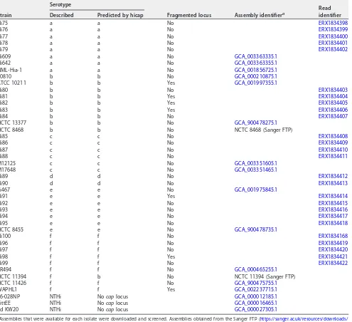
TABLE 2Strains used in the validation of the hicap method, with predicted serotype and fragmentation status of thecaplocus as determined by hicap
Strain
Serotype
Fragmented locus Assembly identifiera
Read identifier Described Predicted by hicap
Hi75 a a No ERX1834398
Hi76 a a No ERX1834399
Hi77 a a No ERX1834400
Hi78 a a No ERX1834401
Hi79 a a No ERX1834402
Hi609 a a No GCA_003363335.1
Hi642 a a No GCA_003363355.1
NML-Hia-1 a a No GCA_001856725.1
10810 b b No GCA_000210875.1
ATCC 10211 b b Yes GCA_001997355.1
Hi80 b b No ERX1834403
Hi81 b b Yes ERX1834404
Hi82 b b Yes ERX1834405
Hi83 b b Yes ERX1834406
Hi84 b b No ERX1834407
NCTC 13377 b b No GCA_900478275.1
NCTC 8468 b b No NCTC 8468 (Sanger FTP)
Hi85 c c No ERX1834408
Hi86 c c No ERX1834409
Hi87 c c No ERX1834410
Hi88 c c No ERX1834411
M12125 c c No GCA_003351605.1
M17648 c c No GCA_003351465.1
Hi89 d d No ERX1834412
Hi90 d d No ERX1834413
hi467 e e No GCA_001975845.1
Hi91 e e Yes ERX1834414
Hi92 e e No ERX1834415
Hi93 e e No ERX1834416
Hi94 e e No ERX1834417
Hi95 e e No ERX1834418
NCTC 8455 e e No GCA_900478735.1
Hi100 f f No ERX1834168
Hi96 f f No ERX1834419
Hi97 f f No ERX1834420
Hi98 f f Yes ERX1834421
Hi99 f f No ERX1834422
KR494 f f No GCA_000465255.1
NCTC 11394 f b No NCTC 11394 (Sanger FTP)
NCTC 11426 f f No GCA_900475755.1
WAPHL1 f f Yes GCA_002237715.1
86-028NP NTHi Nocaplocus GCA_000012185.1
PittEE NTHi Nocaplocus GCA_000016465.1
Rd KW20 NTHi Nocaplocus GCA_000027305.1
aAssemblies that were available for each isolate were downloaded and screened. Assemblies obtained from the Sanger FTP (https://sanger.ac.uk/resources/downloads/
bacteria/nctc) were additionally converted from GFF3 to FASTA format. Where an assembly was not available for an isolate, read sets were downloaded and assem-bled using SPAdes (as described in Materials and Methods) before screening. All assemblies used for testing are available through FigShare (https://doi.org/10.26180/ 5c352c5110712).
on May 17, 2020 by guest
http://jcm.asm.org/
(Table 2). The results show that hicap robustly identifies the
H. influenzae cap
locus even
in highly discontiguous assemblies. For each
cap
locus, the completeness, presence of
truncated genes, duplication, contiguity, and serotype were correctly reported.
Capsule loci were detected by hicap in 41/41 genomes with reported serotypes and
in 0/3 serologically determined NTHi genomes (Table 2). The predicted serotype
matched the reported serotype in 40/41 cases (98%) (Table 2). We found that hicap
yielded accurate predictions even from draft genomes where the
cap
locus was
fragmented across multiple contigs (observed in 7 genomes from the validation set).
Examples of the
cap
loci identified and visualized using hicap are shown in Fig. 3.
The single genome with a discrepancy between predicted and reported serotype,
NCTC 11394, is described as Hif in the National Collection of Type Cultures (NCTC) but
was confidently assigned Hib by hicap analysis of the completed PacBio genome
assembly (Fig. 3b). Manual assessment of the
cap
locus in the NCTC 11394 genome
assembly additionally confirmed the presence of a complete
cap
-b locus
(uninter-rupted, in a single contig) and the absence of any
cap
-f region II genes, with all
expected
cap
-b protein-coding genes present at
ⱖ
95% coverage and
ⱖ
84% homology
to those annotated in the
cap
-b reference sequence (excluding a truncated
bexA
gene).
The standard slide agglutination test classically used for serological typing of
H.
influenzae
has been shown to lack specificity and has been estimated to yield incorrect
results at a rate of 17.5% (34). It therefore appears likely this discordance is due to
inaccuracies in the described serotype rather than misidentification by hicap.
cap
locus distribution and variation.
To demonstrate the utility of
in silico
sero-type prediction with hicap, we used it to investigate all publicly available
H. influenzae
genomes in GenBank that passed quality filtering criteria (
n
⫽
691; see Table S1 in the
supplemental material). hicap identified a complete
cap
locus in 95/691 (13.7%)
ge-nomes (8
cap
-a, 54
cap
-b, 4
cap
-c, 1
cap
-d, 20
cap
-e, and 8
cap
-f). All genomes
contained either zero or one
cap
locus type, but duplication events were observed in
15/95 (15.8%)
cap
-positive genomes (14
cap
-b and 1
cap
-a).
Duplication of the
cap
-b locus has been frequently reported and is associated with
enhanced virulence, conferred by an increased ability to produce capsule (12, 35). This
duplication is thought to be driven by copies of IS
1016
flanking the capsule locus in
some isolates. A common variant of the duplicated
cap
-b locus involves the deletion of
1.2 kbp in one copy of region I, resulting in the truncation of
bexA
and IS
1016
. We
observed this duplication deletion variant in 14/54 (26.0%) predicted Hib genomes.
Complete
cap
-b duplication without truncation of
bexA
was not observed. In addition,
hicap identified a single isolate (NML-Hia-1) containing a tandem duplication of the
cap
-a locus. Strains identified to be carrying
cap
loci were not assessed for capsule
production; however, several of these strains are known to synthesize capsule (e.g.,
10810 and NML-Hia-1).
To examine the distribution of
cap
loci in the
H. influenzae
population, we
con-structed a whole-genome phylogeny (Fig. 4) and inferred STs according to the
H.
influenzae
MLST scheme (Table 3). We observed a high degree of exclusivity for STs in
regard to predicted capsular serotypes, with each ST containing zero or one
cap
locus
serotype.
The whole-genome phylogeny confirmed that encapsulated strains are relatively
clonal and are generally restricted to serotype-specific monophyletic clades (Fig. 4a),
suggesting that each serotype emerged once within the
H. influenzae
population. This
is consistent with earlier studies based on electrophoretic typing (36, 37), 16S rRNA (38),
MLST (27), and WGS (39). Here, the whole-genome phylogeny resolves the
monophy-letic nature of each capsule locus with respect to phylogenetic lineage on a larger scale
and in greater detail.
hicap did not detect
cap
loci in a small number of isolates within these
serotype-specific clades, indicating occasional capsule loss. For example, P590-8360 clustered
with the Hie clade (Fig. 4), but no
cap
locus was identified by hicap or by manual
inspection of the assembly data. The high nucleotide identity between P590-8360 and
on May 17, 2020 by guest
http://jcm.asm.org/
the
cap
-e-positive strain P589-8275 along the rest of the genome (Fig. 4a) suggests that
loss of the
cap
-e locus in the P590-8360 genome is the mostly likely explanation.
Indeed, the loss of capacity to synthesize capsule has previously been observed to
occur by partial or complete deletion of the
cap
locus (39), and the rate of spontaneous
FIG 4Whole-genome neighbor-joining phylogeny inferred from MASH distances of assemblies in the GenBank data set. Isolates are annotated with the respective serotype as predicted by hicap. (a) Distribution of capsular serotypes in the complete data set. (b) The phylogeny subtree including only isolates that contained acaplocus, additionally annotated with the sequence type.
on May 17, 2020 by guest
http://jcm.asm.org/
[image:7.585.45.471.69.639.2]capsule loss is estimated to occur at a frequency of 0.1 to 0.3% (40). Our data are
consistent with deletion of the
cap
locus being a cause of this phenomenon.
Interest-ingly one
cap
-a genome (M21384) falls within the
cap
-e serotype-specific clade,
sug-gesting possible recombination in this strain (Fig. 4) (further evidence for this is
discussed below).
The serotype-specific clades cluster into two superclades within the
H. influenzae
phylogeny: one containing
cap
loci of Hia, Hib, Hic, and Hid and the other containing
Hie and Hif
cap
loci (Fig. 4). Individual gene trees for the region I (
bex
) and III (
hcs
) genes
show the same two-clade structure (Fig. 5) as the core genomes of their host strains.
This observation is consistent with diversification of these
cap
locus regions
in situ
within their host chromosomes following introduction into two distinct
H. influenzae
superclade ancestors. While there is a general lack of homology between region II
genes (Fig. 1), two of the three pairs that do show a measure of homology (
cap
-c/
cap
-f
and
cap
-d/
cap
-e) span both superclades; hence, the evolutionary history of region II
(and thus the distinct capsular serotypes) remains cryptic.
Variation in each region I or III gene was associated with serotype, suggesting that
the sequence of any could potentially be used to predict capsule type with a relatively
high degree of certainty (Fig. 5). Indeed, both
bexA
and
bexB
have been proposed and
used in single-gene PCR assays for the purpose of serotyping (41, 42). Here the gene
bexB
showed the greatest differentiation between serotype-specific alleles (0.63% to
17.71% median pairwise nucleotide divergence; see Fig. S1 in the supplemental
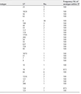
ma-TABLE 3Sequence types associated with each serotype in the GenBank data set
Serotype ST No.
Frequency (%) of serotype within ST
a 23 3 100
2
1828 1 100
56 1 100
62 1 100
b 6 38 100
190 3 100
44 2 100
53 2 100
117 1 100
119 1 100
1756 1 100
282 1 100
54 1 100
55 1 100
637 1 100
724 1 100
95 1 100
c 1870 1 100
51 1 100
7 1 100
9 1 100
d 10 1 100
e 18 7 87.5
66 6 100
2
1336 2 100
121 1 100
27 1 100
386 1 100
f 124 7 87.5
123 1 100
on May 17, 2020 by guest
http://jcm.asm.org/
[image:8.585.47.371.82.458.2]FIG 5Phylogenies of all completecaplocus region I (a) and III (b) genes identified in the GenBank data set. FastTree was used to recover phylogenies from MAFFT gene nucleotide sequence alignments, and isolates were annotated using the serotype as predicted by hicap.
on May 17, 2020 by guest
http://jcm.asm.org/
[image:9.585.39.544.63.680.2]terial) and contained only serotype-specific monophyletic clades in the gene tree.
These data suggest
bexB
to be the most suitable single marker gene for use in PCR or
sequenced-based prediction of serotype. In contrast,
bexA
showed less differentiation
then
bexB
, particularly between serotypes a, b, c, and d (0.17% to 0.85% median
pairwise nucleotide divergence).
The exceptions to the general association between region I/III genes and predicted
serotype were two isolates, M21384 and NCTC 11426 (labeled in Fig. 5). hicap predicted
isolates M21384 and NCTC 11426 to be of serotype a and serotype f, respectively.
However, both carry
cap
region I and/or III gene sequences distinct from other strains
sharing the same
cap
II region type (and thus the same predicted serotype), indicative
of recombination involving the
cap
locus within these isolates. Thus, there is evidence
for occasional recombination within the
cap
locus between the different
serotype-specific variants, which would limit the accuracy of any single marker gene-based
approach to serotype prediction.
Recombination affecting the
cap
locus.
The isolate M21384 was the only
excep-tion to clonal clustering by serotype in the whole-genome phylogeny (Fig. 4). While this
isolate is predicted to be Hia based on the presence of
cap
-a region II genes, the
genome falls outside the Hia clade and within the Hie/Hif superclade (Fig. 4b). In all
gene trees, M21384 also did not cluster in the expected Hia serotype clade, suggesting
that there has been recombination within the
cap
locus of this isolate (see Fig. 5).
Similarly in the
hcsA
and
hcsB
gene trees, the isolate NCTC 11426 did not cluster with
the expected serotype Hif clade (Fig. 5b). Given the phylogenetic relation of M21384
and NCTC 11426 to other capsular serotypes, it was suspected that these two strains
result from recombination events affecting the
cap
locus (representing a 2%
recombi-nation rate involving the
cap
locus).
To better understand the recombinant
cap
loci in isolates M21384 and NCTC
11426, we first examined their positions in the whole-genome phylogeny (Fig. 4)
and the
cap
locus gene trees (Fig. 5) and then compared the full-length
cap
locus
sequences of both isolates to reference
cap
locus sequences (Fig. 6). NCTC 11426
(predicted to be Hif) belongs to the Hif clade in the whole-genome tree and carries
typical
cap
-f regions I and II but contains region III genes more similar to those from
cap
-e (Fig. 6a). Hence, it appears that the
cap
locus of NCTC 11426 has resulted from
a small recombination event between a Hif clade strain and the
cap
locus from a Hie
clade strain. M21384 clusters within the Hie/Hif superclade of the whole-genome
phylogeny and carries
cap
-f-like region I genes (Fig. 5a and 6b). However, this
isolate carries
cap
-a-like region II genes with a
cap
-b-like region III gene (
hcsA
) (Fig.
5b and 6b). The gene content of the M21384
cap
locus suggests at least one
recombination event involving import of foreign
cap
locus DNA into a Hif strain. It
would be interesting to ascertain whether the isolates with recombinant
cap
loci
described here do in fact express capsule and, if so, to then establish the serological
phenotype. However, to our knowledge serotyping has not been performed for
either strain, or the data are not publicly available.
Conclusion.
The need for new tools and methods that leverage WGS continues to
become increasingly pivotal with the adoption of WGS by public health laboratories. In this
study, we validated and demonstrated the robustness of hicap for prediction of
H.
influ-enzae
serotype and capsule locus structure. The application of hicap to WGS enables rapid
and accurate acquisition of capsule information to aid genomic studies at both individual
and population scales. We were also able to explore the diversity and distribution of
cap
loci
in the
H. influenzae
population at unprecedented nucleotide resolution, identifying a likely
misreported serotype in NCTC and describing two novel
H. influenzae cap
locus
recombi-nants. The resurgence of disease caused by encapsulated
H. influenzae
and the potential for
further antigenic diversification through recombination present a potential public health
issue. An important question is whether the geographically disparate reports of increasing
cases of infection with non-Hib encapsulated strains reflect the emergence and wide
dissemination of a small number of highly successful disease-causing subclones (i.e., a rare
on May 17, 2020 by guest
http://jcm.asm.org/
but worrying event) or multiple independent events reflecting sporadic but localized
outbreaks of non-Hib disease. hicap will facilitate extracting answers to these and other
questions from genomic surveillance data.
SUPPLEMENTAL MATERIAL
Supplemental material for this article may be found at
https://doi.org/10.1128/JCM
.00190-19
.
SUPPLEMENTAL FILE 1
, PDF file, 0.5 MB.
SUPPLEMENTAL FILE 2
, XLSX file, 0.03 MB.
ACKNOWLEDGMENTS
This work was supported by the Bill & Melinda Gates Foundation, Seattle, WA, and
the by Australian Government Research Training Program. K.E.H. is supported by a
Senior Medical Research Fellowship from the Viertel Foundation of Victoria.
We declare that there are no conflicts of interest.
REFERENCES
1. Fleischmann RD, Adams MD, White O, Clayton RA, Kirkness EF, Kerlavage AR, Bult CJ, Tomb JF, Dougherty BA, Merrick JM. 1995. Whole-genome random sequencing and assembly ofHaemophilus influenzaeRd. Science 269:496 –512.https://doi.org/10.1126/science.7542800.
2. Pittman M. 1931. Variation and type specificity in the bacterial species
Hemophilus influenzae. J Exp Med 53:471– 492.https://doi.org/10.1084/ jem.53.4.471.
3. Kroll JS, Loynds B, Brophy LN, Moxon ER. 1990. Thebexlocus in encapsu-latedHaemophilus influenzae: a chromosomal region involved in capsule
polysaccharide export. Mol Microbiol 4:1853–1862.https://doi.org/10.1111/ j.1365-2958.1990.tb02034.x.
4. Sukupolvi-Petty S, Grass S, St Geme JW. 2006. The Haemophilus influenzaetype bhcsAandhcsBgene products facilitate transport of capsular polysaccharide across the outer membrane and are essential for virulence. J Bacteriol 188:3870 –3877.https://doi.org/10.1128/JB .01968-05.
5. Follens A, Veiga-da-Cunha M, Merckx R, van Schaftingen E, van Eldere J. 1999.acs1ofHaemophilus influenzaetype a capsulation locus region II
FIG 6Homology plots generated using R and genoPlotR, showing thecaploci of two isolates which appear to have been subject to recombination. Different regions of the NCTC 11426 (a) and M21384 (b)caploci show varying homology to different referencecaploci, suggesting a recombinogenic ancestry.
on May 17, 2020 by guest
http://jcm.asm.org/
[image:11.585.40.508.69.400.2]encodes a bifunctional ribulose 5-phosphate reductase-CDP-ribitol pyro-phosphorylase. J Bacteriol 181:2001–2007.
6. Van Eldere J, Brophy L, Loynds B, Celis P, Hancock I, Carman S, Kroll JS, Moxon ER. 1995. Region II of theHaemophilus influenzaetype be cap-sulation locus is involved in serotype-specific polysaccharide synthesis. Mol Microbiol 15:107–118.https://doi.org/10.1111/j.1365-2958.1995.tb02225.x. 7. Lâm T-T, Claus H, Frosch M, Vogel U. 2011. Sequence analysis of
serotype-specific synthesis regions II ofHaemophilus influenzaeserotypes c and d: evidence for common ancestry of capsule synthesis inPasteurellaceaeand
Neisseria meningitidis. Res Microbiol 162:483– 487.https://doi.org/10.1016/j .resmic.2011.04.002.
8. Giufrè M, Cardines R, Mastrantonio P, Cerquetti M. 2010. Genetic char-acterization of the capsulation locus ofHaemophilus influenzaeserotype e. J Clin Microbiol 48:1404 –1407.https://doi.org/10.1128/JCM.01721-09. 9. Satola SW, Schirmer PL, Farley MM. 2003. Genetic analysis of the capsule locus ofHaemophilus influenzaeserotype f. Infect Immun 71:7202–7207.
https://doi.org/10.1128/IAI.71.12.7202-7207.2003.
10. Corn PG, Anders J, Takala AK, Käyhty H, Hoiseth SK. 1993. Genes involved in Haemophilus influenzaetype b capsule expression are frequently amplified. J Infect Dis 167:356 –364.https://doi.org/10.1093/infdis/167.2 .356.
11. Kroll JS, Hopkins I, Moxon ER. 1988. Capsule loss inH. influenzaetype b occurs by recombination-mediated disruption of a gene essential for polysaccharide export. Cell 53:347–356.https://doi.org/10.1016/ 0092-8674(88)90155-9.
12. Kapogiannis BG, Satola S, Keyserling HL, Farley MM. 2005. Invasive infec-tions withHaemophilus influenzaeserotype a containing an IS1016-bexA
partial deletion: possible association with virulence. Clin Infect Dis 41: e97– e103.https://doi.org/10.1086/498028.
13. Bijlmer HA. 1991. World-wide epidemiology of Haemophilus influenzae
meningitis; industrialized versus non-industrialized countries. Vaccine 9:S5–S9. (Discussion, 9:S25.https://doi.org/10.1016/0264-410X(91)90172-3. 14. Peltola H. 2000. WorldwideHaemophilus influenzaetype b disease at the
beginning of the 21st century: global analysis of the disease burden 25 years after the use of the polysaccharide vaccine and a decade after the advent of conjugates. Clin Microbiol Rev 13:302–317.https://doi.org/10 .1128/CMR.13.2.302.
15. Ulanova M. 2013. Global epidemiology of invasiveHaemophilus influen-zaetype a disease: do we need a new vaccine? J Vaccines 2013:14.
https://doi.org/10.1155/2013/941461.
16. Eton V, Schroeter A, Kelly L, Kirlew M, Tsang RSW, Ulanova M. 2017. Epidemiology of invasive pneumococcal and Haemophilus influenzae
diseases in Northwestern Ontario, Canada, 2010-2015. Int J Infect Dis 65:27–33.https://doi.org/10.1016/j.ijid.2017.09.016.
17. Puig C, Grau I, Marti S, Tubau F, Calatayud L, Pallares R, Liñares J, Ardanuy C. 2014. Clinical and molecular epidemiology ofHaemophilus influenzae
causing invasive disease in adult patients. PLoS One 9:e112711.https://doi .org/10.1371/journal.pone.0112711.
18. Revez J, Espinosa L, Albiger B, Leitmeyer KC, Struelens MJ, ECDC National Microbiology Focal Points and Experts Group. 2017. Survey on the use of whole-genome sequencing for infectious diseases surveillance: rapid expansion of European national capacities, 2015-2016. Front Public Health 5:347.https://doi.org/10.3389/fpubh.2017.00347.
19. Besser J, Carleton HA, Gerner-Smidt P, Lindsey RL, Trees E. 2018. Next-generation sequencing technologies and their application to the study and control of bacterial infections. Clin Microbiol Infect 24:335–341.
https://doi.org/10.1016/j.cmi.2017.10.013.
20. Grad YH, Lipsitch M. 2014. Epidemiologic data and pathogen genome sequences: a powerful synergy for public health. Genome Biol 15:538.
https://doi.org/10.1186/s13059-014-0538-4.
21. Hyatt D, Chen G-L, Locascio PF, Land ML, Larimer FW, Hauser LJ. 2010. Prodigal: prokaryotic gene recognition and translation initiation site identification. BMC Bioinformatics 11:119.https://doi.org/10.1186/1471 -2105-11-119.
22. Camacho C, Coulouris G, Avagyan V, Ma N, Papadopoulos J, Bealer K, Madden TL. 2009. BLAST⫹: architecture and applications. BMC Bioinfor-matics 10:421.https://doi.org/10.1186/1471-2105-10-421.
23. Cock PJA, Antao T, Chang JT, Chapman BA, Cox CJ, Dalke A, Friedberg I, Hamelryck T, Kauff F, Wilczynski B, de Hoon M. 2009. Biopython: freely available Python tools for computational molecular biology and bioinformatics. Bioinformatics 25:1422–1423.https://doi.org/10 .1093/bioinformatics/btp163.
24. Bankevich A, Nurk S, Antipov D, Gurevich AA, Dvorkin M, Kulikov AS, Lesin VM, Nikolenko SI, Pham S, Prjibelski AD, Pyshkin AV, Sirotkin AV,
Vyahhi N, Tesler G, Alekseyev MA, Pevzner PA. 2012. SPAdes: a new genome assembly algorithm and its applications to single-cell se-quencing. J Comput Biol 19:455– 477. https://doi.org/10.1089/cmb .2012.0021.
25. Ondov BD, Treangen TJ, Melsted P, Mallonee AB, Bergman NH, Koren S, Phillippy AM. 2016. Mash: fast genome and metagenome distance esti-mation using MinHash. Genome Biol 17:132.https://doi.org/10.1186/ s13059-016-0997-x.
26. Kim D, Song L, Breitwieser FP, Salzberg SL. 2016. Centrifuge: rapid and sensitive classification of metagenomic sequences. Genome Res 26: 1721–1729.https://doi.org/10.1101/gr.210641.116.
27. Meats E, Feil EJ, Stringer S, Cody AJ, Goldstein R, Kroll JS, Popovic T, Spratt BG. 2003. Characterization of encapsulated and noncapsulated
Haemophilus influenzaeand determination of phylogenetic relationships by multilocus sequence typing. J Clin Microbiol 41:1623–1636.https:// doi.org/10.1128/JCM.41.4.1623-1636.2003.
28. Guy L, Roat Kultima J, Andersson S. 2010. genoPlotR: comparative gene and genome visualization in R. Bioinformatics 26:2334 –2335.https://doi .org/10.1093/bioinformatics/btq413.
29. R Core Team. 2019. R: a language and environment for statistical com-puting. R Foundation for Statistical Computing, Vienna, Austria. 30. Yu G, Smith DK, Zhu H, Guan Y, Lam TT-Y. 2017. ggtree: an R package for
visualization and annotation of phylogenetic trees with their covariates and other associated data. Methods Ecol Evol 8:28 –36.https://doi.org/ 10.1111/2041-210X.12628.
31. Katoh K, Standley DM. 2013. MAFFT multiple sequence alignment soft-ware version 7: improvements in performance and usability. Mol Biol Evol 30:772–780.https://doi.org/10.1093/molbev/mst010.
32. Price MN, Dehal PS, Arkin AP. 2010. FastTree 2—approximately maximum-likelihood trees for large alignments. PLoS One 5:e9490.https://doi.org/10 .1371/journal.pone.0009490.
33. Paradis E, Claude J, Strimmer K. 2004. APE: analyses of phylogenetics and evolution in R language. Bioinformatics 20:289 –290.https://doi.org/10 .1093/bioinformatics/btg412.
34. Satola SW, Collins JT, Napier R, Farley MM. 2007. Capsule gene analysis of invasiveHaemophilus influenzae: accuracy of serotyping and preva-lence of IS1016 among nontypeable isolates. J Clin Microbiol 45: 3230 –3238.https://doi.org/10.1128/JCM.00794-07.
35. Kroll JS, Moxon ER, Loynds BM. 1993. An ancestral mutation enhancing the fitness and increasing the virulence ofHaemophilus influenzaetype b. J Infect Dis 168:172–176.https://doi.org/10.1093/infdis/168.1.172. 36. Musser JM, Granoff DM, Pattison PE, Selander RK. 1985. A population
genetic framework for the study of invasive diseases caused by serotype b strains ofHaemophilus influenzae. Proc Natl Acad Sci U S A 82:5078 –5082.
https://doi.org/10.1073/pnas.82.15.5078.
37. Musser JM, Kroll JS, Granoff DM, Moxon ER, Brodeur BR, Campos J, Dabernat H, Frederiksen W, Hamel J, Hammond G. 1990. Global genetic structure and molecular epidemiology of encapsulatedHaemophilus influenzae. Rev Infect Dis 12:75–111.https://doi.org/10.1093/clinids/12.1.75.
38. Sacchi CT, Alber D, Dull P, Mothershed EA, Whitney AM, Barnett GA, Popovic T, Mayer LW. 2005. High level of sequence diversity in the 16S rRNA genes ofHaemophilus influenzaeisolates is useful for molecular subtyping. J Clin Microbiol 43:3734 –3742.https://doi.org/10.1128/JCM .43.8.3734-3742.2005.
39. De Chiara M, Hood D, Muzzi A, Pickard DJ, Perkins T, Pizza M, Dougan G, Rappuoli R, Moxon ER, Soriani M, Donati C. 2014. Genome sequencing of disease and carriage isolates of nontypeableHaemophilus influenzae
identifies discrete population structure. Proc Natl Acad Sci U S A 111: 5439 –5444.https://doi.org/10.1073/pnas.1403353111.
40. Hoiseth SK, Connelly CJ, Moxon ER. 1985. Genetics of spontaneous, high-frequency loss of b capsule expression inHaemophilus influenzae. Infect Immun 49:389 –395.
41. Davis GS, Sandstedt SA, Patel M, Marrs CF, Gilsdorf JR. 2011. Use ofbexB
to detect the capsule locus inHaemophilus influenzae. J Clin Microbiol 49:2594 –2601.https://doi.org/10.1128/JCM.02509-10.
42. Falla TJ, Crook DW, Brophy LN, Maskell D, Kroll JS, Moxon ER. 1994. PCR for capsular typing ofHaemophilus influenzae. J Clin Microbiol 32:2382–2386.
43. Su Y-C, Hörhold F, Singh B, Riesbeck K. 2013. Complete genome se-quence of encapsulatedHaemophilus influenzaetype f KR494, an inva-sive isolate that caused necrotizing myositis. Genome Announc 1:e00470-13.https://doi.org/10.1128/genomeA.00470-13.
44. Staples M, Graham RMA, Jennison AV. 2017. Characterisation of invasive clinicalHaemophilus influenzaeisolates in Queensland, Australia using
on May 17, 2020 by guest
http://jcm.asm.org/
whole-genome sequencing. Epidemiol Infect 145:1727–1736.https://doi .org/10.1017/S0950268817000450.
45. Pittman M. 1942. Antibacterial action of several sulfonamide compounds onHemophilus influenzaetype B. Public Health Rep 57:1899 –1910. 46. Giufrè M, Cardines R, Cerquetti M. 2017. First whole-genome sequence
of aHaemophilus influenzaetype e strain isolated from a patient with
invasive disease in Italy. Genome Announc 5:e00059-17.https://doi.org/ 10.1128/genomeA.00059-17.
47. Dobson SRM, Kroll JS, Moxon ER. 1992. Insertion sequence IS1016and absence ofHaemophilus capsulation genes in the Brazilian purpuric fever clone ofHaemophilus influenzaebiogroup aegyptius. Infect Immun 60:618 – 622.