R E S E A R C H
Open Access
Deep sequencing reveals clonal evolution
patterns and mutation events associated
with relapse in B-cell lymphomas
Yanwen Jiang
1,2, David Redmond
1, Kui Nie
3, Ken W Eng
1, Thomas Clozel
4, Peter Martin
2, Leonard HC Tan
5,
Ari M Melnick
2, Wayne Tam
3*and Olivier Elemento
1*Abstract
Background:Molecular mechanisms associated with frequent relapse of diffuse large B-cell lymphoma (DLBCL) are poorly defined. It is especially unclear how primary tumor clonal heterogeneity contributes to relapse. Here, we explore unique features of B-cell lymphomas - VDJ recombination and somatic hypermutation - to address this question. Results:We performed high-throughput sequencing of rearranged VDJ junctions in 14 pairs of matched diagnosis-relapse tumors, among which 7 pairs were further characterized by exome sequencing. We identify two distinctive modes of clonal evolution of DLBCL relapse: an early-divergent mode in which clonally related diagnosis and relapse tumors diverged early and developed in parallel; and a late-divergent mode in which relapse tumors developed directly from diagnosis tumors with minor divergence. By examining mutation patterns in the context of phylogenetic information provided by VDJ junctions, we identified mutations in epigenetic modifiers such as KMT2D as potential early driving events in lymphomagenesis and immune escape alterations as relapse-associated events.
Conclusions:Altogether, our study for the first time provides important evidence that DLBCL relapse may result from multiple, distinct tumor evolutionary mechanisms, providing rationale for therapies for each mechanism. Moreover, this study highlights the urgent need to understand the driving roles of epigenetic modifier mutations in lymphomagenesis, and immune surveillance factor genetic lesions in relapse.
Background
Diffuse large B-cell lymphoma (DLBCL) is an aggressive form of non-Hodgkin’s lymphoma in which one-third of patients either do not respond to initial therapy or re-lapse after standard therapy, such as dose-dense or standard immunochemotherapy with rituximab and cyclophosphamide, doxorubicin, vincristine, and prednisone (R-CHOP) [1]. Although relapses normally occur early (first 2 to 3 years), some do occur after 5 years [2]. Treatment options for relapse and refractory DLBCLs are limited and only 10% of the relapsed patients achieve 3-year progression-free survival following these treatments,
underlying the urgent need for novel approaches to treat DLBCL relapse [3,4]. Unfortunately, our current understanding of the molecular mechanisms associated with DLBCL relapse is limited. It is, for example, unclear whether genetic mutations present in the tumor at diag-nosis or acquired after treatment help certain DLBCL cell populations to acquire resistance to treatment. More gen-erally, it is presently unclear whether clonal heterogeneity in primary tumors plays a role in DLBCL relapse.
Previously, Ding et al. [5] examined genetic changes associated with acute myeloid leukemia relapse using whole-genome sequencing and used patterns of mutational abundances to infer clonal evolution patterns. However, due to the lack of tractable markers on myeloid cells, it is difficult to precisely and definitely deduce clonal evolution patterns between diagnosis and relapse acute myeloid leu-kemias. The situation is potentially different in lymphoid malignancies originating from mature B cells, since the
* Correspondence:[email protected];[email protected] 3
Department of Pathology and Laboratory Medicine, Weill Cornell Medical College, New York, NY 10021, USA
1
Institute for Computational Biomedicine, Weill Cornell Medical College, New York, NY 10021, USA
Full list of author information is available at the end of the article
latter harbor a natural clonality marker in the form of VDJ (or VJ) junctions. Indeed, in order to generate a diversified repertoire of antibodies, each B cell undergoes somatic immunoglobulin heavy chain (IGH) VDJ recombination at the pro B-cell stage to create a single productive VDJ junction from a large pool of VH(variable), D (diversity),
and JH(joining) segments. In addition, during this process,
non-templated nucleotides (N-bases) may be added at the junctions, and a small part of the VH, D, JHgermline
sequences may be deleted, resulting in a unique VDJ rearrangement, which effectively tags each B cell and its progeny [6]. In the subsequent germinal center (GC) reac-tion, B cells further introduce point mutations into the recombined VDJ sequences, a process known as somatic hypermutation (SHM), to enhance antibody affinity [7,8]. Because DLBCLs arise from GC or post-GC B cells, we hypothesized that their clonal populations can be tracked by their VDJ and SHM patterns to reveal valu-able information regarding intra-tumor heterogeneity and clonal evolution of the disease. To test this hypothesis, we performed high-throughput sequencing of rearranged VDJ junctions in 14 pairs of matched primary diagnosis-relapse DLBCLs and discovered two distinct clonal evolutionary scenarios of DLBCL relapse. Furthermore, in conjunction with exome sequencing on several diagnosis-relapse pairs, we identified mutations within histone-modifying enzymes as candidate early drivers in DLBCL lymphomagenesis, and genetic lesions in immune surveillance genes as potential facilitators in DLBCL relapse.
Results
Deep VDJ sequencing revealing clonal heterogeneity of DLBCL
To trace the clonal identities of the diagnosis-relapse DLBCL pairs (N = 14, patient information summarized in Additional file 1), we performed Illumina MiSeq PE 2 × 150 bp sequencing on the 300 to 350 bp PCR products targeting the genomic IGH VDJ sequences (see Materials and methods). We reasoned that long reads generated using this approach should enable phylogenetic analysis based on SHM mutation patterns. We generated 0.38 to 1.42 million paired-end reads/sample (average 0.75 ± 0.26 million; Additional file 2). We developed a custom bioinformatics pipeline to align each paired-end read to germline immunoglobulin V, D, and J sequences in the IMGT database [9] and annotate the recombined VDJ junctions (see Materials and methods). The average align-ment rate was 67.7 ± 14.1% (Additional file 2). In total, we identified 0.28 to 0.93 million (average 0.49 ± 0.15 million) VHDJH junctions per sample (Additional file 2), and
the numbers of aligned reads were comparable be-tween diagnosis and relapse samples within each pair (Figure S1 in Additional file 3).
We first sought to examine the VHDJHheterogeneity of
each individual tumor (diagnosis or relapse) and counted how many times each combination of VH, D, and JHwas
found. We found, on average, 7.4 distinct VHDJH
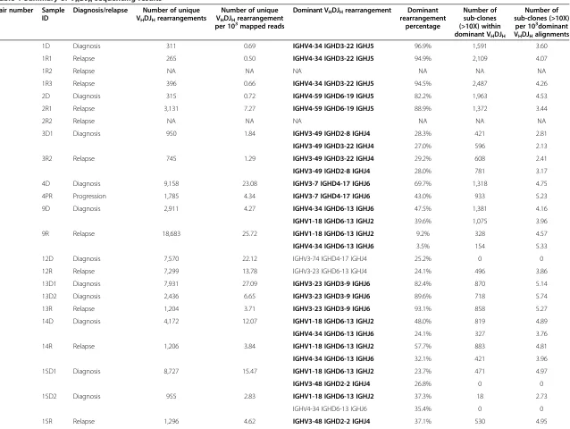
junc-tions per thousand mapped paired-end reads per sample (Table 1). However, the number of unique rearrangements per sample ranged broadly from 0.5 to 27.1 per thousand mapped paired-end reads, indicating high VDJ hetero-geneity within DLBCL samples, similar to what has been reported in acute B lymphoblastic leukemia (B-ALL) [10]. There was no significant difference in number of unique rearrangements per thousand mapped paired-end reads between the paired diagnosis and relapse samples (6.6 ± 7.5 versus 6.2 ± 6.9,P= 0.85, pairedt-test; orP= 0.80, Wilcoxon matched pairs test; Figure S2 in Additional file 3). For each sample, there was either one or two dominant VHDJH rearrangements that together account for 65%,
on average, of all the identified VHDJHsequences, likely
representing the productive and nonproductive alleles of the major tumor subclone [11] (Table 1). The other less abundant VHDJHrearrangements were likely representing
either VHDJH sequences with high SHM rates that could
not be readily mapped to major tumor VHDJHsequences,
or benign B-cell infiltration within the DLBCL tumors. To further address this question, we decided to compare the clonal distribution frequency between the tumor and nor-mal B-cell populations by performing VDJ sequencing on a bone marrow (BM) B-cell sample and tumor sample from one DLBCL patient. Although tumor cell infiltration was observed in the BM sample (approximately 1.1% of the total VHDJH sequences), most VHDJH sequences did
not match the tumor VHDJHand therefore correspond to
normal non-malignant B cells. We observed that the major dominant VHDJHrearrangements only accounted for
1.5% in the BM sample, similar to what has been reported previously for normal B cells [12]. Thus, the dominant VHDJH rearrangements we observed in tumor samples
are not compatible with normal B-cell clonal expansion and indeed represent the malignant B-cell populations in these tumors. Furthermore, when we excluded the dominant VHDJH rearrangements from the tumor
sam-ple (the tumor rearrangement) and then compared the distribution of the remaining VHDJHrearrangements of
the tumor sample to the BM sample, we observed similar distribution frequencies (Figure S3 in Additional file 3), indicating that these minor VHDJHrearrangement clones
were indeed likely representing infiltrating non-malignant normal B-cell populations (VHDJH sequence mapping
errors due to high SHM rates cannot be completely ex-cluded). The diagnosis and relapse tumors of all but one pair (pair 6) harbored the same major VHDJH
rearrange-ment, demonstrating that they were clonally related regard-less of the length of time it took the relapse to develop (Table 1). Accordingly, the relapse tumors of late-relapse
Jianget al. Genome Biology2014,15:432 Page 2 of 17
per 10 mapped reads percentage (>10X) within dominant VHDJH
per 10dominant VHDJHalignments
1 1D Diagnosis 311 0.69 IGHV4-34 IGHD3-22 IGHJ5 96.9% 1,591 3.60
1R1 Relapse 265 0.50 IGHV4-34 IGHD3-22 IGHJ5 94.9% 2,109 4.07
1R2 Relapse NA NA NA NA NA NA
1R3 Relapse 396 0.66 IGHV4-34 IGHD3-22 IGHJ5 94.5% 2,487 4.26
2 2D Diagnosis 315 0.72 IGHV4-59 IGHD6-19 IGHJ5 82.2% 1,963 4.53
2R1 Relapse 3,131 7.27 IGHV4-59 IGHD6-19 IGHJ5 88.9% 1,372 3.44
2R2 Relapse NA NA NA NA NA NA
3 3D1 Diagnosis 950 1.84 IGHV3-49 IGHD2-8 IGHJ4 28.3% 421 2.81
IGHV3-49 IGHD3-22 IGHJ4 27.0% 596 2.13
3R2 Relapse 745 1.29 IGHV3-49 IGHD3-22 IGHJ4 29.2% 608 2.41
IGHV3-49 IGHD2-8 IGHJ4 28.0% 781 3.17
4 4D Diagnosis 9,158 23.08 IGHV3-7 IGHD4-17 IGHJ6 69.7% 1,318 4.75
4PR Progression 1,785 4.34 IGHV3-7 IGHD4-17 IGHJ6 43.0% 933 5.23
5 9D Diagnosis 2,911 4.27 IGHV4-34 IGHD6-13 IGHJ6 47.5% 1,381 4.16
IGHV1-18 IGHD6-13 IGHJ2 39.6% 1,075 3.96
9R Relapse 18,683 25.72 IGHV1-18 IGHD6-13 IGHJ2 9.2% 328 4.57
IGHV4-34 IGHD6-13 IGHJ6 3.5% 154 5.33
6 12D Diagnosis 7,570 22.12 IGHV3-74 IGHD4-17 IGHJ4 25.2% 0 0
12R Relapse 7,299 13.78 IGHV3-23 IGHD6-13 IGHJ4 24.1% 496 3.86
7 13D1 Diagnosis 7,931 27.09 IGHV3-23 IGHD3-9 IGHJ6 82.4% 870 5.14
13D2 Diagnosis 2,436 6.65 IGHV3-23 IGHD3-9 IGHJ6 89.6% 718 5.74
13R Relapse 1,204 3.71 IGHV3-23 IGHD3-9 IGHJ6 93.1% 858 5.27
8 14D Diagnosis 4,172 12.07 IGHV1-18 IGHD6-13 IGHJ2 48.0% 819 4.89
IGHV4-34 IGHD6-13 IGHJ6 24.1% 327 3.76
14R Relapse 1,206 3.84 IGHV1-18 IGHD6-13 IGHJ2 57.7% 883 4.81
IGHV4-34 IGHD6-13 IGHJ6 32.1% 421 3.96
9 15D1 Diagnosis 8,727 15.47 IGHV1-18 IGHD6-13 IGHJ2 23.7% 471 4.97
IGHV3-48 IGHD2-2 IGHJ4 26.8% 0 0
15D2 Diagnosis 955 2.83 IGHV1-18 IGHD6-13 IGHJ2 37.3% 18 2.73
IGHV4-34 IGHD6-13 IGHJ6 35.4% 0 0
15R Relapse 1,296 4.62 IGHV3-48 IGHD2-2 IGHJ4 37.1% 530 4.95
Genome
Biology
2014,
15
:432
Page
3
o
f
1
7
mebiology
.com/2014/
[image:3.794.67.705.63.543.2]Table 1 Summary of VHDJHsequencing results(Continued)
IGHV1-18 IGHD6-13 IGHJ2 33.5% 471 5.00
10 16D Diagnosis 11,303 12.10 IGHV1-18 IGHD6-13 IGHJ2 55.2% 867 1.61
16R Relapse 1,875 2.67 IGHV1-18 IGHD6-13 IGHJ2 44.8% 1,079 3.42
11 F6D Diagnosis 786 1.04 IGHV3-23 IGHD3-22 IGHJ4 72.5% 991 4.22
F6PR Progression 2,135 4.03 IGHV3-23 IGHD3-22 IGHJ4 61.0% 1,245 5.29
12 F7D Diagnosis 3,491 6.07 IGHV4-34 IGHD3-22 IGHJ6 45.2% 231 0.61
F7R Relapse 6,115 14.58 IGHV4-34 IGHD3-22 IGHJ6 40.0% 82 0.33
13 SPF6-1 Diagnosis 1,160 2.41 IGHV3-7 IGHD3-10 IGHJ4 90.0% 1,691 3.83
SPF6-2 Diagnosis 828 1.75 IGHV3-7 IGHD3-10 IGHJ4 93.1% 1,854 3.93
SPF6-3 Relapse 719 1.54 IGHV3-7 IGHD3-10 IGHJ4 95.8% 1,754 3.85
14 SPF10-1 Diagnosis 1,463 3.17 IGHV3-49 IGHD3-10 IGHJ5 67.8% 1,425 3.65
SPF10-2 Relapse 1,731 3.73 IGHV3-49 IGHD3-10 IGHJ5 30.2% 777 4.37
NA, not available.
Jiang
et
al.
Genome
Biology
2014,
15
:432
Page
4
o
f
1
7
http://geno
mebiology
.com/2014/
patients who developed relapse disease 5 to 10 years after initial diagnosis (pairs 1, 8, 9, 12; Additional file 1) still car-ried the same VHDJH rearrangements as their respective
diagnosis tumors.
Two scenarios of DLBCL relapse clonal evolution
We further focused our analysis on the major VHDJH
rearrangement identified in each tumor. In addition to VHDJH rearrangement, DLBCL cells have undergone
various degrees of SHM within the rearranged VHDJH
se-quences, which can in theory be further utilized to delineate the subclonal population structure of the tumor. Indeed, we identified unique subclones (found at least 10 times) with distinctive SHM patterns within the dominant VHDJH
sequence of each sample (Table 1). Diagnosis samples had, on average, 3.6 ± 1.4 subclones per thousand mapped major VHDJH rearrangements. Relapse tumors had 4.1 ± 1.3
subclones per thousand mapped major VHDJH
rearrange-ments. There was no significant difference in the subclone frequency between the relapse and diagnosis samples (P= 0.07, two-tailed paired t-test; or P= 0.17, Wilcoxon matched pairs test; Figure S4 in Additional file 3). We then observed that eight pairs of samples had increased normalized subclone numbers in relapse samples, while five pairs had decreased normalized subclone numbers in relapse samples compared with their respective diagnosis samples (Figure S4 in Additional file 3; we excluded pair 6 in this and subsequent VDJ subclone analyses due to un-matched dominant VDJ between diagnosis and relapse samples), suggesting that the clonal subpopulation change is a dynamic process during DLBCL relapse.
We then sought to use the subclone SHM information to trace the clonal evolution of each sample pair. As described in Materials and methods, we performed phylogenetic ana-lysis of the SHM profiles of the major VHDJH
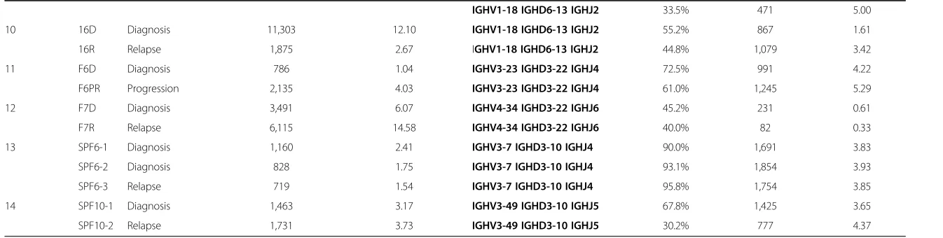
rearrange-ments between each diagnosis and relapse pair using the neighbor joining phylogenetic tree reconstruction approach [13]. This analysis uncovered two distinct scenarios of DLBCL relapse (Figure 1A; Figure S5 in Additional file 3). In one scenario (scenario I, n = 6, pairs 1, 2, 4, 5, 9, 10), the major relapse clones clustered in a separate branch from the diagnosis clones on the phylogenetic tree (Figure 1A; Figure S5A,B,D,E,H,I in Additional file 3). The relapse clones clustered either alone (n = 2, pairs 4 and 9) or together with a highly divergent minor diagnosis clone (n = 4; Figure 1A; see the small blue clone (third bar above the major relapse clone) clustered together with the red clones within the top branch). This particular pattern indicates that, in this scenario, although the relapse and diagnosis tumors were derived from the same B cell (they share the same VHDJH rearrangement and several
somatic hypermutations), their precursors have diverged, acquired different mutations within VHDJH sequences
(Figure 1A, right panel), and expanded at different
times. Moreover, that the major relapse clones frequently cluster with a minor divergent clone found at diagnosis suggest that preexisting, chemoresistant and divergent diagnosis subclones are capable of eventually regenerating entire relapse tumors.
In the second scenario (scenario II, n = 7, pairs 3, 7, 8, 11, 12, 13, 14), the dominant diagnosis and relapse clones clustered together very closely (Figure 1B, left panel), with similar SHM patterns. The dominant clones in the diag-nosis and relapse tumor share the vast majority of the IgVH somatic mutations, with only one or few different mutations in the relapse tumor that could not differentiate them into separate branches (Figure 1B, right panel; Figure S5C,F-G,J-M in Additional file 3). This observa-tion is compatible with a scenario where the relapse tu-mors arise linearly from the major diagnosis clone or from a highly related subclone that appears to be more abundant in the diagnosis tumor compared with the former scenario.
Due to unequal starting amounts of DNA, our VDJ sequencing libraries produced variable numbers of reads as indicated in the previous section. To make sure that VDJ sequencing depth would not affect our clonal evolution analysis, we sequenced two pairs of samples (one from scenario I (pair 10) and one from scenario II (pair 3)) to a very high depth (approximately two million paired reads per sample) compared with our original analysis, and then systematically subsampled the reads with decreasing rates (1/10th each round) to assess the VDJ recombination composition. We found that although the major VDJ frequency remained the same across all sample rates and for each sample (Figure S6A in Additional file 3), we did observe increased numbers of subclones with increasing sequencing depth (Figure S6B in Additional file 3). When we constructed phylogenetic trees of the subclones discovered at different subsampling levels for these two samples, we found that the relapse clonal evolution patterns did not change with increased number of subclones between the range of 10,000 to 1,000,000 reads (Figure S6C in Additional file 3), within which our original VDJ sequencing depth occurred, sug-gesting that the slight difference in VDJ sequencing depth across our samples would not undermine the accuracy of our relapse clonal evolution analysis.
scenario pairs (P= 0.81, Wilcoxon matched pairs test; Figure 1C).
Moreover, when we traced the fate of the major diagnosis subclone within the relapse tumor, we found that they
[image:6.595.57.539.88.591.2]almost disappeared in the relapse tumors of the more diver-gent mode pairs (scenario I; Figure 1D). On the contrary, the major diagnosis subclone maintained the same relative abundance in the relapse tumors of the less divergent cases
Figure 1Two scenarios of DLBCL relapse. (A,B)Phylogenetic analysis of the SHM profiles of sample pair 1 showing the early-divergent relapse mode(A), and sample pair 3 showing the late-divergent relapse mode(B). The length of the blue and red lines in the phylogenetic trees indicates the number of VDJ sequences with a particular SHM profile. The SHM profiles of the major VDJ subclones of the diagnosis and relapse samples are shown on the right with color tickers representing the mutation status.(C)Mutational distance between the major clones within the diagnosis and the relapse pairs.(D)The frequencies of the major diagnosis subclone in the diagnosis sample and the respective relapse sample of each sample.(E)Empirical entropy for the major VHDJHwas calculated for each diagnosis tumor sample, and the average of the estimated entropy was compared between the divergent mode samples and relapse mode samples.
Jianget al. Genome Biology2014,15:432 Page 6 of 17
(scenario II; Figure 1D). Surprisingly, we noticed a couple of the less divergent cases (scenario II) behaved similarly to the more divergent cases (scenario I), namely sample pairs 7 and 13 (Figure 1D). We plotted the relapse subclone frequencies according to their distance from the major diagnosis subclones (number of SHMs different from the major diagnosis subclone) and found that, in these two pairs, the entire major relapse subclones differed in only one or two mutations compared with their respect-ive diagnosis subclones (Figure S7 in Additional file 3). Therefore, the abundance of the major diagnosis sub-clone decreased to almost 0 at relapse in these samples. We performed the same analysis on the more divergent sample pairs (scenario I), and found that the difference in SHM pattern between the major diagnosis and relapse subclones was much greater (pairs 2 and 9, for example; Figure S7 in Additional file 3).
Based on these VDJ SHM analyses, two patterns emerge that differ in the degree of divergence (from the major diagnosis subclones) of the tumor clones from which the relapse tumors were derived. The first mode (scenario I) demonstrates a distinct divergence; the diverged subclone is present as a very minor subclone in the diagnosis tumor in some cases and appears to arise earlier in the diagnosis tumor during tumor evolution. We termed scenario I the early-divergent mode. The second mode (scenario II) does not show a distinct divergence and suggests that the relapse tumor may be derived from a subclone that is more closely related to the major diagnosis subclone (often more abundant) at a relatively late stage during tumor evolution. We therefore named scenario II the late-divergent mode. The presence of two modes of relapse, early-divergent (scenario I) or late-divergent (scenario II), can be further confirmed by whole exome sequencing (see below).
Diagnosis tumors with early-divergent evolution to relapse show increased clonal heterogeneity compared with tumors with late-divergent evolution
We examined whether the two relapse modes identified in this study were correlated with clinical features of DLBCL. By performing immunohistochemistry on BCL6, CD10, and MUM1 (Hans classification), we were able to determine the subtypes of these samples (GCB-DLBCL versus non-GCB-DLBCL; Additional file 1). We found the time to relapse was significantly higher in GCB-DLBCL compared with non-GCB-DLBCL (P= 0.02, Mann– Whitney test). However, when we examined the relation-ship between disease subtypes and relapse modes, we found that both subtypes were evenly distributed between the two relapse modes (chi-square test P= 0.55), indicating that there is no correlation between DLBCL subtypes and re-lapse clonal evolution. We also compared the average time to relapse and did not find significant difference
between the two modes of relapse (3.2 ± 1.5 years versus 3.0 ± 1.0 years, late-divergent versus early-divergent, P= 0.74, Mann–Whitney test). We then investigated whether clonal heterogeneity at diagnosis could predict which scenario towards relapse (early or late divergent) a patient was most likely to follow. We examined the pat-tern of subclones with distinctive SHM patpat-terns within the dominant VHDJH sequence of each sample, and used
the frequency with which each subclone appeared to calculate the empirical entropy for the major VHDJHof
each diagnosis tumor sample. Empirical entropy mea-sures the diversity of SHM patterns found in each sample, with higher entropy corresponding to higher diversity (see Materials and methods). We found that although entropy did not correlate with time to relapse (Figure S8 in Additional file 3), the early-divergent samples had, on average, statistically significantly higher entropy than the late-divergent cases at diagnosis (Figure 1E; P= 0.005, Student’st-test; orP= 0.02, Mann–Whitney test). These analyses indicate that, overall, diagnosis tumors that evolve according to the early-divergent scenario have a more diverse pattern of subclones at diagnosis. This observation is compatible with the observation made above that a minor, highly divergent subclone likely gives rise to the relapse tumor in the early-divergent scenario. It does suggest, however, that in such diver-gent scenarios, many other diverdiver-gent clones exist at diagnosis besides the one that gives rise to relapse.
Distinctive genetic evolution patterns of the two relapse scenarios
sequencing on somatic control DNA (2C of sample pair 2, 3C of sample pair 3, and 13C of sample pair 7; Additional file 2), and found that 90.4% of gained and lost coding region nonsynonymous SNVs identified in their respective tumor samples were not mutated in the somatic control DNA. Of note is that our analysis focused on SNVs whose allelic frequency changes significantly be-tween diagnosis and relapse (in either direction). We used Fisher exact with 10% false discovery rate control to deter-mine significance. This analysis does not remove variants whose allelic frequency changes significantly between diagnosis and relapse but that are nonetheless present in germline. Since it is not impossible that these vari-ants contribute to some aspects of the disease, we left them in the analysis. In addition, we performed Sanger sequencing on the remaining three sample pairs whose somatic control DNA was in low quantities that it was not suitable for exome study (1C of sample pair 1, 2C of sample pair 2, and 15C of sample pair 9). We ran-domly selected 37 SNVs from these three samples to validate and 94.6% them (35 out of 37) were confirmed to be somatic (Additional file 5). These results validate our approach as a robust method to identify specific disease-associated mutations in tumor samples even in the absence of germline controls. We found that overall re-lapse samples gained coding region nonsynonymous SNVs in 305 genes, among which only 71 were mutated in other previously reported primary DLBCL genomic sequencing studies, includingBCL2,EP300,KMT2D,MYC,TET2, and TNFRSF14[17-20]. We also found one late-divergent mode patient (pair 8, sample ID 14R) with relapse-specific EZH2Y641 mutation (Figure 2A), indicating that relapse
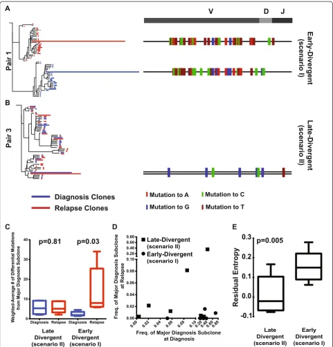
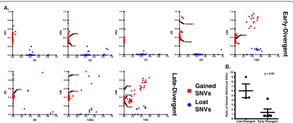
DLBCL can also acquire newEZH2mutations and that, in addition to its roles in initiating and maintaining DLBCL [21], mutant EZH2 may also contribute to disease relapse. For patient 1, we were able to obtain biopsies from three different relapse sites (Additional file 1). We compared the coding region nonsynonymous SNVs gained in these three relapse samples with the original diagnosis sample and found all of them gained mutations inBET1L,GNAS, and UBR4 genes, suggesting these mutations may be respon-sible for the initial transformation to relapse disease. Inter-estingly, relapse tumors 1R2 and 1R3 had more overlapping mutations, indicating that these two relapse tumors were more clonally related than they were to 1R1. The numbers of gained and lost SNVs between diagnosis and relapse samples were variable among patients (Additional file 4). On average, the late-divergent relapse samples gained 44.3 ± 17.6 SNVs and lost 7.5 ± 1.9 SNVs compared with their diagnosis samples; in contrast, the early-divergent relapse samples gained 32.0 ± 5.6 and lost 35.8 ± 6.6 SNVs. The late-divergent scenario relapse samples gained roughly four times more coding region nonsynonymous SNVs than they lost compared with their respective diagnosis samples (5.2 ± 1.9-fold; Figure 2B), represent-ing the continuous alteration of the tumor genome with additional mutations acquired to achieve relapse. On the contrary, the early-divergent scenario relapse samples gained and lost approximately equal numbers of coding region nonsynonymous SNVs compared with the diagnosis samples (1.4 ± 0.7-fold; Figure 2B). The coding region nonsynonymous SNV spectrums of the relapse-diagnosis pairs confirm the two tumor evolu-tion modes uncovered by VDJ sequencing. Tumors in A.
0.0 0.2 0.4 0.6 0.8 1.0 0.0 0.2 0.4 0.6 0.8 1.0 1D 1R 1
0.0 0.2 0.4 0.6 0.8 1.0 0.0 0.2 0.4 0.6 0.8 1.0 B2M TET2 1D 1R 2
0.0 0.2 0.4 0.6 0.8 1.0 0.0 0.2 0.4 0.6 0.8 1.0 1D 1R 3
0.0 0.2 0.4 0.6 0.8 1.0 0.0 0.2 0.4 0.6 0.8 1.0 TNFAIP3 EP300 2D 2 R
0.0 0.2 0.4 0.6 0.8 1.0 0.0 0.2 0.4 0.6 0.8 1.0 BRD4 15D 1 5R B.
Late Divergent Early Divergent 0 1 2 3 4 5 6 7 8 9
10 p = 0.03
Ratio of Gained SNVs/Lost SNVs
0.0 0.2 0.4 0.6 0.8 1.0 0.0 0.2 0.4 0.6 0.8 1.0 MYC 3D 3R
0.0 0.2 0.4 0.6 0.8 1.0 0.0 0.2 0.4 0.6 0.8 1.0 STAT3 BCL2 13D2 13R
[image:8.595.58.538.88.290.2]0.0 0.2 0.4 0.6 0.8 1.0 0.0 0.2 0.4 0.6 0.8 1.0 B2M EP300 EZH2 14D 14 R Late-Divergent t n e gr e vi D-yl r a E Gained SNVs Lost SNVs
Figure 2Different patterns of SNV gain and loss between the early and late-divergent modes. (A)SNV profile of each sample pair.(B)The ratio of the numbers of gained and lost SNVs between each relapse and diagnosis sample pair was calculated. The average and standard error of the ratio for each relapse mode are presented.
Jianget al. Genome Biology2014,15:432 Page 8 of 17
the early-divergent scenario undergo parallel evolution of their diagnosis and relapse DLBCL genome early during tumor development and therefore acquire new genomic mutations independently and share fewer mutations in common (that is, appear to lose more SNVs). Whereas in the late-divergent scenario, relapse tumors originate from subclones generated during the late stage of tumor evolu-tion and therefore share more mutaevolu-tions in common with the diagnosis tumors (that is, appear to lose fewer SNVs). The presence of 'lost' SNVs in the relapsed tumor com-pared with the diagnosis tumors within the late-divergent category argues against a 'direct evolution' scenario in which the relapse tumors are generated directly from the major diagnosis subclone. If that had been the case, the relapsed tumors should have shown no lost SNVs.
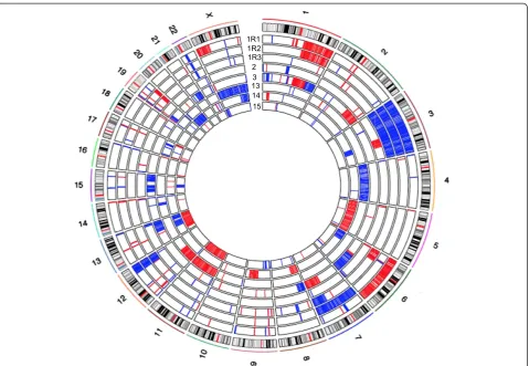
Immune surveillance genes are specifically targeted by indels and deletions in relapse
In addition to coding region nonsynonymous SNVs, we identified 39 small indels gained in relapse samples that targeted the coding regions of 36 genes (Additional file 6). On average, each relapse sample gained 5.2 ± 3.8 indels. Moreover, we identified CNAs by comparing each relapse exome with its respective diagnosis exome. We identified CNA segments specific to each relapse sample by using a previously widely used and validated R package, DNAcopy [22,23] applied to sequencing depth-adjusted relapse versus diagnosis read count log ratios (Figure S9 in Additional file 3; see Materials and methods). Some CNAs span large chromosome regions, while some are focal alterations (Figure 3; Additional file 7). On average, relapse tumors acquired 55.6 ± 6.8 CNAs, including 25.0 ± 5.0 amplifications and 30.6 ± 3.5 deletions. We did not detect CNAs that were common to all the relapse samples. However, we observed several loci that were deleted in multiple relapse samples compared with their respective diagnosis samples (Table 2). We validated two such loci by TaqMan copy number assays, both of which showed loss of genetic material in the relapse samples compared with their respective diagnosis samples (Figure S10 in Additional file 3). These commonly de-leted regions harbor genes that are potentially important for B-cell development and malignant transformation (Table 2). For example, three relapse samples (1R3, 3R, 14R) had a relapse-specific deletion spanning CD58, a gene that has been shown to be genetically altered in a subset of DLBCLs [24]. Furthermore, 14R had a relapse specific frameshift indel within the remaining allele of theCD58 gene (Additional file 6). Alteration of this gene may help tumor B cells to escape immune surveillance mecha-nisms [24]. In addition to CD58deletion in these three relapse samples, we also found that samples 1R2 and 14R gained coding region nonsynonymous SNVs in B2M(Additional file 4), another gene that is involved in
immune surveillance escape [24]. There was also a relapse-specific frameshift indel withinB2Min sample 15R (Additional file 6). Overall, in five out of seven patients we sequenced, there were coding region nonsynonymous SNVs, frameshift indels, and gross chromosomal deletions targeting CD58 and B2M genes, suggesting that escaping immune surveillance via mutations in key genes, such as CD58andB2M, may represent a common relapse strategy. Two other deleted genes, ARHGEF7 and PLCB2, are in-volved in RAC1 activation and downstream effects [25-27]. Deletions of these two genes may impair RAC1-mediated B-cell receptor signaling [28]. In addition, deletion ofIL9R may affect JAK-STAT signaling in response to IL9 [29], which is another pathway important in normal B cells and lymphomas [30].
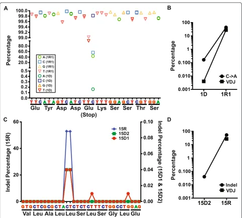
Ultra-deep target resequencing of relapse-specific mutations in diagnostic DLBCL with early-divergent mode of relapse identified minor subclones
By tracing VDJ SHM, we identified two clonal evolution scenarios of DLBCL relapse. We reasoned that the evo-lution of genetic events in DLBCL should follow similar patterns. In the case of relapse with an early-divergent mode, it is expected based on VDJ SHM analysis that a small subclone exists within the diagnosis tumor that carries a portion of relapse-specific SNVs or indels, which were not detected by whole exome sequencing but may be identified through deeper sequencing. To test this idea, we performed targeted re-sequencing of a number of SNVs and indels in early-divergent pairs. We obtained roughly two million sequencing reads for each amplicon, which allowed us to detect minor allele frequency of 0.001% with 20X sequencing depth. Indeed, targeted re-sequencing revealed that relapse-specific SNVs and indels could be found in a small number of cells at diagnosis in the early-divergent cases. For example, patient 1 gained at relapse a non-synonymous SNV in theUBR4 gene (chr1:19519971, C > A) that produced a premature stop codon. Targeted re-sequencing of this locus confirmed this SNV in the relapse sample of this patient (1R1) (Figure 4A). Interestingly, we observed a very small portion of the tumor cells at diagnosis (1D) also car-ried this SNV (Figure 4A). This small clone accounted for 0.16% of the total diagnosis tumor population (background C > A conversion within this amplicon was 0.03% ± 0.03%). In addition, the genetic evolution pattern of this SNV during relapse of patient 1 mir-rored this patient’s VHDJH clonal evolution pattern
the exact same frameshift indel (Figure 4C, 15D1 and 15D2, right y-axis; background indel rate 0.004 ± 0.010%). Although we did not observe any subclone in the diag-nosis sample carrying the same major relapse VHDJH
sequence, probably due to the difficulty of aligning VHDJH sequencing reads (Figure 4D), ultra-deep
tar-get amplicon re-sequencing allowed us to detect this relapse-causing subclone. Taken together, our results further confirmed the existence of small relapse-causing subclones in the diagnosis tumors of the early-divergent relapse scenario.
Coding-region SNV changes reveal potential mechanisms of DLBCL relapse
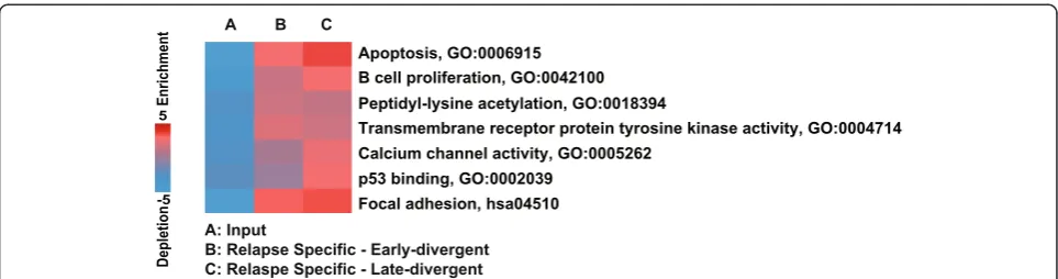
[image:10.595.60.540.87.418.2]To further explore the potential mechanisms of DLBCL relapse, we performed pathway enrichment analysis [31] on genes where we observed gain of coding region nonsynonymous SNVs in relapse samples (false discovery rate <10%; Figure 5). Pathway analysis revealed several interesting characteristics of DLBCL relapse. First, relapse samples in both groups gained additional mutations in genes that regulate apoptosis (GO:0006915). Many of these additional mutated genes are pro-apoptotic, such as
Figure 3Summary of copy number alteration information.The inner circles represent the CNA of each sample pair. Red indicates copy number gain, and blue indicates copy number loss.
Table 2 Summary of common deleted regions in relapse samples
Chromosome Common deleted region coordinates
Sample ID Genes Function in B cells
1 117064566-117131520 1R3, 3R, 14R CD58 Genetically inactivated in DLBCL to escape immune recognition [24]
4 961371-967030 1R1, 2R, 13R DGKQ Not known
13 111885580-111996434 1R1, 2R, 3R, 14R ARHGEF7 Guanine nucleotide exchange factor for RAC1 GTPases that
plays an important role in mature B-cell development [25]
15 40594724-40628804 1R1, 3R, 15R PLCB2 Activated by RAC1 to modulate Ca2+signaling [26]
X 155003966-155250615 3R, 13R, 15R VAMP7, IL9R IL9R activates JAK-STAT signaling in response to IL9 [29]
Jianget al. Genome Biology2014,15:432 Page 10 of 17
[image:10.595.55.541.631.733.2]CASP8, BID, and SIAH1, suggesting that for relapse tumors to develop, tumor cells might need to acquire extra survival advantages. Second, relapse samples gained mutations in transmembrane receptor tyrosine kinases (GO:0004714), among which wereEPHB2 and EPHB6, family members of the ephrin receptors, which are involved in cell-cell signaling. Third, late-divergent relapse samples gained more mutations in genes that are involved in calcium channel activity (GO:0005262) and p53 binding (GO:0002039). Inter-estingly, calcium signals play a critical role in B-cell
[image:11.595.57.539.85.519.2]development and functions, and are regulated by many signaling pathways, including B-cell receptor signaling [32]. Our data further suggest that DLBCL tumors with defects in calcium flux regulation may be resistant to current therapy and prone to relapse. Finally, a recent report by Xu-Monette et al.[33] demonstrated that patients with p53 mutations had worse overall and progression-free survival compared with those without. Our data now suggest that DLBCL relapse patients with mutations in p53 functional partners may also have simi-lar outcomes.
Mutations within histone modifiers are potential 'driver' mutations of DLBCL
Exome sequencing data on paired diagnosis and relapse samples in the early-divergent evolution mode provide us with an opportunity to study the early events in lym-phomagenesis. We reasoned that mutations that occurred in both diagnosis and relapse tumors have been acquired early in the life of these tumors and could have acted as early 'driver' or 'facilitator' mutations that initiate tumori-genesis. We observed that in all three divergent pairs, histone-modifying enzymes were mutated in both relapse and diagnosis tumors (Additional file 8). For example, we detectedKMT2Dand SETDB1mutations in both relapse and diagnosis tumors of pair 1,KMT2Dmutations in pair 2, andEP300mutations pair 9. The frequencies of all of these SNVs were comparable between the diagnosis and relapse samples within the same pair (Additional file 8). In pair 2, we had limited amounts of germline tissue at our dis-posal and performed Sanger sequencing to confirm that KMT2D mutation is not found in germline DNA (Figure S11 in Additional file 3). These observations suggested that mutations of histone modifiers could act as an early event to establish an aberrant epigenetic landscape in tumor-initiating cells and eventually drive the malignant transformation. Moreover, we observed that re-lapse samples in the early-divergent group gained additional coding region SNVs in epigenetic modifiers; that is, sample 1R2 in pair 1 gained a TET2 SNV, sample 2R of pair 2 gained a SNV inEP300, and sample 15R of pair 9 gained a BRD4SNV (Additional file 4). These findings suggested that epigenetic modifiers were further targeted during the relapse process, resulting in potential chemoresistance and other features that facilitate the development of relapse disease.
Discussion
When cancer patients relapse, their tumors often be-come more aggressive, chemoresistant, and refractory to treatment. The molecular pathogenesis of cancer relapse is largely unknown due to challenges in examining tumor
heterogeneity and clonal evolution between diagnosis and relapse tumors in many types of cancers. Here we sought to investigate the molecular mechanisms of DLBCL relapse, utilizing the ability to track tumor heterogeneity and clonal evolution through examination of VHDJH rearrangements
and SHM, as well as exome sequencing. To exhaustively catalog the VHDJHrepertoires of the tumor samples, we
adapted next-generation sequencing technology to se-quence the IgVDJ rearrangements in great depth. This approach has been successfully used in other studies to examine human antibody repertoires [12,34,35], assess clonal heterogeneity in B-ALL or chronic lymphocytic leukemia [10,36,37], or monitor minimal residual dis-ease in chronic lymphocytic leukemia [38]. We observed a wide range of VDJ heterogeneity within diagnosis DLBCL samples, but no significant differences in this between the paired diagnosis and relapse tumors, suggesting that tumor heterogeneity was preserved after disease progression due to either the incomplete eradication of the diagnosis tumor or ongoing VDJ rearrangement during relapse. However, we could not completely rule out the possibility of non-tumor B-cell contamination.
Because DLBCL is a clonal disease, we decided to focus on the major VHDJHrearrangement clone. We found that,
with one exception, the majority of the diagnosis and tumor pairs had the same major VHDJH rearrangement,
[image:12.595.57.539.88.215.2]indicating they were clonally related, even when the re-lapse tumors developed more than 5 years after the diag-nosis tumor, similar to what has been reported before [39]. We identified two distinct patterns of SHM amongst the diagnosis-relapse sample pairs, suggesting two modes of relapse disease development. In addition, exome se-quencing on several sample pairs with different SHM pat-terns also revealed profound evidence to further support the idea that there are two discrete pathogenesis mecha-nisms of DLBCL relapse. In the late-divergent mode, the diagnosis-relapse pairs exhibited: 1) almost identical SHM frequency and distribution of the subclones that they clustered together in the phylogenetic tree; 2) highly
Figure 5Pathway analysis on genes with coding-region SNVs revealing interesting characteristics of the early- and late-divergent relapse modes.PAGE (Pathway Analysis of Gene Expression) pathway enrichment analysis was performed on gained SNV genes specific to either late-divergent pairs or early-divergent pairs. The enriched functional groups are listed with a heat map representing the degree of enrichment.
Jianget al. Genome Biology2014,15:432 Page 12 of 17
overlapping SNV profiles with additional gained SNVs in the relapse samples; 3) ongoing genomic instability that resulted in CNA in relapse samples. In the early-divergent mode, the diagnosis-relapse pairs exhibited: 1) vastly different distribution of SHM, which led to segregation in the phylogenetic tree; and 2) less over-lapping SNV profiles in which relapse tumors 'gained' and 'lost' similar numbers of SNVs compared with diagnosis tumors. Taken together, our results suggest that DLBCL relapse develops either from a late sub-clone within the diagnosis tumor, which shares the ma-jority of the mutations with the major diagnosis clone (late-divergent mode), or from a highly diverged, rela-tively early minor subclone within the diagnosis tumor that may share some tumor-initiating (driver) muta-tions with the major diagnosis clone but may acquire additional independent facilitating (facilitator) muta-tions important for lymphomagenesis at a later time point during relapse (early-divergent mode) (Figure 6). Indeed, by performing ultra-deep targeted re-sequencing of SNVs and indels gained in relapse samples, we con-firmed in the diagnostic tumors the presence of minor subclones that already carry some of the same mutations as the relapse tumors in the early divergent scenario. The frequency of the diagnosis tumor cells harboring relapse-specific SNVs and indels was similar to the frequency of diagnosis cells carrying the same major relapse VHDJH
se-quences, implying that these subclones indeed are the pre-cursors of the final relapse tumors. Taken together, our data suggest that a small subpopulation of the lymphoma cells may survive chemotherapy, possibly due to acquisi-tion of addiacquisi-tional mutaacquisi-tions that confer chemotherapy re-sistance or 'hiding' of the tumor cells in protective cellular milieu (for example, BM). This subclone may then acquire additional facilitator mutations important for full gener-ation of relapse at a later time point. A similar scenario is likely to operate in the late-divergent scenario. Ultra-deep targeted re-sequencing of relapse-specific alterations will be useful to further investigate the size of relapse precur-sor subclones present in the diagnostic tumors in those cases. Recently, Pasqualucciet al.[40] demonstrated that follicular lymphomas (FLs) and transformed FLs evolve via linear or divergent evolution patterns by examining FL exomes. By characterizing both the immunoglobulin gene and the rest of the genome, we showed similar evolution patterns for DLBCL relapse, suggesting that these evolu-tion patterns may be common to lymphoid malignancies.
In recent years, several studies have examined primary (non-relapse) DLBCL exomes and discovered many re-current mutations [17-20,41]. However, with one excep-tion, that is, EZH2 [21], the lack of knowledge about how these mutations lead to disease development hinders the development of effective targeted therapies. To date, it is still unclear whether these mutations are 'driver'
Activated B-cell-like DLBCL [46], it would be interesting to further investigate the role of this pathway in the DLBCL relapse process and develop targeted therapy against this pathway to treat relapse DLBCL. Indeed, targeting B-cell receptor signaling components has already been shown to be beneficial for relapsed/refractory B-cell malignancies [47].
Conclusions
Using ultra-deep sequencing of the rearranged IgH locus and the exomes of diagnosis and relapse DLBCL tumor pairs, we identified two distinct evolutionary scenarios that lead to relapse. In one scenario, the relapse clone evolves directly from the main diagnosis clone via the acquisition of additional relapse-driving mutations. We termed this scenario the 'late-divergent' mode. In the other scenario, diagnosis and relapse evolve in parallel from a common, early progenitor cell and carry very different patterns of somatic mutations. We named this scenario the 'early-divergent' mode. Our data fur-ther suggest that mutations within epigenetic modifiers could occur early in lymphomagenesis and act as the driving events. We also identified frequent genetic alter-ations in immune surveillance genes (B2M and CD58), suggesting immune escape contributes to lymphoma relapse. Therefore, our study presents important evidence for the first time that DLBCL relapse may result from multiple different pathogenesis mechanisms, providing
rationales for the design of distinct therapies for each mechanism. Moreover, this study highlights the urgent need for understanding the roles of epigenetic modifier and immune escape mutations in driving lymphoma-genesis and the relapse phenotype.
Materials and methods
Case selection
Diagnosis and relapse DLBCL cases were selected from a search of the database of the Department of Pathology and Laboratory Medicine at Weill Cornell Medical College and Department of Pathology at Singapore General Hospital. Information regarding clinical history and presentation, therapy and follow-up was obtained from electronic clinical records. All patients provided written consent for use of tis-sues samples for research, in accordance with the Declar-ation of Helsinki regulDeclar-ations of the protocols approved by the Institutional Review Board of Weill Cornell Medical College, New York, USA (IRB # 0107004999), and by SingHealth Services, Singapore (IRB # 2006/036/B).
DNA extraction
[image:14.595.60.540.89.295.2]DNA was extracted from either frozen solid tissue tions or formalin-fixed paraffin-embedded tissue sec-tions. Frozen tissue samples were first digested overnight with 0.5 mg/ml Proteinase K and 0.625% SDS in 4 ml nucleic lysis buffer at 37°C. After digestion, 1 ml of satu-rated NaCl was added to the samples and samples were
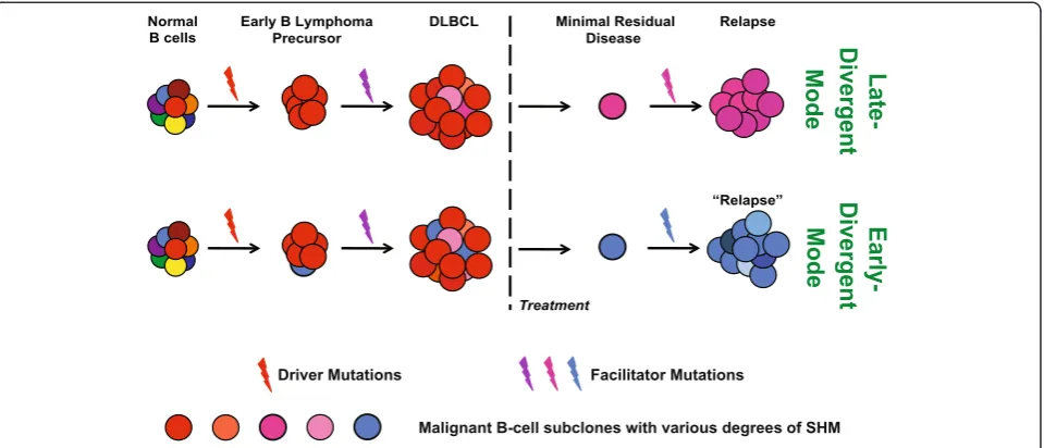
Figure 6A model of early- and late-divergent modes of DLBCL relapse.In both modes, the early B lymphoma precursors arise from normal B cells that sustain key genetic lesions, such as mutations occurring at epigenetic modifying enzymes, that is,EP300,KMT2D, andSETDB1. In the late-divergent mode (top panel), after acquiring additional facilitator mutations, early B lymphoma precursors develop into DLBCL-containing subclones that have similar but slightly different SHM profiles (indicated by different shades of red) due to ongoing SHM. After treatment, one or few subclones survive and develop into relapse disease, potentially by acquiring additional mutations. In the early-divergent mode (bottom panel), early B lymphoma precursor cells progress into initial DLBCL-containing subclones that have similar SHMs (indicated by different shades of red) and one or few minor subclones (depicted by blue) that have unique SHM profiles vastly different from the subclones of the major diagnostic clone (the red ones), indicating divergence during clonal expansion of the tumor. This minor clone later survives or escapes chemotherapy, and develops into a relapse tumor that has a diverged subclonal origin from the diagnosis tumor (blue versus red).
Jianget al. Genome Biology2014,15:432 Page 14 of 17
shaken vigorously for 15 s before being spun at 2,500 rpm for 15 minutes. Supernatant was transferred to a new tube and mixed with two volumes of room temperature 100% ethanol. DNA was precipitated by centrifugation at max-imum speed for 30 minutes, washed twice with 70% etha-nol, and finally dissolved in TE or nuclease-free water overnight at room temperature. Formalin-fixed paraffin-embedded samples were de-paraffined first by incubating in Xylene at room temperature for 10 minutes twice followed by incubating with 100% ethanol at room temperature for 10 minutes twice. Samples were then allowed to air-dry and then incubated with 0.5 mg/ml Proteinase K in 1X PCR buffer overnight at 37°C followed by 95°C for 10 minutes to heat inactivate Proteinase K.
VHDJHsequencing
The IgVHFR1 VDJ junctions were amplified using mix 2 of the Somatic Hypermutation Assay from InvivoScribe Technologies (San Diego, CA, U.S.A.). The IgVHFR2 VDJ junctions were amplified using Tube B of IGH Gene Clonality Assay from InvivoScribe Technologies. PCR products were resolved on 2% agarose gel and the major product of the appropriate size was purified by using a QIAGEN MiniElute Gel Extraction kit (Valencia, CA, U.S.A.). Sequencing libraries were constructed from the purified PCR product by using Illumina TruSeq DNA Sample Preparation Kit v2 (San Diego, CA, U.S.A.). Each sample was tagged with a unique index. For each MiSeq sequencing run, five VDJ samples were mixed together at 7μM with 50% PhiX spike-in to ensure the complexity of the run.
Exome sequencing
Exome sequencing samples were prepared using the Aglient SureSelectXT Human All Exon 50 MB Target Enrichment System for Illumina Paired-End Sequencing Library kit. PE75 sequencing was performed on Illumina HiSeq 2000. For sample 1, we sequenced all three relapse samples that were obtained at three separate biopsy sites, and analyzed them independently.
VHDJHanalysis pipeline
Paired-end sequence reads were mapped against a human IGH reference database available from the IMGT website [9] using a modified nucleotide blast search. Sequences without a hit in all three V, D and J regions were filtered out and reads with all three regions present were counted for the number of each unique rearrangement. These counts were then ranked for each sample and the major rearrangements were then aligned against their corresponding dominant VHDJHsequences. To perform
phylogenetic analysis, within each sample we selected the subclones of the major VHDJH rearrangement that
had at least 10 sequencing counts and a minimum 80%
sequence similarity to the reference germline VDJ sequence. We then ranked these subclones in the diagnosis sample based on their similarity to the SHM profile of the respective relapse sample, and vice versa. The top 10 ranked subclones along with the 10 most abundant clones and a random selection of minor subclones from each diagnosis and re-lapse samples within each pair were then re-aligned using the multiple sequence alignment tool Clustalw to build a neighbor-joining tree by using the R package 'ape' [13]. The tree coordinates alongside the corresponding frequencies for each alignment were drawn using a custom script.
Exome analysis pipeline
SNV discovery
Short sequencing reads were aligned to human genome assembly GRCh37/hg19 using the BWA aligner [48]. Duplicated paired reads were filtered and variant detection was performed as previously described [14-16]. Novel cod-ing region SNVs (not present in SNP132) were further fil-tered according to sequencing depth (≥20X) and variant percentage (≥25%). To analyze the mutational status change (gain or loss) within each diagnosis and relapse pair, we compared the variant ratio of each novel coding SNV between the diagnosis and relapse samples and estimated the statistical significance of the difference by using a Chi-square tested corrected with multiple hypothesis testing (Benjamini-Hochberg corrected P< 0.1). To obtain the list of common novel coding region SNVs, we took those SNVs that did not show a signifi-cant different variant ratio between the diagnosis and relapse samples (corrected P> 0.1) and selected the ones that were mutated in at least two non-Hodgkin’s lymphoma patients in other studies.
CNA segment calling
In order to call CNA segments, aligned short sequencing reads were used to generate log2 ratios between the diagnosis and relapse samples for each patient using an in-house program, CNVseeqer. The log2 ratios represent the number of reads mapped to an exon in the relapse sample compared with a diagnosis sample. Log2 ratios of exons that had more than 100 total reads between both the diagnosis and relapse samples were smoothed and then used for segmentation identification using a circular binary segmentation algorithm DNAcopy in R [22]. Segments with a standard deviation >1.5 were defined as CNA segments. Segments with a mean log2 ratio >0.3 were categorized as copy number gain loci, while segments with a mean log2 ratio <−0.3 were categorized as copy number loss loci.
Data deposition
All targeted sequencing raw data files, including VDJ-sequencing and targeted reVDJ-sequencing of the UBR4 and B2M loci, have been deposited into NBCI database BioProject PRJNA240336 [50].
Additional files
Additional file 1: Table S1.Clinical information of the patient samples used in this study.
Additional file 2: Table S2.Sequencing run metrics.
Additional file 3:Supplementary figures.
Additional file 4: Table S3.Coding region nonsynonymous SNVs gained or lost in relapse samples.
Additional file 5: Table S4.Sanger validation results on selected nonsynonymous SNVs gained in relapse samples.
Additional file 6: Table S5.Coding region indels gained in relapse samples.
Additional file 7: Table S6.Gained copy number alterations in relapse samples.
Additional file 8: Table S7.Sequencing results of epigenetic modifier mutations in sample pairs 1, 2, and 9.
Abbreviations
B-All:acute B lymphoblastic leukemia; BM: bone marrow; bp: base pair; CNA: copy number alteration; DLBCL: diffuse large B-cell lymphoma; FL: follicular lymphoma; GC: germinal center; IGH: immunoglobulin heavy chain; IL: interleukin; PCR: polymerase chain reaction; SHM: somatic hypermutation; SNP: single nucleotide polymorphism; SNV: single nucleotide variant.
Competing interests
The authors declare that they have no competing financial interests.
Authors’contributions
YJ, WT, and OE conceived the study. TC, PM, LHCT, and WT collected samples. YJ and KN performed the experiments. YJ, DR, and KWE performed data analyses. YJ, AMM, WT, and OE wrote the manuscript. All authors have read and approved the manuscript. WT and OE are co-senior authors.
Acknowledgements
We would like to thank Dr Rita Shaknovich and members of Melnick lab, Tam lab, and Elemento lab for thoughtful discussions. We would like to thank the Genomics Resources Core Facility at Weill Cornell Medical College for performing the VDJ and exome sequencing. YJ is supported by ASH Scholar Award. WT and OE are supported by Weill Cornell Cancer Center Pilot Grant. OE is supported by the NSF CAREER award, the Starr Cancer Consortium and the Hirschl Trust. We would also like to thank Katherine Benesch, JD, MPH for her generous support to this project.
Author details
1Institute for Computational Biomedicine, Weill Cornell Medical College, New
York, NY 10021, USA.2Department of Medicine, Weill Cornell Medical College, New York, NY 10021, USA.3Department of Pathology and Laboratory Medicine, Weill Cornell Medical College, New York, NY 10021, USA.4Hematology, Lymphoid Unit, Hôpital Henri Mondor, Creteil 94010, France.5Department of Pathology, Singapore General Hospital, Singapore 169608, Singapore.
Received: 22 July 2014 Accepted: 7 August 2014 Published: 15 August 2014
References
1. Coiffier B, Lepage E, Briere J, Herbrecht R, Tilly H, Bouabdallah R, Morel P, Van Den Neste E, Salles G, Gaulard P, Reyes F, Lederlin P, Gisselbrecht C:CHOP chemotherapy plus rituximab compared with CHOP alone in elderly patients with diffuse large-B-cell lymphoma.N Engl J Med2002,346:235–242.
2. Larouche JF, Berger F, Chassagne-Clement C, Ffrench M, Callet-Bauchu E, Sebban C, Ghesquieres H, Broussais-Guillaumot F, Salles G, Coiffier B:Lymphoma recurrence 5 years or later following diffuse large B-cell lymphoma: clinical characteristics and outcome.J Clin Oncol2010,28:2094–2100.
3. Gisselbrecht C, Glass B, Mounier N, Singh Gill D, Linch DC, Trneny M, Bosly A, Ketterer N, Shpilberg O, Hagberg H, Ma D, Briere J, Moskowitz CH, Schmitz N:
Salvage regimens with autologous transplantation for relapsed large B-cell lymphoma in the rituximab era.J Clin Oncol2010,28:4184–4190. 4. Friedberg JW:Relapsed/refractory diffuse large B-cell lymphoma.
Hematology Am Soc Hematol Educ Program2011,2011:498–505. 5. Ding L, Ley TJ, Larson DE, Miller CA, Koboldt DC, Welch JS, Ritchey JK,
Young MA, Lamprecht T, McLellan MD, McMichael JF, Wallis JW, Lu C, Shen D, Harris CC, Dooling DJ, Fulton RS, Fulton LL, Chen K, Schmidt H, Kalicki-Veizer J, Magrini VJ, Cook L, McGrath SD, Vickery TL, Wendl MC, Heath S, Watson MA, Link DC, Tomasson MH,et al:Clonal evolution in relapsed acute myeloid leukaemia revealed by whole-genome sequencing.Nature2012,481:506–510. 6. Jung D, Alt FW:Unraveling V(D)J recombination; insights into gene
regulation.Cell2004,116:299–311.
7. Berek C, Berger A, Apel M:Maturation of the immune response in germinal centers.Cell1991,67:1121–1129.
8. Jacob J, Kelsoe G, Rajewsky K, Weiss U:Intraclonal generation of antibody mutants in germinal centres.Nature1991,354:389–392.
9. Lefranc MP, Giudicelli V, Ginestoux C, Jabado-Michaloud J, Folch G, Bellahcene F, Wu Y, Gemrot E, Brochet X, Lane J, Regnier L, Ehrenmann F, Lefranc G, Duroux P:
IMGT, the international ImMunoGeneTics information system.Nucleic Acids Res
2009,37:D1006–D1012.
10. Gawad C, Pepin F, Carlton VE, Klinger M, Logan AC, Miklos DB, Faham M, Dahl G, Lacayo N:Massive evolution of the immunoglobulin heavy chain locus in children with B precursor acute lymphoblastic leukemia.
Blood2012,120:4407–4417.
11. Yancopoulos GD, Alt FW:Regulation of the assembly and expression of variable-region genes.Annu Rev Immunol1986,4:339–368.
12. Arnaout R, Lee W, Cahill P, Honan T, Sparrow T, Weiand M, Nusbaum C, Rajewsky K, Koralov SB:High-resolution description of antibody heavy-chain repertoires in humans.PLoS One2011,6:e22365.
13. Saitou N, Nei M:The neighbor-joining method: a new method for reconstructing phylogenetic trees.Mol Biol Evol1987,4:406–425. 14. Jiang Y, Soong TD, Wang L, Melnick AM, Elemento O:Genome-wide detection
of genes targeted by non-Ig somatic hypermutation in lymphoma.PLoS One
2012,7:e40332.
15. Wacker SA, Houghtaling BR, Elemento O, Kapoor TM:Using transcriptome sequencing to identify mechanisms of drug action and resistance.
Nat Chem Biol2012,8:235–237.
16. Rajadhyaksha AM, Elemento O, Puffenberger EG, Schierberl KC, Xiang JZ, Putorti ML, Berciano J, Poulin C, Brais B, Michaelides M, Weleber RG, Higgins JJ:Mutations in FLVCR1 cause posterior column ataxia and retinitis pigmentosa.Am J Hum Genet2010,87:643–654.
17. Lohr JG, Stojanov P, Lawrence MS, Auclair D, Chapuy B, Sougnez C, Cruz-Gordillo P, Knoechel B, Asmann YW, Slager SL, Novak AJ, Dogan A, Ansell SM, Link BK, Zou L, Gould J, Saksena G, Stransky N, Rangel-Escareno C, Fernandez-Lopez JC, Hidalgo-Miranda A, Melendez-Zajgla J, Hernandez-Lemus E, Schwarz-Cruz y Celis A, Imaz-Rosshandler I, Ojesina AI, Jung J, Pedamallu CS, Lander ES, Habermann TM,et al:Discovery and prioritization of somatic mutations in diffuse large B-cell lymphoma (DLBCL) by whole-exome sequencing.Proc Natl Acad Sci U S A2012,109:3879–3884.
18. Pasqualucci L, Trifonov V, Fabbri G, Ma J, Rossi D, Chiarenza A, Wells VA, Grunn A, Messina M, Elliot O, Chan J, Bhagat G, Chadburn A, Gaidano G, Mullighan CG, Rabadan R, Dalla-Favera R:Analysis of the coding genome of diffuse large B-cell lymphoma.Nat Genet2011,43:830–837. 19. Morin RD, Mendez-Lago M, Mungall AJ, Goya R, Mungall KL, Corbett RD,
Johnson NA, Severson TM, Chiu R, Field M, Jackman S, Krzywinski M, Scott DW, Trinh DL, Tamura-Wells J, Li S, Firme MR, Rogic S, Griffith M, Chan S, Yakovenko O, Meyer IM, Zhao EY, Smailus D, Moksa M, Chittaranjan S, Rimsza L, Brooks-Wilson A, Spinelli JJ, Ben-Neriah S,et al:Frequent mutation of histone-modifying genes in non-Hodgkin lymphoma.Nature2011,476:298–303. 20. Zhang J, Grubor V, Love CL, Banerjee A, Richards KL, Mieczkowski PA,
Dunphy C, Choi W, Au WY, Srivastava G, Lugar PL, Rizzieri DA, Lagoo AS, Bernal-Mizrachi L, Mann KP, Flowers C, Naresh K, Evens A, Gordon LI, Czader M, Gill JI, Hsi ED, Liu Q, Fan A, Walsh K, Jima D, Smith LL, Johnson AJ, Byrd JC, Luftig MA,et al:Genetic heterogeneity of diffuse large B-cell lymphoma.
Proc Natl Acad Sci U S A2013,110:1398–1403.
Jianget al. Genome Biology2014,15:432 Page 16 of 17
21. Beguelin W, Popovic R, Teater M, Jiang Y, Bunting KL, Rosen M, Shen H, Yang SN, Wang L, Ezponda T, Martinez-Garcia E, Zhang H, Zheng Y, Verma SK, McCabe MT, Ott HM, Van Aller GS, Kruger RG, Liu Y, McHugh CF, Scott DW, Chung YR, Kelleher N, Shaknovich R, Creasy CL, Gascoyne RD, Wong KK, Cerchietti L, Levine RL, Abdel-Wahab O,et al:EZH2 is required for germinal center formation and somatic EZH2 mutations promote lymphoid transformation.Cancer Cell2013,23:677–692.
22. Olshen AB, Venkatraman ES, Lucito R, Wigler M:Circular binary segmentation for the analysis of array-based DNA copy number data.
Biostatistics2004,5:557–572.
23. Lonigro RJ, Grasso CS, Robinson DR, Jing X, Wu YM, Cao X, Quist MJ, Tomlins SA, Pienta KJ, Chinnaiyan AM:Detection of somatic copy number alterations in cancer using targeted exome capture sequencing.
Neoplasia2011,13:1019–1025.
24. Challa-Malladi M, Lieu YK, Califano O, Holmes AB, Bhagat G, Murty VV, Dominguez-Sola D, Pasqualucci L, Dalla-Favera R:Combined genetic inactivation of beta2-Microglobulin and CD58 reveals frequent escape from immune recognition in diffuse large B cell lymphoma.Cancer Cell
2011,20:728–740.
25. Manser E, Loo TH, Koh CG, Zhao ZS, Chen XQ, Tan L, Tan I, Leung T, Lim L:
PAK kinases are directly coupled to the PIX family of nucleotide exchange factors.Mol Cell1998,1:183–192.
26. Harden TK, Hicks SN, Sondek J:Phospholipase C isozymes as effectors of Ras superfamily GTPases.J Lipid Res2009,50:S243–S248.
27. Dumont C, Henderson R, Tybulewicz VL:Characterization of the roles of Rac1 and Rac2 GTPases in lymphocyte development.Methods Enzymol
2008,439:235–254.
28. Johmura S, Oh-hora M, Inabe K, Nishikawa Y, Hayashi K, Vigorito E, Kitamura D, Turner M, Shingu K, Hikida M, Kurosaki T:Regulation of Vav localization in membrane rafts by adaptor molecules Grb2 and BLNK.Immunity2003,
18:777–787.
29. Demoulin JB, Uyttenhove C, Van Roost E, DeLestre B, Donckers D, Van Snick J, Renauld JC:A single tyrosine of the interleukin-9 (IL-9) receptor is required for STAT activation, antiapoptotic activity, and growth regulation by IL-9.
Mol Cell Biol1996,16:4710–4716.
30. O’Shea JJ, Holland SM, Staudt LM:JAKs and STATs in immunity, immunodeficiency, and cancer.N Engl J Med2013,368:161–170. 31. Goodarzi H, Elemento O, Tavazoie S:Revealing global regulatory perturbations across human cancers.Mol Cell2009,36:900–911. 32. Young RM, Staudt LM:Targeting pathological B cell receptor signalling in
lymphoid malignancies.Nat Rev Drug Discov2013,12:229–243. 33. Xu-Monette ZY, Wu L, Visco C, Tai YC, Tzankov A, Liu WM, Montes-Moreno S,
Dybkaer K, Chiu A, Orazi A, Zu Y, Bhagat G, Richards KL, Hsi ED, Zhao XF, Choi WW, Zhao X, van Krieken JH, Huang Q, Huh J, Ai W, Ponzoni M, Ferreri AJ, Zhou F, Kahl BS, Winter JN, Xu W, Li J, Go RS, Li Y,et al:
Mutational profile and prognostic significance of TP53 in diffuse large B-cell lymphoma patients treated with R-CHOP: report from an International DLBCL Rituximab-CHOP Consortium Program Study.
Blood2012,120:3986–3996.
34. Freeman JD, Warren RL, Webb JR, Nelson BH, Holt RA:Profiling the T-cell receptor beta-chain repertoire by massively parallel sequencing.Genome Res2009,19:1817–1824.
35. Boyd SD, Gaeta BA, Jackson KJ, Fire AZ, Marshall EL, Merker JD, Maniar JM, Zhang LN, Sahaf B, Jones CD, Simen BB, Hanczaruk B, Nguyen KD, Nadeau KC, Egholm M, Miklos DB, Zehnder JL, Collins AM:Individual variation in the germline Ig gene repertoire inferred from variable region gene rearrangements.J Immunol2010,184:6986–6992.
36. Campbell PJ, Pleasance ED, Stephens PJ, Dicks E, Rance R, Goodhead I, Follows GA, Green AR, Futreal PA, Stratton MR:Subclonal phylogenetic structures in cancer revealed by ultra-deep sequencing.Proc Natl Acad Sci U S A2008,105:13081–13086.
37. Boyd SD, Marshall EL, Merker JD, Maniar JM, Zhang LN, Sahaf B, Jones CD, Simen BB, Hanczaruk B, Nguyen KD, Nadeau KC, Egholm M, Miklos DB, Zehnder JL, Fire AZ:Measurement and clinical monitoring of human lymphocyte clonality by massively parallel VDJ pyrosequencing.Sci Transl Med2009,1:12ra23.
38. Logan AC, Gao H, Wang C, Sahaf B, Jones CD, Marshall EL, Buno I, Armstrong R, Fire AZ, Weinberg KI, Mindrinos M, Zehnder JL, Boyd SD, Xiao W, Davis RW, Miklos DB:High-throughput VDJ sequencing for quantification of minimal residual disease in chronic lymphocytic leukemia and immune reconstitution assessment.Proc Natl Acad Sci U S A2011,108:21194–21199.
39. de Jong D, Glas AM, Boerrigter L, Hermus MC, Dalesio O, Willemse E, Nederlof PM, Kersten MJ:Very late relapse in diffuse large B-cell lymphoma represents clonally related disease and is marked by germinal center cell features.Blood2003,102:324–327.
40. Pasqualucci L, Khiabanian H, Fangazio M, Vasishtha M, Messina M, Holmes AB, Ouillette P, Trifonov V, Rossi D, Tabbo F, Ponzoni M, Chadburn A, Murty VV, Bhagat G, Gaidano G, Inghirami G, Malek SN, Rabadan R, Dalla-Favera R:
Genetics of follicular lymphoma transformation.Cell Rep2014,6:130–140. 41. Morin RD, Johnson NA, Severson TM, Mungall AJ, An J, Goya R, Paul JE,
Boyle M, Woolcock BW, Kuchenbauer F, Yap D, Humphries RK, Griffith OL, Shah S, Zhu H, Kimbara M, Shashkin P, Charlot JF, Tcherpakov M, Corbett R, Tam A, Varhol R, Smailus D, Moksa M, Zhao Y, Delaney A, Qian H, Birol I, Schein J, Moore R,et al:Somatic mutations altering EZH2 (Tyr641) in follicular and diffuse large B-cell lymphomas of germinal-center origin.
Nat Genet2010,42:181–185.
42. Green MR, Gentles AJ, Nair RV, Irish JM, Kihira S, Liu CL, Kela I, Hopmans ES, Myklebust JH, Ji H, Plevritis SK, Levy R, Alizadeh AA:Hierarchy in somatic mutations arising during genomic evolution and progression of follicular lymphoma.Blood2013,121:1604–1611.
43. Mar BG, Bullinger LB, McLean KM, Grauman PV, Harris MH, Stevenson K, Neuberg DS, Sinha AU, Sallan SE, Silverman LB, Kung AL:Lo Nigro L, Ebert BL, Armstrong SA: Mutations in epigenetic regulators including SETD2 are gained during relapse in paediatric acute lymphoblastic leukaemia.
Nat Commun2014,5:3469.
44. Mullighan CG, Zhang J, Kasper LH, Lerach S, Payne-Turner D, Phillips LA, Heatley SL, Holmfeldt L, Collins-Underwood JR, Ma J, Buetow KH, Pui CH, Baker SD, Brindle PK, Downing JR:CREBBP mutations in relapsed acute lymphoblastic leukaemia.Nature2011,471:235–239.
45. Oricchio E, Nanjangud G, Wolfe AL, Schatz JH, Mavrakis KJ, Jiang M, Liu X, Bruno J, Heguy A, Olshen AB, Socci ND, Teruya-Feldstein J, Weis-Garcia F, Tam W, Shaknovich R, Melnick A, Himanen JP, Chaganti RS, Wendel HG:The Eph-receptor A7 is a soluble tumor suppressor for follicular lymphoma.
Cell2011,147:554–564.
46. Davis RE, Ngo VN, Lenz G, Tolar P, Young RM, Romesser PB, Kohlhammer H, Lamy L, Zhao H, Yang Y, Xu W, Shaffer AL, Wright G, Xiao W, Powell J, Jiang JK, Thomas CJ, Rosenwald A, Ott G, Muller-Hermelink HK, Gascoyne RD, Connors JM, Johnson NA, Rimsza LM, Campo E, Jaffe ES, Wilson WH, Delabie J, Smeland EB, Fisher RI,et al:Chronic active B-cell-receptor signalling in diffuse large B-cell lymphoma.Nature2010,463:88–92.
47. Fowler N, Davis E:Targeting B-cell receptor signaling: changing the paradigm.
Hematology Am Soc Hematol Educ Program2013,2013:553–560.
48. Li H, Durbin R:Fast and accurate short read alignment with Burrows-Wheeler transform.Bioinformatics2009,25:1754–1760.
49. NCBI database BioProject PRJNA240335.[http://www.ncbi.nlm.nih.gov/ bioproject/?term=PRJNA240335]
50. NBCI database BioProject PRJNA240336.[http://www.ncbi.nlm.nih.gov/ bioproject/?term=PRJNA240336]
doi:10.1186/s13059-014-0432-0
Cite this article as:Jianget al.:Deep sequencing reveals clonal evolution patterns and mutation events associated
with relapse in B-cell lymphomas.Genome Biology201415:432.
Submit your next manuscript to BioMed Central and take full advantage of:
• Convenient online submission
• Thorough peer review
• No space constraints or color figure charges
• Immediate publication on acceptance
• Inclusion in PubMed, CAS, Scopus and Google Scholar
• Research which is freely available for redistribution