http://dx.doi.org/10.4236/jct.2014.58086
Gastrointestinal Stromal Tumors
in the 21
st
Century
Rani Kanthan
1, Jenna-Lynn Senger
1, Shahid Ahmed
2, Selliah Chandra Kanthan
3 1Department of Pathology & Laboratory Medicine, University of Saskatchewan, Saskatoon, Canada 2Divison of Medical Oncology, Department of Medicine, University of Saskatchewan, Saskatoon, Canada 3Department of Surgery, University of Saskatchewan, Saskatoon, CanadaEmail: rani.kanthan@saskatoonhealthregion.ca
Received 12 May 2014; revised 10 June 2014; accepted 3 July 2014
Copyright © 2014 by authors and Scientific Research Publishing Inc.
This work is licensed under the Creative Commons Attribution International License (CC BY). http://creativecommons.org/licenses/by/4.0/
Abstract
Gastrointestinal stromal tumours (GISTs) are rare mesenchymal lesions accounting for only 0.2% of all gastrointestinal neoplasms. These tumors arise from the interstitial cells of Cajal, with muta-tions described in proto-oncogenes such as KIT, PDGFRA, DOG-1, and SDH. The majority of these lesions are asymptomatic, thus the true incidence remains unknown. While patients typically un-dergo initial endoscopy, CT scan and/or MRI, findings are often nonspecific and require a biopsy to identify the tumor. As such, immunohistochemical evaluation is the gold-standard for the accu-rate diagnosis of GIST. Though surgical excision remains the gold-standard for curative manage-ment, the discovery of imatinib, a tyrosine kinase inhibitor (TKI), has revolutionized the
treat-ment of GIST in the 21st century as a “prototype” of molecular targeted therapy for solid tumors.
Risk assessment for recurrence divides these tumors into low and high-risk categories. In the lat-ter, a role for adjuvant therapy with TKI confers a significantly better prognosis than previously observed. However, secondary mutations conferring drug resistance remain an ongoing challenge for management, as few alternative treatment options are available for patients intolerant/re- fractory to TKI therapy. In this review, we summarize the epidemiology, molecular pathogenesis, clinical presentation, diagnosis, pathology features, management options, and prognostic features of GISTs.
Keywords
Gastrointestinal Stromal Tumor, Targeted Therapy, Drug Resistance, Histopathology, Surgery
1. History & Evolution of the Terminology “GIST”
intestinal tract. In the past, there has been considerable discord regarding its nomenclature, cellular origin, diag-nosis, treatment, and prognosis [1]. These lesions were originally thought to be smooth muscle/spindle cell neoplasms and were thus labelled as leiomyomas, leiomyoblastomas, leiomyosarcomas, or schwannomas [1] [2]. These tumors have also been referred to as “gastrointestinal autonomic nerve tumors” (GANT) [3]. In the 1960s, with the advent of electron microscopy, some of these gastric tumors demonstrated a lack of the typical smooth muscle differentiation [4].
The term “GIST” was first introduced in 1983 by Mazur and Clark to describe tumors lacking smooth muscle differentiation or immunohistochemical features of Schwann cells [4]. In 1998, Kindblom et al. described GISTs as originating from pluripotent mesenchymal stem cells programmed to differentiate into the interstitial cells of Cajal which are considered to act as the gastrointestinal pacemaker cell [5]. It is now recognized that these tu-mors arise from the cells of Cajal in the muscular layer of the gastrointestinal tract, anywhere from the foregut to the hindgut [6]. In the same year, Hirota et al. discovered a functional mutation in the c-kit proto-oncogene [5] [7]. KIT is a transmembrane receptor tyrosine kinase (TK) protein that is a product of the KIT proto-oncogene. The CD117 molecule is part of the KIT (c-kit) receptors and most GISTs stain positive for CD117. The use of molecular targeted therapy against c-kit with agents such as imatinib mesylate, a tyrosine kinase inhibitor (TKI), has revolutionized the management of GIST in the 21st century. Detection of this proto-oncogene protein by immunohistochemistry is thus a pivotal diagnostic and therapeutic target. Thus, gastrointestinal stromal tumor (GIST), as defined by their molecular signature, is the accepted nomenclature of this family of tumors.
The following is a comprehensive review that provides a discussion on the epidemiology, molecular patho-genesis, clinical presentation, diagnosis, pathology, management, and prognostic features of GIST.
2. Methodology
A systematic review of the published medical literature for English-language articles using PubMed and Med-line was carried out using the search terms: “gastrointestinal stromal tumor” and “GIST” with a special empha-sis on review articles. Secondary references obtained from these publications were also reviewed as appropriate. Selected relevant abstracts from key oncology meetings were also reviewed. We limited our search to reports published since January 1, 2000 as GIST was not a generally recognized tumor entity prior to this time.
3. Epidemiology
GISTs are rare, accounting for only 1% to 3% of all malignant gastrointestinal tumors and only 0.2% of all GI tract neoplasms [8] [9]. The annual incidence of GIST is 11 - 14/million annually and the median age of diagno-sis is 60 years (range 51 - 60) [1] [10]. These tumors are very rare in the pediatric population, with <1% occur-ring prior to age 21 [11]. This number may be an underestimation, as it is likely that many smaller GISTs go undetected as they are asymptomatic [12]. The diagnosis of GIST has increased 25-fold in the past 2 decades which is likely related to incidental discoveries with increased use of many advanced imaging modalities and reclassification of many GI smooth muscle tumors as GISTs [13]. While some articles have reported no gender predisposition, others have indicated a distinct male preponderance [12]. GISTs are typically solitary tumors; however, there are published reports of synchronous GISTs in the stomach and jejunum. Familial GISTs how-ever are a notable exception to this observation, wherein multiple GISTs are more frequently encountered [14].
3.1. Anatomical Location: GIST vs. EGIST
GISTs may arise anywhere along the gastrointestinal tract, with the most common sites including wherever the interstitial cells of Cajal are present including the stomach (60%), small bowel (30%), duodenum (5%), colorec-tum (4%) and esophagus [1] [15]. While rare, extra-gastrointestinal GIST (eGIST) are most common (80%) in the omentum and mesentery; however, these have also been reported in the pleura, pancreas, abdominal wall, liver, rectovaginal septum, gallbladder, and urinary bladder. eGISTs are morphologically and immunopheno-typically similar to gastrointestinal GISTs; however, the pathogenesis and their exact cell of origin remains con-troversial. It is postulated that these lesions likely arise from multipotential mesenchymal stem cells, or possibly represent extramural outgrowths of GISTs that have lost contact with the muscular wall of the GI tract [15] [16].
3.2. Biological Behavior
minal spread is common in tumors with potentially aggressive biological behavior. However, lymph node dis-semination is extremely uncommon in adults. Up to 30% of all GISTs will exhibit malignant behavior including metastasis and infiltration [1]. Whether the benign vs. malignant GIST represents two distinct entities or a con-tinuing spectrum remains undetermined; however, it is suggested clonal expansion may be responsible for this transformation. This theory is supported by a case report that showed two GISTs in a single stomach, one of low malignant potential and the other with high malignancy [17]. GISTs have also been reported to arise synchron-ously with adenocarcinomas of the jejunum, colon and stomach [14] [18] [19].
4. Molecular Pathogenesis
4.1. KIT-Mutant GIST
In 1986 the oncogene v-KIT was identified encoded in the viral genome of HZ4-FeSV (Hardy-Zuckerman 4 fe- line sarcoma virus). V-KIT was subsequently found to encode a transmembrane tyrosine kinase receptor, KIT, and it was determined that the KIT receptor is expressed by the interstitial cells of Cajal (ICCs), the pacemaker cells of the gastrointestinal tract found in the myenteric plexus [1] [2] [20]. ICCs are able to transdifferentiate into smooth muscle cells and thus have stem-cell-like characteristics [11]. It is suggested that hyperplasia of the ICCs may be a precursor lesion to the development of GIST [21]. It has been shown that microscopic foci of KIT-positive spindle hyperplasia is common in patients with KIT/PDGFRA mutations; however, <1% will progress to a clinically significant GIST [22].
Normally KIT is autoinhibited, unless bound by a stem cell factor (SCF), which causes KIT receptor homo- dimerization. This subsequently leads to activation of KIT tyrosine kinase activity, affecting intracellular signal transduction [1]. Downstream pathways including mitogen-activated protein kinase and phosphatidylinositol 3-kinase (PI3K) are activated [23]. Additionally, the RAS-RAF-MAPK pathway is also implicated [24]. It is recognized that products of RAS are downstream of KIT and PDGFRA and thus likely play a role as an onco-genic driver mutation in GISTs. This signal transduction is involved in the regulation of cellular proliferation, differentiation, and anti-apoptotic signaling [1].
KIT mutations have been identified in GISTs, wherein they play a central role in its pathogenesis [1]. This is thought to be an early event in the tumorigenesis of GIST, particularly involving exon 11 [25]. KIT mutations release the autoinhibition such that the receptor remains in a constitutively active state [26]. Mutations may be further classified into primary vs. secondary mutations, the latter developing through exposure to tyrosine kinase inhibitors leading to drug resistance [4]. Primary mutations include in-frame deletions or insertions, point muta-tions, and duplications [4] [20]. The majority of these mutations occur in exon 11, and cause disruption of the juxtamembrane scaffolding, thus preventing the kinase activation loop from alternating into an active configura-tion [13] [24]. In-frame deletions are the most common (60% - 70%) mutation at this site followed by missense point mutation (20% - 30%) [24]. Less commonly exons 9 (extracellular domain), 13, and 17 may be involved [6]. Mutations of the extracellular domain of KIT, predominately exon 9, and in kinase I and II domains (exons 13 and 17) may also be present [20].
In the presence of a mutation, when c-KIT is autophosphorylated, ligand-independent tyrosine kinase activity leads to uncontrolled cellular proliferation. As SCF-KIT interactions are involved in the normal development of melanocytes, erythrocytes, germ cells, mast cells, and ICCs, mutations cause defects in melanogenesis, hemato-poiesis, gametogenesis, and in the interstitial cells of Cajal [1]. Secondary mutations occur in KIT kinase do-mains during or after imatinib treatment, leading to drug resistance [4].
4.2. PDGFRA-Mutant GIST
exclusive [22] [29].
4.3. Wild-Type GIST
Approximately 15% of GISTs have no KIT or PDGFRA mutations and are thus classified as “wild-type tumors”. Nevertheless, these tumors stain positively for KIT on immunohistochemistry [13]. Many of these wild-type tumors have BRAF V600E mutations at exon 15 [22]. This mutation is thought to arise in early-stage incidental GISTs, and confer a low risk for malignant potential. Tumors with BRAF mutations most commonly arise in the small bowel [30]. Mutations of NF1, BRAF, NRAS, and KRAS have also been described [6] as well as variants in CYP1B1, RAD23B, GSTM1, and ERCC2 [31]. A germline alteration of the BAX gene, which encodes a BCL2- associated protein that is directly activated by p53, has also been reported in GISTs [32].
A subset of KIT/PDGFRA wild-type GISTs are characterized by germline/somatic mutations of succinate de- hydrogenase (SDH) subunits A, B, C, D (SDHA, SDHB, SDHC, SDHD). Only 7.5% of GISTs are driven by SDH deficiency rather than the typical KIT/PDGFRA mutation, of which half are due to mutations of the SDH- subunit genes and half are attributable to epigenetic silencing. SDH deficient cells accumulate succinate which leads to overexpression of aberrant transcription factors resulting in hypoxia-associated tumorigenesis and an-giogenesis, referred to as “hypoxic drive” or “pseudohypoxia signaling”. SDH-deficient GISTs are also asso-ciated with over expression of Insulin-Like-Growth Factor-1 Receptor (IGF1) possibly by gene amplification. Thus, high expression of IGF-1 protein detected immunohistochemically is a strong surrogate marker for the diagnosis of SDH-deficient GISTs in the appropriate clinical scenario. SDH-mutated GISTs are more frequent in a younger population, encompassing the majority of pediatric GISTs as well as GISTs associated with Carney- Stratakis syndrome, with a greater prevalence in females, and arise exclusively in the stomach, typically in a multinodular pattern. On histopathological examination, cells typically have an epithelioid morphology with eo- sinophilic cytoplasm, contrasting the typical spindle-cells and pale cytoplasm of KIT-mutant GISTs. These GISTs express KIT equally strong as KIT-mutant GISTs and are also uniformly positive for DOG1/Anocta- min-1 by immunohistochemistry. SDHA mutation has an excellent correlation with immunohistochemical loss of SDHA expression and is thus an excellent screening tool and surrogate marker for SDHA mutation analysis. Up to 50% of patients present with lymphovascular invasion. Despite the presence of node positivity or liver metastases, SDH-mutated GISTs usually have an indolent course with patients living many years after identifi-cation of metastases. A minority of SDH-silenced GISTs are fatal within a few years of diagnosis (up to 15%), while others will metastasize >10 years after the initial diagnosis. Delayed recurrences up to 42 years have also been described [33] [34].
4.4. Chromosomal Alterations
Typical chromosomal alterations in GISTs include losses at 1p, 14q, 15q, and 22q [4]. Loss of 9p is associated with an aggressive behavior, corresponding to loss of CDKN2A. Similarly, aggressive/metastatic behavior is as-sociated with gains in chromosomes 5p, 20q, 8q, and 17q [22].
4.5. Epigenetics
While KIT or PDGFRA mutations are present in the majority of cases, it is speculated that epigenetic alterations may also contribute to the malignant transformation. Hypomethylation of LINE1 correlating with the aggres-siveness of GISTs, is proportional to the total number of chromosomal aberrations, and may be a marker for risk assessment. Hypomethylation of SATA and NBL2 additionally are associated with high-risk GIST. Hypermethy-lation has been detected in several genes, the most common being MGMT (47%), P16/INK4A (45%), RASSFIA (40%), E-Cadherin (37%), HMLH1 (34%), and APC (31%) [6]. GISTs may also have di- or tri-methylation of lysine 4 on histone H3. The active histone H3K4me3 marks multiple genes including HOTAIR in malignant GISTs.
4.6. MicroRNA
proteins. Upregulation of these microRNAs is associated with downregulation of ETV1 protein [35]. Other mi-croRNAs that have been implicated include miR-494, -132, -504, -134/170, -196a, and 133b [6]. miR-494 is re-ported to target downstream KIT, thus inhibiting GIST cell growth while inducing apoptosis [35].
5. Clinical Presentation
Only 70% of patients with GISTs are symptomatic, with 20% being asymptomatic incidentalomas and 10% be-ing detected at autopsy. Presentbe-ing signs and symptoms depend on the site of involvement; however, abdominal discomfort is the most common symptom (60% - 70%) followed by gastrointestinal bleeding due to erosion into the lumen (30% - 40%). If bleeding is intraperitoneal, patients may present as an acute abdomen whereas he-morrhage into the lumen may present more chronically with hematemesis, melena, or anemia [1]. Anemia is present in ~50% of patients with GIST [36]. Bleeding is more common as a clinical presentation in duodenal GISTs (75%) compared with gastric (54%) or ileojejunal (28%) [37]. Many patients may have vague complaints including nausea, vomiting, weight loss, or early satiety [1] [2]. Bowel obstruction is uncommon in GISTs as it, like other sarcomas, displaces rather than invades adjacent structures [2]. More rarely, symptoms such as pelvic pain, pleuritic chest pain, and dysuria may be present [24]. Site-specific symptoms may include dysphagia (eso-phagus), biliary obstruction (ampulla of Vater), or intussusception (small bowel) [1] [38]. Rare presentations in-clude perforation, and hypoglycemia secondary to paraneoplastic production of insulin-like-growthfactor-II [8].
When <1.0 cm, these tumors are commonly referred to as “microGIST” and are most often incidental findings. These lesions are present in 10% - 22.5% of stomachs in middle-aged to elderly patients [20]. This high preva-lence suggests that the majority of KIT-positive microGISTs do not progress to malignant behavior [39]. Such microGISTS are most typically found at the GE junction or proximal stomach. MicroGISTs generally are im-munoreactive for KIT and contain KIT or PDGFRA mutations [40].
5.1. Familial GIST
Familial GISTs are the result of an autosomal dominant mutation of KIT exon 8, 11, 13, or 17. Such individuals are at risk of developing multiple GISTs as early as age 18. This germline mutation has 100% penetrance [41]. These patients may present with pigmented macules on the perineum, axilla, hands, and face as well as urticaria pigmentosa [20]. Morphologically, these GISTs are indistinguishable from sporadic tumors. This patient popu-lation is thought to confer a high risk of metastatic spread irrespective of tumor size and mitotic activity [22]. The worse prognosis in this patient population is, however, primarily due to local complications such as he-morrhage, perforation, or surgical complications due to the large number of multiple GISTs [41].
5.2. Carney’s Triad
Carney’s triad is a rare non-heritable syndrome mainly in young females that includes gastric GIST, paragan-glioma, and pulmonary chondroma. These lesions tend to be of the epithelioid subtype and have a higher risk of metastatic spread including lymphadenopathy. Tumors usually do not have KIT or PDGFRA mutations, with some occurring in the context of SDH germline mutation (SDH-deficient GIST) [34]. These GISTs are thought to be sporadic, non-familial, growing more slowly with frequent metastases, and have minimal or equivocal re-sponse to imatinib therapy [22].
5.3. Carney-Stratakis Syndrome
Carney-Stratakis syndrome occurs secondary to germline mutation of succinate dehydrogenase (SDH) that pre-disposes individuals to multifocal GISTs, paragangliomas, and pheochromocytomas [22]. Recently esophageal leiomyoma and adrenal cortical adenoma have been added as potential components to this syndrome [42]. These tumors are negative for KIT or PDGFRA mutations yet have loss of expression of succinate dehydrogenase subunit B. This syndrome is most commonly seen in young females, with >80% presenting prior to the age of 30. Unlike sporadic GISTs, these tumors more commonly have lymph node metastases [43].
5.4. Type I Neurofibromatosis
younger population than sporadic GISTs [44]. One autopsy series revealed up to one-third of patients with NF1 had undiagnosed GISTs [11]. These tumors are typically small, multifocal, and are usually KIT and PDGFRA- negative [43] [44].
5.5. Other Associations
Additionally, GISTs are thought to have a relationship with desmoid tumors. It is not yet understood whether the presence of a GIST predisposes to the development of a desmoid, or vice versa [43].
5.6. Pediatric GISTs
GISTs in children are rare, accounting for ~1% of these tumors [43]. Pediatric GISTs are most common in the stomach of females in the 2nd decade of life, and are often multifocal with a multinodular pattern of growth [42] [43]. These lesions are distinct in their pathogenesis and clinical behavior and are thus considered separate from their adult counterpart. Many of the clinical features of pediatric GIST are similar to tumors associated with Carney’s triad [22]. Children most often present with symptoms of anemia, pallor, fatigue, vertigo, vomiting, abdominal pain, distension, and/or intestinal obstruction (intussusception) [38]. Up to 20% of pediatric patients will present with metastases at diagnosis, typically to the liver, lymph nodes and peritoneum [3]. They uniformly express KIT and Dog1, though rarely have detectable mutations in KIT or PDGFR. Generally, pediatric GISTs follow an indolent course and some argue that treatment with imatinib therapy should be reserved only for pa-tients with metastatic disease or restricted to papa-tients in whom the tumor is unresectable [22].
6. Diagnosis
The majority of GISTs are detected by imaging. The radiologic appearance of GISTs is highly variable, compli-cating their accurate detection and diagnosis. Tumors may have intraluminal, intramural, or external components with/without pedunculated extramural or cystic appearance [11]. These tumors arise within the muscularis pro-pria and appear as endophytic or exophytic nodules with a mean size of 3 - 5 cm in diameter and are usually highly vascular and friable lesions [45].
6.1. Endoscopy
Upper GI endoscopy is normally the first investigation chosen, which typically reveals a smooth lesion protrud-ing into the bowel lumen with normal-appearprotrud-ing overlyprotrud-ing mucosa associated with or without superimposed ul-ceration [45]. Upper endoscopy with biopsy has a low diagnostic yield of only 17% - 42% [46]. Additionally, GI endoscopy is unable to determine the accurate size of the lesion due to the submucosal location of GISTs [45].
Endoscopic ultrasound (EUS) may be used to assess the depth of invasion and to obtain a tissue sample for histopathological confirmation. Features of a GIST on EUS include irregular extraluminal borders, heterogene-ous echo patterns, and the presence of cystic spaces and echogenic foci [1] [39] [47]. Lesions are typically in the fourth sonographic wall layer corresponding to the muscularis propria. This technique is limited by inter-obser- ver variability resulting in a diagnostic accuracy as low as 43%. By contrast, other studies have found a 100% positive predictive value when EUS features include a) irregular extraluminal margins, b) cystic spaces, and c) lymph nodes with malignant pattern [46].
6.2. CT Scan
6.3. MRI
Magnetic resonance imaging (MRI) has a comparable diagnostic yield to CT scans and is the preferred choice for the evaluation of GISTs in specific locations such as the rectum or liver [8]. It is additionally indicated when CT scanning is inconclusive or contraindicated [45]. GISTs appear as hypointense lesions on T1-weighted im-aging (T1WI) and hyperintense on T2-weighted imim-aging (T2WI) [48].
6.4. Biopsy
Biopsies are not routinely recommended for GISTs due to fear of intraabdominal seeding; however, it is a useful technique in the diagnosis of an unidentified abdominal neoplasm [39]. Percutaneous biopsy is not recommend-ed due to a high risk of inducing tumor rupture, and endoscopic modalities have supplantrecommend-ed this option [3]. En-doscopic biopsy specimens are, however, often unable to obtain sufficient tissue to make a diagnosis, as they are only successful in 20% - 30% of cases [11]. As GISTs are submucosal, routine forceps biopsy technique is not reliable [3]. Core-needle biopsy with a 19gauge Tru-cut needle may improve yield rates; however, this technique is limited to tumors in the fundus, antrum, and duodenal bulb [39]. Lesions not amenable to endoscopic biopsy may require a laparoscopic or open biopsy [46]. The choice of which biopsy technique to pursue depends on the anatomic location of the lesion, the patient’s physiological status, and the equipment and resources available [3].
Recently, endoscopic ultrasound-guided fine needle aspiration (EUS-FNA) has been reported to have an ac-curacy rate of 80% - 85% and is thus the procedure of choice to obtain a tissue diagnosis when indicated [1]. However, EUS-FNA fails to yield an adequate tissue sample in up to one-third of specimens [39]. Factors that affect the diagnostic yield include: site, size and characteristics of the tissue; technical and procedural factors; endosonographer and cytopathologist expertise [52]. Larger tumors have a higher diagnostic accuracy, with re-ported rates for tumours of 2 cm, 2 - 4 cm, and 4+ cm at 71%, 86%, and 100% respectively [53]. A similar pro-cedure, endoscopic ultrasound-guided tru-cut biopsy (EUS-TCB) which uses a stiffer device, has additionally been described as rapid and safe; however, no standards for its use have been determined [52].
6.5. PET Scan
Positron emission tomography (PET) can be used to detect GISTs because the receptor tyrosine kinase increases glucose transport protein signaling. This imaging modality is used to identify small metastases not detectable on CECT and to differentiate active tumor vs. necrotic/scar tissue [1]. The reported sensitivity of PET scanning is 86% - 100% [8]. PET scans may also be used to assess early response to tyrosine kinase inhibitor therapy, as this treatment creates changes at the metabolic level prior to detectable anatomic changes which are then de-tectable by CECT [1].
6.6. Miscellaneous
Video capsule endoscope (VCE) and double balloon enteroscopy (DBE) have both been used successfully in the detection of GISTs, with detection rates of 80% and 60% respectively [36].
The use of fluoroscopy has been previously described in the detection of GISTs. A recent study has success-fully used fluorescent anti-KIT antibodies to label GIST as an in vivo imaging modality in transgenic mice. Flu-orophore-conjugated antibodies against KIT are effective in labeling KIT-positive GISTs for the detection of primary and metastatic disease. Thus similar to CT scans, fluoroscopy may be used in the initial diagnosis and treatment planning as well as monitoring disease response to therapy [51].
7. Pathology
7.1. Gross Features
7.2. Cytopathology
Cytopathological findings obtained in FNA specimens suggestive of a GIST may include spindle shaped cells with scant cytoplasm and elongated-to-oval nuclei [52]. Cellularity is typically moderate to high. The presence of cohesive and tight fascicular bundles with minimal pleomorphism is suggestive of this neoplasm. Chromatin is typically fine, and nuclei are most often indistinct. The diagnosis of GIST can, however, only be suspected on cytology and confirmatory immunostaining is necessary for definitive diagnosis [57].
7.3. Histopathology
Three distinct histopathological subtypes have been described: spindle, epithelioid, and mixed [8] [20]. The spindle-type is the most common phenotype seen in 70% of GISTs, and fascicles of uniform spindled neoplastic cells with minimal nuclear atypia and perinuclear vacuoles can be visualized [23]. Extracellular dense collagen deposits (skeinoid fibres) may be seen [40]. Individual cells may have ill-defined cell borders with ovoid nuclei, fine nuclear chromatin, and inconspicuous nucleoli. The cytoplasm is often pale, eosinophilic, with a fibrillary quality. Nuclear palisading reminiscent of Antoni A regions of schwannoma may be observed. The epithelioid- type is characterized by the presence of round cells in sheets or nests [22]. Cytoplasm may be clear, rhabdoid, plasmacytoid retracted spider-like, or vacuolated [58]. The mixed-type contains elements of both the spindle and epithelioid morphology in varying proportions.
Certain morphological patterns have a predilection in relation to the anatomical location. On histopathology, common features of gastric GISTs include cells with a sclerosing matrix, perinuclear vacuolization, and nuclear palisading, with an epithelioid or sarcomatoid, mitotically active morphology [11]. Gastric GISTs appear as sol-id/nested tumors with an epithelioid morphology and a stroma showing myxoid change whereas small bowel tumors are spindle-type with a paragangliomatous architectural pattern [1]. Small bowel GISTs are more homo-genous than gastric GISTs, and contain extracellular collagen globules and Verocay bodies or neuropil-like ma-terial [11][56]. Colonic GISTs typically mirror their small bowel counterpart, and histologic features of rectal GISTs include features of both gastric and small bowel [56].
7.4. Immunohistochemistry
The immunohistochemistry of GISTs has been well researched and remains the only true diagnostic test for its accurate and definitive diagnosis. The vast majority (>95%) of GISTs are positive for CD117, with diffuse strong expression in the spindle sub-type and focal expression in the epithelioid-type [8]. KIT expression is the most specific marker for these tumors; however, the absence of CD117 (KIT) expression does not rule out the diagnosis [47]. Though staining by CD117 is sensitive, it is not specific for GISTs as other tumors such as me-tastatic melanoma, pulmonary small cell carcinoma, other types of carcinomas, angiosarcoma, Ewing’s sarcoma, and seminoma can also be KIT positive [4].
Additionally, the majority (60% - 70%) of GISTs stain positive for CD34 (60% - 70%), smooth muscle actin (30% - 40%), S100 (5%), vimentin and rarely desmin [2][59]. Immunostaining with PDGFRA is paranuclear in the majority of cases [58]. Caldesmon immunoreactivity is present in >66% of GISTs [40].
Accurate identification of CD117-negative GISTs remains a challenge. Protein kinase C-θ (PKC-θ) has been reported to be a useful biomarker for such tumors, as it is a downstream effector in the kit signaling pathway [24] [60]. DOG-1 (Discovered on GIST-1), also known as anoctamin 1, can assist in identifying some of these lesions as it holds a high specificity, possibly even higher than that of CD117, for the accurate diagnosis of GIST [8] [56] [60] [61]. Other tumors that may express DOG-1 include leiomyomas and certain GI carcinomas, especially squamous cell [56]. DOG-1 is a chloride channel composed of eight transmembrane domains that is activated by calcium. This channel is also referred to as FLJ10261, TMEM16A, and ANO1. Sensitivity of DOG-1 staining in GISTs is 75% - 100% [11]. The presence or absence of DOG1 expression has not been correlated with any cli-nicopathological characteristics [61]. Additionally, histopathology features do not reliably relate to the immu-nophenotype or the molecular genetics of the lesion [8] ie: there is no strong genotype/phenotype correlation.
8. Management Principles
En-doscopic mucosal resection is more challenging for tumors >2 cm or those arising from the muscularis propria [1]. This conclusion is supported by the observation of Lim et al. who reported an increase in size of only 8/252 gastric tumors over 59 months [39].
8.1. Surgical Management
8.1.1. General PrinciplesWhenever possible, surgical management is indicated as it is the only potentially curative option and the most important factor with regard to overall survival (OS) regardless of the approach chosen or the technique em-ployed [62]. Surgical resection with negative margins is the gold-standard treatment for all resectable GISTs [13]. Surgery may be positioned as the initial step in management, following neoadjuvant therapy or as a de-bulking procedure for symptomatic relief in advanced metastatic disease [27]. Ideally, the pseudocapsule should remain intact [1]. The surgical procedure chosen must minimize the risk of tumor rupture, as it is a highly unfa-vorable prognostic factor that may lead to peritoneal seeding [62] [63]. Excessive tumor manipulation increases the risk of bleeding and intraperitoneal dissemination [2].
Four classes of surgical approaches have been described for the treatment of GISTs [45]: 1) Endoscopy (risk of positive margins),
2) Laparoscopy (risk of technical challenges with large tumors), 3) Laparoendoscopy (evidence limited to case reports),
4) Laparotomy (more invasive).
Segmental or wedge resection aimed at histologically negative margins is ideal. Wide excision with multivis-ceral and radical en bloc excisions should be avoided [1] as GISTs do not typically spread intramurally though a clear margin of 1 - 3 cm has been recommended by some authors [9] whereas others suggest that microscopi-cally free millimeter margin is sufficient in gastric GISTs [13]. Incomplete tumor excision is, however, asso-ciated with a decreased 5-year survival of 8% - 9% (compared with 42% in complete resection) [5].
8.1.2. Laparoscopic Surgery Principles
Laparoscopic treatment is reported to confer decreased recurrence rates, shorter hospital stays, and a lower mor-bidity. This treatment strategy may be indicated in certain anatomic locations including the anterior wall of the stomach, jejunum, and ileum [1]. Typically GISTs do not invade adjacent organs and as such the tumor can be lifted away, making laparoscopic surgery a viable preferred modality [4]. The reported success rates are excel-lent, from 92% - 96% [11]. There are, however, concerns about the oncologic safety and technical feasibility of laparoscopic resection, predominately related to tumor size, risk of tumor rupture, and adequacy of the resection margins. The National Comprehensive Cancer Network Clinical Practice Guidelines for Optimal Management of Patients with GIST states that a laparoscopic approach should be reserved for tumors <2 cm. However, stu-dies report this technique being used for tumors 2.7 - 6.0 cm with low recurrence rates and margin negativity suggests that a large tumor size should not be an absolute contraindication to laparoscopic resection [64]. For GISTs > 5 cm, the use of an endobag may be used to protect the exophytic lesion from lacerations [65]. Spe-cially designed techniques such as a “non-touch lesion-lifting” method have been described in the treatment of GISTs [66].
The subsequent steps in the event of a microscopically positive margin post-resection are controversial. If the original margin can be located, re-excision is a possibility with low associated morbidity [13].
8.1.3. Localized GISTs 1) Gastric GISTs
In gastric GISTs, wedge resection is often possible, particularly for tumors of the greater curvature, with seg-mental or total gastrectomy reserved for large tumors [13]. GISTs at the gastroesophageal junction or pylorus may not be amenable to wedge resection and require a proximal/distal gastrectomy. If the tumor infiltrates the surrounding organs, a complete en bloc multivisceral resection including the adjacent liver, spleen, pancreas and/or colon should be considered [39]. Lymph node involvement is rare and therefore routine lymphadenecto-my is not indicated unless locoregional nodes are enlarged. Additionally, as GIST does not typically exhibit an intramural mode of spread, wide margins of resection are deemed unnecessary [2].
gas-tric GISTs [64]. As would be expected, a higher proportion of high-risk tumors and gastrectomies were per-formed by open technique in comparison with laparoscopic gastric resection. The precise laparoscopic surgical techniques for GISTs located at different locations within the stomach have been previously reported [39]; however, novel techniques continue to emerge. Gasless laparoscopic surgery has recently been described to de-crease the postoperative hospital stay, wound length, white blood cell count on post-op day 1, and peak daily body temperatures when compared with open surgery [67]. Single-incision laparoscopic surgery (SILS) is another new safe and feasible procedure for treating gastric GISTs. In the hands of an experienced laparoscopic surgeon, SILS allows for improved direct visualization and better control of surgical margins [68]. In general, laparoscopic surgery was found to confer a lower risk of intraoperative blood loss, less minor complications, a shorter postoperative hospital stay, and less time in resuming oral intake. Operation time, major complication rate, margin positivity, local recurrence, and recurrence-free survival were however found to be comparable between laparoscopic vs. open procedures. These results were influenced by selection/publication bias as tech-nically more complex cases were chosen for an open approach whereas the simpler cases were completed lapa-roscopically [69].
Laparoscopic and endoscopic cooperative surgery (LECS) is a procedure in which the tumor is resected with minimal surgical margins [1]. This procedure allows for intraoperative definition of the tumor size and addition-al information required to inform operative decision making [21]. This technique is particularly useful for tu-mors at the esophagogastric junction [66].
2) Duodenal GISTs
GISTs of the small intestine are typically resected [20]. Surgical treatment of duodenal GISTs can be catego-rized as a limited resection (LR, wedge and segmental resection) or extended resection (ER, pancreaticoduode-nectomy) [70]. Factors influencing choice of surgical procedure include the size, location and proximity of the GIST to the duodenal papilla [59] [71]. Wedge resection is recommended for GISTs up to 1cm located over 2 cm from the ampulla of Vater while segmental duodenectomy is the preferred choice for larger tumors not in-volving the ampulla and extended resection is reserved for tumors inin-volving the ampulla. Whether limited duo-denal resection is a better option for tumors close to the ampulla of Vater remains controversial [55]. Segmental duodenectomy typically has good oncologic outcomes and is associated with a lower morbidity and mortality. Limited resections do, however, carry a higher risk of anastomotic leakage, stenosis and/or recurrence [59]. Larger tumors involving the antimesenteric border of the second and third part of the duodenum should be treated with a partial duodenectomy with Roux-en-Y duodenojejunostomy [37].
When the tumor specifically involves the papilla of Vater, extensive resection such as a Whipple’s is the standard treatment of choice [37] [71] [72]. Limited resection, such as a pancreas-sparing duodenectomy with reimplantation of the major papilla may be an option; however, this technique is only viable if the tumor does not invade adjacent organs/lymph nodes and if the papilla itself is preservable [73]. A study by Colombo et al. compared patients who underwent pancreaticoduodenectomy vs. limited resection and found no difference in overall outcome [74].
3) Colorectal GISTs
The traditional treatment of colonic GIST is segmental colectomy as skip metastases is extremely rare [75]. Regarding rectal GISTs, there is debate whether conservative therapy is equal to abdominoperineal resection, as survival and the rate of distant metastasis is reported to be similar [76]. Other authors suggest that all rectova-ginal GISTs require a biopsy with excision regardless of the size due to the risk of increased mortality [45]. In the case of small tumors, transanal or trans-sphincteric methods may be attempted [13]. Rectal GISTs < 3 cm may be treated with local excision providing clear margins are possible [75].
8.1.4. Risk Assessment of Recurrence
Surgery is the mainstay treatment for localized GIST as previously discussed. However, a significant number of patients develop disease recurrence within 5 years of surgery. The median time to recurrence following surgery of the primary tumor ranges from 18 to 24 months [77]. A population based study involving 2560 patients dem-onstrated that 5- and 15-year recurrence-free survival (RFS) rates for patients with GISTs treated with surgery alone were 70.5% and 59.9%, respectively [78].
Health (NIH) in 2002 which used tumor size and mitotic rate to group GISTs into very low, low, intermediate, or high-risk tumors; however, this criterion was criticized for not including tumor site [79] [80]. The Armed Forces Institute of Pathology led by Miettinen in 2006 responded by the creation of their own criteria that in-cluded mitoses, size, and location to create eight prognostic groups. A second NIH classification was then created to include these three factors as well as tumor rupture. The prognostic accuracy is, however, thought to be equal in all three schemes [4] [79]. Using these same parameters, a “rule of 5s” has been proposed to deter-mine low vs. higher risk that incorporates the three main risk factors of tumor size, proliferation, and location. In this “rule”, intermediate-to high risk gastric GISTs are both >5 cm and have >5 mitoses/50HPF. Non-gastric GISTs are considered high-grade if they are either >5 cm in size or >5 mitoses/50HPF [81].
Two prognostic normograms have been created by Gold et al. and Rossi et al., both aimed at predicting the likelihood of recurrence. Gold’s normogram estimates outcome at 2 years and 6 years post-resection based on a continuous spectrum of tumor sizes and categorical mitotic count [22] [79]. By contrast the Rossi normogram measures tumor size and mitotic count both as continuous variables; however, this normogram is only applicable to patients <65 years and is predictive for 10-year overall survival [79].
Prognostic heat maps are a risk stratification tool that uses tumor size, tumor site, mitotic count, and rupture status to depict recurrence risk with different colors. These maps are reported to be more accurate than NIH or AFIP criteria [79].
Moving away from these traditional prognostic parameters, the CINSARC (Complexity Index in SARComas) evaluates 67 genes involved in maintaining chromosome integrity and mitotic control to differentiate GISTs into low- and high-risk groups. The Genomic Index (GI), calculated by determining the ratio of the square of the to-tal number of genomic alterations vs. the number of chromosomes involved, is used to segregate interme-diate-risk GISTs into two prognostic groups. The true prognostic value of genomic profiling is, however, unes-tablished to date [79].
8.2. Advanced GIST
Surgery for advanced GIST, defined as locally invasive or metastatic disease, may have a role to play in a) al-leviation of symptoms for acute surgical complications such as bowel obstruction, tumor rupture, or GI hemorr-hage [20], or b) debulking or curative resection. Cytoreductive surgery can be used in a) patients with stable disease or who respond to TKI therapy, b) resecting clones of disease with drug resistance, and c) surgical emer- gencies such as perforation or abscess [47]. The true survival benefit of cytoreductive surgery for recurrences or metastases however, is largely undetermined [82].
Often recurrent GIST is multifocal within the peritoneal cavity and surgery alone may thus not be a viable curative option. Such patients may undergo downsizing of the tumor with TKI in order to increase the likelihood of surgical success as part of a multidisciplinary approach. In these instances, the optimal time for surgical re-section is within a 6 - 12 month drug treatment interval, and the drug should be continued postoperatively. If, however, the multi-site tumor is not responsive to drug therapy, there is generally no benefit observed from sur-gery [13]. In the presence of peritoneal and/or liver metastases, resection should be considered for all surgical candidates in order to eliminate the drug-resistant clones such that imatinib-therapy may be continued [1]. Each case must, however, be considered individually.
8.2.1. Targeted Therapy in GIST
Conventional chemotherapy and radiation therapy have limited value in GIST. Hence, until the advent of tar-geted therapy, there was no effective treatment available for advanced GISTs. Over the past decade there has been rapid progress in the management of GIST. The discovery of oncogenic kinase mutations in the vast ma-jority of these tumors and the introduction of specific targeted molecular therapies that inhibit the molecular de-fect have revolutionized the management and prognosis of GIST. These specific agents block signaling of KIT or PDGFRA by binding to the ATP-binding pocket essential for phosphorylation and activation of the receptor.
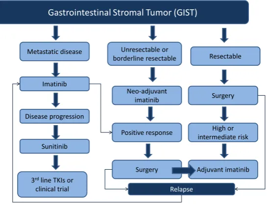
Figure 1 summarizes the role of systemic therapy in the management of resectable, unresectable/borderline
re-sectable and metastatic GISTs.
8.2.2. First Line Treatment of Advanced GISTs 1) Imatinib
Gastrointestinal Stromal Tumor (GIST)
Resectable Unresectable or
borderline resectable Metastatic disease
Surgery
High or intermediate risk
Adjuvant imatinib Positive response
Neo-adjuvant imatinib
Surgery Imatinib
Disease progression
Sunitinib
3rdline TKIs or
[image:12.595.178.451.86.294.2]clinical trial Relapse
Figure 1. Framework of management of gastrointestinal stromal tumors (GIST). This flowchart provides a summarized framework for the management of re-sectable, unresectable/borderline rere-sectable, and metastatic gastrointestinal stro- mal tumors.
bit the intracellular kinases ABL and BCR-ABL fusion protein in chronic myeloid leukemia (CML) cells. It was subsequently found that imatinib can induce dramatic and rapid clinical benefit in GIST as well [83]. A formal phase I study involving 40 patients demonstrated clinical benefit in 91% patients (54% partial response and 37% stable disease) [84]. Subsequently, several phase 2 trials assessed efficacy of imatinib at varying doses from 400 to 800 mg per day in patients with GIST and confirmed the benefit reported in the phase 1 trial [85]-[87]. For example in a phase 2 study, 147 patients were randomly assigned to receive 400 mg or 600 mg of imatinib daily. Overall, 54% patients had a partial response within six months of treatment. Early resistance to imatinib was noted in 14% patients. Therapy was well tolerated, although mild-to-moderate edema, diarrhea, and fatigue were common side effects [85]. In a long-term follow-up of a cohort of 56 patients who continued to take imatinib beyond three years, 26 (18% of the initial cohort) remained on imatinib at a median follow-up period of 9.4 years [88].
The median time to objective response is about 12 - 14 weeks; however, some patients have a dramatic im-provement in symptoms and signs within days of therapy initiation. Duration of imatinib therapy, in advanced GISTs, should be as long as possible, providing that the patient is still responding to the treatment. Following the introduction of imatinib, the median survival of patients with advanced GIST increased from an average of 18 to 57 months in an extended follow-up trial. Nearly 50% of patients with advanced GIST who were treated with imatinib mesylate survived for more than 5 years regardless of imatinib dose (400 mg/d vs. 600 mg/d) [89].
There have been two large randomized controlled phase III trials assessing the optimum dosage of imatinib [86] [90]. In the European trial 946 patients were randomly assigned to imatinib 400 mg or 800 mg daily. Pa-tients who were assigned to 400 mg, on progression were offered the option of crossover. Overall, 5% paPa-tients achieved a complete response, 47% a partial response, and 32% stable disease, with no difference in response rate between the groups. However, at median follow up period of 760 days, 56% of patients who received imati-nib 400 mg daily developed disease progression compared with 50% of patients who were assigned 800 mg (HR: 0.82 [95% CI 0.69 - 0.98]). Overall survival was 85% at 1 year and 69% at 2 years for the 400 mg group and 86% at 1 year and 74% at 2 years for the 800 mg group [86].
The meta-analysis of two large randomized studies revealed that after a median follow up of 45 months, a small but significant progression free survival advantage was seen in the high dose arm. Overall survival was identical in the two arms (median survival 4.08 years in the high dose group versus 4.05 years in the standard dose group, HR 1.00). The presence of a KIT exon 9 mutation was the predictive marker for benefit from higher doses. Patients with an exon 9 mutation had a significantly better response rate (47 versus 21%) and progression free survival [PFS] (HR 0.58, 95% CI 0.38 - 0.91) with high-dose therapy. However, no difference was observed between the treatment arms in the absence of such mutations [91].
2) Continuous vs. Interrupted Imatinib
A small phase III French trial assessed optimal schedule of imatinib and compared continuous with inter-rupted imatinib therapy beyond 1 year of treatment in patients with advanced GIST. Of 58 eligible patients, do-cumented progression was seen in 8 of 26 patients in the continuous group and 26 of 32 patients in the inter-rupted group (p < 0.001). Although there were no differences in overall survival, imatinib resistance or quality of life between the two groups, due to rapid progression after treatment interruption, this study supports the use of continuous imatinib [92].
3) Imatinib Dose Escalation
The standard dose of imatinib is 400 mg daily. Clinical data from the EORTC and North American Intergroup studies, where patients were allowed to cross over to high-dose imatinib upon progression, revealed that ap-proximately one third of these patients were able to regain disease control [86] [89]. Hence, imatinib dose esca-lation can occasionally control advanced GISTs that progress on imatinib [93]. The standard approach as rec-ommended by the European Society of Medical Oncology (ESMO) Clinical Practice Guidelines in the case of tumor progression on 400 mg is to increase the imatinib dose to 800 mg daily. Dose escalation is particularly useful in the case of a KIT exon 9 mutated GIST [94] [95].
4) Combination Strategy
Combination strategies have focused on targeting upregulated signaling pathways in imatinib-resistant pa-tients or on reducing the stability of activated signaling proteins [93]. Agents such as everolimus targeting the PI3K/Akt/mTOR signaling pathway and heat shock protein 90 and histone deacetylase inhibitors in combination with imatinib have been studied [96]-[98]. However, disappointing efficacy or toxicity results have precluded their use in routine clinical practice.
8.2.3. Response Evaluation
Measurement of lesion density is important for response assessment. Both tumor size and tumor density on CT scan, or consistent changes on MRI, should be considered as criteria for tumor response [94] [95]. An FDG-PET scan has proved to be highly sensitive in early assessment of tumor response and may be useful in doubtful cases, or when early prediction of the response is highly useful [99]. In one study, more than 10% reduction in lesion size on CT, or a greater than 15% reduction in density had 97% sensitivity and 100% specificity for identifica-tion of patients who showed a response on PET scan [100].
The measure of tumor response can be classified into four categories based on tumor size, degree, extent of enhancement, and the presence/absence of intra-tumoral solid nodules. The categories are as follows [41]:
• Category 1—Complete response: the original lesion previously identified by imaging is no longer present,
• Category 2—Partial response: two subcategories include: a) Continuous regression throughout treatment,
b) Initial regression followed by stabilization without further change,
• Category 3—Stable disease: tumor size and enhancement remains unchanged,
• Category 4—Progressive disease: two subcategories include:
a) Initial regression followed by development of new lesions and increased size/enhancement, b) Continuous progression of increasing tumor size/enhancement throughout treatment.
The Choi criterion is an alternative set of response evaluation criteria utilizing tumor size and density and predicts response to therapy better than other criteria. A 10% decrease in unidimensional tumor size or a 15% decrease in tumor density on contrast-enhanced CT scans correlates well with PET scan findings [100]. Howev-er, research continues in determining additional such criteria for accurate response evaluation.
8.2.4. Preoperative (Neoadjuvant) Treatment
[101]. Although level I evidence is lacking, non-randomized studies have demonstrated that preoperative imati-nib can reduce tumor bulk in GIST, and permit staged resection of initially unresectable or borderline resectable disease [102]-[106]. For instance, in a retrospective study of 46 patients, 11 with a locally advanced primary tumor, and 35 with recurrent or metastatic disease, all patients were treated with imatinib. Eleven patients who were treated for a locally advanced disease underwent surgical resection. At a median follow-up of 19.5 months, 10 patients were recurrence free and all were alive. Among the 35 patients who were treated for metastatic dis-ease, 11 patients were able to undergo a compete resection. Of 11 patients 6 developed recurrences at median follow up period of 15.1 months [103].
In a prospective phase II trial, 63 patients with KIT-positive GIST, with either a resectable primary greater than 5 cm, or resectable recurrent disease were treated with preoperative imatinib 600 mg daily for 8 to 12 weeks. Following surgery, all patients received at least two additional years of postoperative imatinib. At a me-dian follow-up of 5.1 years, the estimated five-year progression-free and disease-specific survival rates for pa-tients presenting with localized primary disease were 57% and 77%, respectively [107].
Consensus guidelines for preoperative or neoadjuvant therapy includes 1) unresectable or borderline resecta-ble primary tumor, 2) potentially resectaresecta-ble tumor that requires extensive surgery, 3) local recurrence of locally advanced disease, or 4) selected patients with limited potentially resectable metastatic disease. The ESMO and National Comprehensive Cancer Network (NCCN) guidelines support the use of preoperative imatinib in cases of limited disease if it would facilitate less extensive surgery with organ sparing [94] [95]. Imatinib is usually administered for 2 - 6 months before surgery. Tumor mutation analysis and longitudinal imaging should be done to identify patients who do not benefit from preoperative imatinib. The most appropriate method to assess re-sponse to targeted therapies is controversial. PET scan using fluorodeoxyglucose (FDG-PET) is highly sensitive for disease with a high glucose metabolism [108]. Evidence suggests that radiographic response and tumor cell apoptosis occur within the first week of imatinib therapy.
8.2.5. Adjuvant Treatment
The high risk of recurrence following surgery has led to exploration of the role of adjuvant imatinib in patients with completely resected GIST. Several single-arm and phase II studies assessed adjuvant imatinib for 12 - 26 months duration and suggested benefit [109].Three major phase III trials have evaluated the benefit of adjuvant imatinib in GIST after surgery [110]-[112]. The American College of Surgeons Oncology Group Z9000 pivotal trial revealed that 1 year of adjuvant imatinib therapy provides significantly superior RFS in patients with GIST after surgical resection, when compared to placebo. In this trial, 713 patients with a completely resected 3+ cm GIST were randomly assigned to one year of adjuvant imatinib (400 mg daily) versus placebo. At median fol-low-up of 19.7 months 30 (8%) patients in the imatinib group and 70 (20%) in the placebo group developed re-currence or died. The one-year RFS rate was 98% in imatinib group versus 83% in the placebo group (Hazard ratio [HR]: 0.35, 95% CI 0.22 to 0.53) [109] [110] [113]. In an unplanned subset analysis, tumor size, particu-larly over 10 cm, was an independent predictor of benefit from adjuvant imatinib.
The Scandinavian Sarcoma Group compared 36 versus 12 months of adjuvant imatinib in 400 patients with high-risk GIST with at least one of the following: tumor size greater than 10 cm, mitotic count greater than10 per 50 high-power fields (hpf), tumor size greater than 5 cm with mitotic rate greater than 5 per hpf, or tumor rupture. The median follow-up time was 54 months. Patients assigned for 36 months of imatinib had longer RFS compared with those assigned for 12 months (HR, 0.46; 95% CI, 0.32 - 0.65). Patients who received 36 months of therapy had a significantly better 5-year RFS (65.6% vs. 47.9%) and overall survival (HR, 0.45; 95% CI, 0.22 - 0.89) with 5-year survival of 92% compared with 81.7% in group treated with 12 months of imatinib [111].
The Intergroup European Organization for Research and Treatment of Cancer (EORTC) 62024 trial randomly assigned 908 patients with intermediate or high risk GIST to two years of imatinib versus observation alone. The preliminary report revealed that at a median follow-up of 4.7 years, five-year imatinib free survival (IFS) was 87% in the imatinib arm compared with 84% percent in the control arm (HR 0.80, 95% CI 0.51 - 1.26), and three-year RFS was 84 versus 66% [112].
8.2.6. Surveillance
Although most recurrences are noted within 2 years of curative surgery, late relapses and delayed recurrences up to 42 years [34] are not uncommon for intermediate and high risk GISTs thereby necessitating lifelong surveil-lance. The availability of effective targeted therapy and feasibility of salvage surgery in selected patients makes it important to detect early recurrences. The goals of post-treatment surveillance are 1) early recognition and treatment of potentially curable disease recurrences, 2) recognition of therapy-related complications, and 3) de-tection of symptoms consistent with metastatic disease. In patients treated with curative intent, follow up in-cludes a careful history and physical examination with periodic postoperative radiologic surveillance for recur-rent disease [94] [95] [117]. Details explaining the best-practice post-operative follow-up remain ill-defined. It is however recommended that patients at high- and intermediate-risk perhaps should undergo a CT or MRI q 3 - 6 months while undergoing adjuvant therapy, then every 3 months after cessation of therapy for the following 2 years, then 2 times per year for the next 5 years. For low-risk patients, 1 - 2 scans/year for 5 years is generally acknowledged to be acceptable though its usefulness remains unknown [39]. Recently Plumb et al. further de-fined follow-up in GIST patients into high-risk (CT every six months ×3 years then annually ×2 years), interme-diate risk (CT at 9 months then annually ×4 years), low-risk (CT at 1 year post-op) and very low risk (no further imaging). This being said, the clinical relevance of early detection remains unclear, as there is no evidence that earlier detection with treatment initiation leads to improved outcomes [118]. However, as 20% - 35% of GISTs recur during the first 2 years after completion of adjuvant imatinib, more frequent imaging at intervals of 3 - 4 months during this period is not unreasonable [78].
Site specific GISTs may need alternative surveillance regimens due to their natural history, i.e.: duodenal GISTs have been reported to recur even ten years after the initial excision, and as such longer follow-up may be necessary in these cases [119]. Additionally, as delayed recurrences of GIST up to 30 years have also been re-ported in the literature, some authors recommend lifelong clinical follow-up.
8.3. Recurrence & Metastases
Approximately 40% of GISTs localized at detection will develop metastases, and 10% - 20% of patients will present initially with metastatic disease. The most common sites of spread include the liver, omentum, and peri-toneum; extraabdominal metastases are uncommon [20]. Spread to the lung, bone, and other soft tissue sites generally only occurs in late-stage disease [45]. GISTs have been reported to metastasize to the lung, pleura, bone, subcutaneous tissues, and central nervous system [42]. The rectovaginal space, urinary bladder, pharynx, and gallbladder have additionally been reported to be involved [4]. Lymph node metastases are more common with epithelioid GISTs compared with the spindle type [42]. Two main routes of metastases have been reported: intraperitoneal dissemination and hematogenous spread predominately to the liver [120]. Delayed metastases may be seen over five years after the initial surgery [40].
8.3.1. Liver Metastases
experiences [1]. While hepatic artery embolization and debulking surgery have been attempted, results were not promising [2]. Enucleation or shellout procedures are not recommended due to the risk of peritoneal dissemina-tion secondary to disrupdissemina-tion of the pseduocapsule and/or tumor rupture [39] [63].
8.3.2. Non-Hepatic Metastases
Surgery for metastatic disease often requires multivisceral resections as many of the recurrences are diffuse. In well selected patients with stable or responsive disease on imatinib, a benefit has been reported from such exten-sive surgical resections which may include cytoreductive surgery ± multivisceral resection. Omentectomy and peritoneal stripping are often required as, in contrast to primary tumors, metastatic deposits often adhere to sur-rounding structures and can seldom be peeled off with negative margins. Though formal lymphadenectomy is not required, all enlarged nodes encountered during surgery should be removed. In summary, elective surgery can be considered as part of the multidisciplinary approach in the management of carefully selected patients with metastatic disease; while, emergency surgeries may be required in situations such as perforation, high- grade obstruction, or refractory bleeding [121].
8.4. Treatment of GIST after First Line Therapy
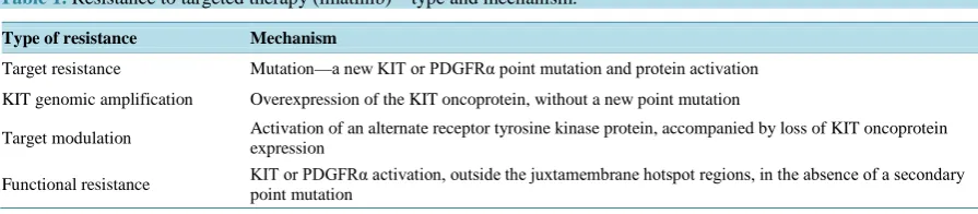
8.4.1. Resistance to ImatinibDespite the effectiveness of imatinib in advanced GISTs, complete responses are uncommon (less than 10 per-cent). Ten to fifteen percent of patients are primarily resistant to treatment with imatinib and most patients who initially respond to imatinib eventually develop resistance via additional mutations in KIT. Resistance to imati-nib can develop due to a new mutation in the target protein, gene amplification, target modulation and functional resistance [124] [125] (Table 1). Moreover, retrospective data suggest that suboptimal plasma levels of imatinib are associated with a worse outcome [95].
Two types of resistance to imatinib have been described; primary and secondary. Primary resistance, occur-ring in 10% patients with GIST, is defined as progression within the first six months of treatment and incidence is related to the causational mutation. The D842V mutation of PDGFRA consistently has a low response rate to imatinib. By contrast, secondary resistance occurs after an initial benefit of >6 months and typically occurs due to acquired mutations in KIT or PDGFRA [26]. Thus, different molecular mechanisms are responsible for pri-mary and secondary imatinibresistance in GISTs.
8.4.2. Imatinib Toxicity
Typically imatinib toxicities are controllable, including edema, rash, diarrhea, nausea, abdominal pain, and fati-gue [77]. Additionally, patient intolerance to imatinib is observed. “Cystic-like” transformation of some hepatic metastases has been noted as a response to imatinib therapy and is a diagnostic challenge to distinguish relapse and regrowth vs. true response/toxicity [99]. Hepatotoxicty has been described as a risk of imatinib therapy, oc-curring in 2% - 5% of treated patients. Though the majority of these present with elevated transaminases within the first 2 - 3 months of therapy which resolve with dose reduction, histopathological findings of periportal ne-crosis and massive hepatic nene-crosis has been reported, as well as five deaths from liver failure [126]. Corticos-teroids may contribute to reversing hepatotoxicity.
8.4.3. Sunitinib
[image:16.595.87.535.625.723.2]Sunitinib (Sutent) is a multi-targeted tyrosine kinase that has demonstrated activity in imatinib-refractory or in-tolerant patients [127]-[131]. In a phase III trial, 312 patients who were resistant to, or intolerant of, previous
Table 1. Resistance to targeted therapy (imatinib)—type and mechanism.
Type of resistance Mechanism
Target resistance Mutation—a new KIT or PDGFRα point mutation and protein activation
KIT genomic amplification Overexpression of the KIT oncoprotein, without a new point mutation
Target modulation Activation of an alternate receptor tyrosine kinase protein, accompanied by loss of KIT oncoprotein expression
treatment with imatinib were randomized to receive sunitinib (50 mg a day, orally, in 6-week cycles, 4 weeks on and 2 weeks off) or placebo [127]. Median time to tumor progression was 27.3 weeks in patients treated with sunitinib compared with 6.4 weeks in the placebo group (hazard ratio 0.33; p < 0.0001). Therapy was reasonably well tolerated and the most common treatment-related adverse events were fatigue, diarrhea, skin discoloration, and nausea. Due to cross-over design there was no statistical difference in overall survival (median 72.7 in suni-tinib arm vs. 64.9 weeks in the placebo arm) [128].
Similar to imatinib, the efficacy of sunitinib is influenced by both primary and secondary mutations in the predominant pathogenic kinases. In a phase I/II trial of sunitinib in 97 patients with metastatic, imatinib-resis- tant/intolerant GIST, clinical benefit with sunitinib was observed for the three most common primary GIST ge- notypes: KIT exon 9 (58%), KIT exon 11 (34%), and wild-type KIT/PDGFRA (56%) [130]. Progression-free survival was significantly longer for patients with primary KIT exon 9 mutations or with a wild-type genotype than for those with KIT exon 11 mutations. Following progression on imatinib, patients with KIT exon 9 muta- tion or a PDGFRA mutation had a median time to progression of 19 months, compared with 5 months for pa- tients with exon 11 mutations. The same pattern was observed for overall survival. Patients with secondary KIT exon 13 or 14 mutations had longer PFS and overall survival compared with those with exon 17 or 18 mutations.
8.4.4. Regorafenib
Regorafenib is a structurally unique inhibitor of multiple cancer-associated kinases, including KIT and PDGFR, with broad-spectrum anticancer activity. Because KIT and PDGFRA remain oncogenic drivers of GIST after re- sistance to imatinib and sunitinib, regorafenib was evaluated in patients who developed resistant to them. In a prospective phase II trial 34 patients received regorafenib, 160 mg daily, on days 1 to 21 of a 28-day cycle. Clinical benefit rate was 79% (95% CI, 61% to 91%) with overall median progression-free survival was 10.0 months [132].
Efficacy of regorafenib was confirmed in a phase III trial in which 199 patients were randomized to regorafe-nib or placebo [133]. Median PFS was 4.8 months for regorafenib compared with 0.9 months for placebo (HR: 0.27, 95% CI 0.19 - 0.39). After progression, 56 patients (85%) assigned placebo crossed over to regorafenib. Drug-related adverse events were reported in 98% of patients assigned to regorafenib and 68% of patients as-signed to the placebo arm. The most common serious adverse effects were hypertension, hand-foot-skin reaction and diarrhea.
8.4.5. Nilotinib & Other Tyrosine Kinase Inhibitors
Nilotinib is a potent second-generation TKI that targets c-KIT and PDGFR and has demonstrated efficacy in pa-tients with imatinib-refractory GISTs [134]-[137]. In an open phase III trial 248 patients with advanced GIST following prior imatinib and sunitinib failure were randomized to nilotinib or best supportive care [137]. Median PFS, based on blinded central radiology review was similar between the groups (109 days versus 111 days; P = 0.56). Local investigator-based intent-to-treat analysis, however, showed a significantly longer median PFS with nilotinib (119 versus 70 days; P = 0.0007). A trend in longer median overall survival was noted with nilotinib (332 versus 280 days; P = 0.29).
Limited data are available on the efficacy of other TKIs in patients with imatinib and sunitinib-refractory GISTs. Sorafenib is a multi-TKI that has demonstrated efficacy in such patients [138]-[140]. Retrospective data demonstrated median PFS of 6.4months and median overall survival of 13.5 months in patients treated with so-rafenib as third- or fourth-line treatment [140]. A phase II trial revealed disease control rate of 68%, and median PFS of 5.2 months. In addition, there are published reports of efficacy of motesanib, dasatinib, and vatalanib in refectory GISTs previously treated with sunitinib and or imatinib [141]-[143]. Masitinib mesylate has shown promising results in phase I and II clinical trials. Currently it is being compared in a first-line setting with imati-nib in a randomized phase III trial [144].
population. Recent development of a monoclonal antibody against KIT studied in vitro and in vivo, a) shows a decrease in proliferation, b) induces KIT downregulation, and c) enhances cell-mediated tumor clearance and thus may be the future drug for the 22nd century [144].
9. Prognostic Features
The three most important prognostic factors predictive of postoperative GIST recurrence are the tumor prolifera-tion rate, tumor size and tumor locaprolifera-tion [79]. Additional prognostic factors have been identified that include the surgical/medical treatment undertaken, tumor location, and other tumor characteristics on gross and microscopic evaluation.
9.1. Surgical Resection
Surgical resection alone is associated with a recurrence rate of 50% and a 5-year survival rate also of 50% [2] [13]. According to some authors, tumor rupture automatically makes a GIST high-risk [63] [79]. The median time for post-resection recurrence is 19 - 25 months. These most commonly occur in the abdomen, involving the peritoneum and/or liver, and are generally non-resectable [13]. Though recurrence is uncommon ten years post-operatively, delayed recurrences up to 25 - 30 years after initial diagnosis have also been reported [79].
9.2. Medical Treatment
GISTs are known to be refractory to conventional chemotherapy and radiation. Since the advent of imatinib in the 21st century, the response rate has increased from 5 to 80%, with an associated increase in median survival from 15 months to 5 years [2]. Survival in advanced GIST has also improved dramatically with the advent of imatinib, from 10 - 20 months to 51 - 57 months [20]. GISTs with an exon 11 mutation which increases the like-lihood of responding to imatinib confers a better prognosis [23]. Though PDGFRA mutations appear to confer resistance to imatinib therapy, the American College of Surgeons Oncology Group Z9000 clinical trial showed this population to have the best outcome, with 90% recurrence-free survival at 3 years [12].
9.3. Tumor Location
Comparable tumors of the small bowel, colon, rectum, or mesentery confer a poorer outcome than if located in the stomach. Half of all duodenal GISTs are malignant of which just under 50% are metastatic at presentation [37]. Characteristics of colonic GIST are similar to rectal GIST and receive a high-risk categorization in Mietti-nen and Lasota system [5]. Among colonic GISTs, small tumors <2 cm with low mitotic rate (<5 mitoses/ 50HPF) do not show evidence of clinical progression. Larger tumors or tumors with a higher mitotic rate (>5/50 HPF) results in a metastatic rate of >50% [4]. Approximately 75% of recurrences are asymptomatic [118].
9.4. Gross Characteristics
The malignant potential of GIST is highly variable, as even those <2 cm with bland histologic features may have malignant potential [8]. The presence of tumor ulceration or necrosis confers a shorter overall survival; however, this relationship has not been shown to be statistically significant [145].