S T U D Y P R O T O C O L
Open Access
Interaction of titanium, zirconia and lithium
disilicate with peri-implant soft tissue: study
protocol for a randomized controlled trial
Katharina Kuhn
1*†, Heike Rudolph
1†, Michael Graf
1, Matthias Moldan
1, Shaoxia Zhou
2, Martin Udart
3,
Andrea Böhmler
3and Ralph G. Luthardt
1Abstract
Background:Against the background of increasing use of dental implants, and thus an increasing prevalence of implant-associated complications, a deeper understanding of the biomolecular mechanisms in the peri-implant tissue is needed. Peri-implant soft tissue is in direct contact with transmucosal dental implant abutments. The aim of this trial is to distinguish the biomolecular and histological interactions of various dental abutment materials with peri-implant soft tissue.
Methods/Design:The study is designed as a prospective, randomized, investigator-initiated clinical pilot trial with blinded assessment. We will ultimately include 24 eligible patients who opt for implant treatment to replace a single missing posterior tooth. Three months after implantation (submerged procedure), the study begins with the second-stage surgery. Each of the 24 patients will be given three different transmucosal abutments (zirconia, lithium disilicate, titanium) consecutively. The sequence in which the three materials are used is randomized. Peri-implant crevicular fluid is sampled weekly around the respective abutment for biomolecular analyses. After one month of wearing time, the stamping press from the second-stage surgery is used to gain a narrow gingival ring biopsy around the abutment for immunohistochemical analyses. The next abutment is then inserted. The same procedure is used for all three abutments. After sampling is completed, the patients will receive a definitive crown. The primary outcome measure of the trial is biomolecular detection of specific markers in the peri-implant crevicular fluid: matrix metalloproteinase 8, interleukin- 1β, polymorphonuclear elastase, and myeloid-related protein MRP8/14 (calprotectin). Secondary outcome measures include immunohistochemical analyses and clinical parameters. Discussion:The study design will allow us to perform correlation analyses between the clinical indices with
biomarkers’expression in the interface of the transmucosal abutments and the peri-implant soft tissue. A deeper understanding of the three abutment materials’interactions with peri-implant soft tissue will help us understand the formation mechanisms of implant-associated complications and then develop prevention strategies.
Trial registration:The trial is registered at the German Clinical Trial Register and the International Clinical Trials Registry Platform by the WHO under DRKS00006555 (Registered on 27 October 2014).
Keywords:Biomolecular, Peri-implant crevicular fluid, Immunohistochemical markers, Lithium disilicate
* Correspondence:katharina.kuhn@uniklinik-ulm.de †Equal contributors
1
Department of Prosthetic Dentistry, Center of Dentistry, Ulm University, Ulm, Germany
Full list of author information is available at the end of the article
Background
The prevalence of biological implant-associated complica-tions rises with the increasing use of dental implants. A common biological complication is peri-implant mucositis [1, 2], with a prevalence of 50 % of all dental implant sites [3]. Biological implant-associated complications usually begin in the peri-implant soft tissue. Both connective tis-sue and epithelium of the peri-implant soft tistis-sue are in direct contact with transmucosal implant abutments. Thus, the interaction between the respective abutment materials with the peri-implant soft tissue may favor, counteract, or not influence at all the development of peri-implant mucositis or of unfavorable mucosal struc-tures such as an apical shift of the barrier epithelium [4, 5]. The particular interaction between abutment materials and soft tissue is likely to be affected by the abutments’properties, such as bacterial adhesion [6–8], surface condition [9–12] (e.g., free surface energy, rough-ness), and soft tissue integration ability [4, 5]. There is only low-level evidence available on the interaction be-tween different abutment materials with peri-implant tis-sues [13, 14]. It has been suggested in histological studies that the abutment materials may have an influence on the stability of peri-implant tissues [4, 5, 15], although clinical studies that compared titanium to aluminum oxide abut-ments [16, 17] and titanium to gold-alloy abutabut-ments [18] found no significant differences in the clinical parameters. Titanium and zirconia are well-established as abutment materials. Lithium disilicate ceramic has recently been in-troduced as abutment material [19, 20]. Accordingly, there is a lack of studies on lithium disilicate compared with ti-tanium and zirconia as dental implant abutment material.
The abutment’s interaction with peri-implant soft tis-sue can be analyzed by quantifying possible inflamma-tion processes. The use of biomolecular analysis of the peri-implant crevicular fluid (PICF) has been established [21, 22]. For the PICF marker analyses the biomarkers matrix metalloproteinase 8 (MMP-8), interleukin-1β (IL-1ß) and polymorphonuclear elastase (PMN-elas-tase) proved to be reliable indicators of peri-implant inflammation [22–28]. They also play important roles in oral wound healing [29–32]. The biomarker myeloid-related protein MRP8/14 (calprotectin) has been used only rarely in PICF marker evaluations [33]. Its correlation with periodontitis has been proven by means of gingival crevic-ular fluid analyses [34–37]. Its release from monocytes is induced by IL-1ß [38].
Only one recently published study [39] compared the interaction of two abutment materials (titanium versus zirconia) with peri-implant tissue by means of PICF marker analyses. No such investigation is available on the biomo-lecular interaction of lithium disilicate ceramic. Overall, lit-tle evidence is available on the biomolecular interactions between different abutment materials and peri-implant soft
tissue. The aim of this study is to analyze the interactions at the interface of peri-implant soft tissue and three dental abutment materials (zirconia, lithium disilicate, titanium). The hypothesis is that this interface differs biomolecu-larly and immunohistochemically depending on the specific abutment material used.
Methods/Design
The study protocol is reported according to the Consoli-dated Standards of Reporting Trials (CONSORT) statement [40]. The study was designed in accordance with the follow-ing guidelines:
World’s Medical Association’s Declaration of Helsinki
Clinical investigation of medical devices for human subjects–Good Clinical Practice (ISO 14155:2011)
Guidelines of Good Clinical Practice (2001/20/EC)
The Ethics Commission of Ulm University approved the study design on 30 July 2013 (processing number 68/13).
Trial design
This clinical trial was designed as a prospective, random-ized investigator-initiated pilot trial to be conducted in one center. In all, 24 patients are intended to participate in the study after giving informed consent. The study de-sign was reevaluated after the first three patients had passed the endpoint of the biomolecular and histological sampling. Since the first three patients required no adjust-ments post-treatment, it is feasible that for the treatment of the remaining 21 patients no further adjustments will be required.
Participants
Participants will be 24 patients with a single missing tooth in the posterior area (premolar or molar) with both adja-cent teeth in situ and who choose to undergo implant re-placement. Patients will be included in the study provided they:
Are between 18 and 75 years of age
Have an edentulous space at least 7 mm in width
Have primary implantation or implantation after two-stage or one-stage augmentation (sinus lift or minor lateral augmentation)
Have gingiva≥3 mm in height
Are in need of prosthetic treatment
Are legally competent
Patients will be excluded from the study if:
one-stage sinus lift or minor lateral augmentation is possible)
They are smokers
Implant insertion using an implant template before the beginning of the study is not possible
The edentulous space < 7 mm in width (conservation of papilla)
The gingiva < 3 mm in height at the lowest point
There is no consent given for study participation
A chronic disease is present
They are pregnant
There is evidence of alcohol or drug abuse
Settings and locations where the data will be collected
The study is taking place at the Department of Pros-thetic Dentistry, Center of Dentistry, Ulm University. Screening began in August 2013. So far, the first eight patients have received a single-tooth implant. Five of the eight have already finished the study. All patients gave informed consent. The biomolecular analyses will take place in the Department of Clinical Chemistry, Ulm University. The immunohistochemical analyses will be carried out at the Institut für Lasertechnologien in der Medizin und Messtechnik, Ulm University. Completion of the study (last patient out) is planned for mid-2016.
Interventions
All 24 patients will have received the three abutments con-secutively, each for a 1-month wearing period. The abut-ments are as follows: zirconia ceramic (Z) (Zenostar MO; Wieland Dental GmbH, Pforzheim, Germany; lot S13270); lithium disilicate ceramic (L) (IPS e.max Press; Ivoclar Vivadent, Schaan, Liechtenstein; lot S44695); titanium
(T) (Zenotec Ti; Wieland Dental GmbH, Pforzheim, Germany; lot 20130305 4012). The sequence of the three materials is randomly assigned. The abutments are manu-factured by luting hollow Z-, L-, or T-cylinders on Vario-bases™(Straumann, Basel, Switzerland; lot: HE661/HK699) by means of Multilink® Hybrid Abutment cement (Ivoclar Vivadent, Schaan, Liechtenstein; lot: T10017). The abut-ments’surface roughness is adjusted by polishing them to ensure conformity. The results are checked by Ra measure-ments of each abutment (Ra < 0.1μm).
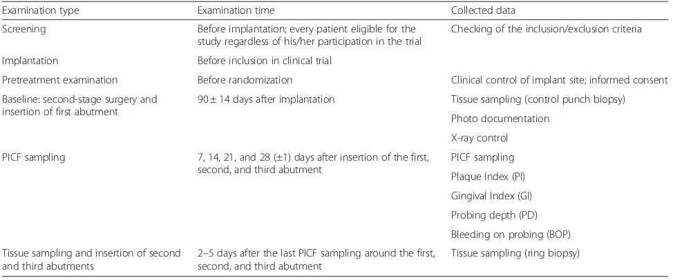
The examinations and specific data collection are shown in Table 1. Screening is performed before implantation to check the inclusion/exclusion criteria. Implantation is per-formed according to the standardized implantation pro-cedure in the Department of Prosthetic Dentistry making use of the Straumann®-guided surgery implantation system (Straumann, Basel, Switzerland) in combination with an implantation template after three-dimensional implant planning (CoDiagnostix; Dental Wings GmbH, Chem-nitz, Germany). Bone-level Straumann implants (SLAc-tive, RC) are submerged.
Informed consent is given during the healing period (90 ± 14 days). Inclusion in the study and randomization takes place prior to the second-stage surgery (baseline). For the second-stage surgery, a stamping press is used, with a punch biopsy serving as a histological control specimen. The first abutment (Z, L, or T) is inserted (20 Ncm) ac-cording to the randomization process. The occlusal screw hole is provisionally closed by means of a piece of dental foam and Luxatemp Inlay (DMG, Hamburg, Germany), which is light-cured. These materials are not in contact with the gingiva.
[image:3.595.55.542.532.735.2]Peri-implant crevicular fluid (PICF) is sampled both buccally and lingually every week using paper strips
Table 1Examinations and collected data
Examination type Examination time Collected data
Screening Before implantation; every patient eligible for the study regardless of his/her participation in the trial
Checking of the inclusion/exclusion criteria
Implantation Before inclusion in clinical trial
Pretreatment examination Before randomization Clinical control of implant site; informed consent Baseline: second-stage surgery and
insertion of first abutment
90 ± 14 days after implantation Tissue sampling (control punch biopsy) Photo documentation
X-ray control PICF sampling 7, 14, 21, and 28 (±1) days after insertion of the first,
second, and third abutment
PICF sampling Plaque Index (PI) Gingival Index (GI) Probing depth (PD) Bleeding on probing (BOP) Tissue sampling and insertion of second
and third abutments
2–5 days after the last PICF sampling around the first, second, and third abutment
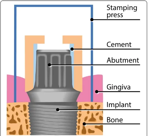
(Periopaper; Oraflow Inc., New York, NY, USA). Before PICF sampling, supragingival plaque is carefully re-moved with cotton balls to eliminate the risk of plaque contamination. Cotton rolls are applied to isolate the sample from saliva. After air-drying the teeth, the paper strips are inserted into the sulcus/pocket until there is mild resistance and then left there for 30 seconds. Samples with visible blood contamination are discarded. In addition to weekly PICF sampling, clinical parameters are recorded for the implant and both adjacent teeth, including the Plaque Index (PI), Gingival Index (GI), probing depth (PD) using an electronic pressure-sensitive probe (Flori-daProbe, Essen, Germany) at four sites (mesiobuccal, disto-buccal, mesiolingual, distolingual), and bleeding on probing (BOP). After a 1-month wearing period, the abut-ment is exchanged for the next one. Before unscrewing the abutment, the stamping press from the second-stage sur-gery is used to gain a narrow (0.6 mm) gingival ring bi-opsy around the abutment for immunohistochemical analyses (Figs. 1 and 2). The same procedure as de-scribed above is repeated for the second and third
abut-ments. After completion of the sampling for
biomolecular and immunohistochemical analyses, the study is completed, and the patient receives a screw-retained lithium disilicate crown.
The three clinical investigators collecting the data re-ceived training before the first study visit to guarantee conformity.
Biomolecular analyses
The paper strips collected for biomolecular analyses are frozen immediately after sampling (−80 °C) and remain
frozen until assayed. For the analyses, the PICF is released from the paper strip by means of analysis buffer. The incu-bation takes place on an orbital shaker on ice, and separ-ation is performed by centrifugsepar-ation. The supernatant is collected into a fresh tube. To exclude erythrocyte contam-ination, all extracts are examined with the Combur-Test® strip (Roche Diagnostics GmbH, Mannheim, Germany).
The PICF extracts are analyzed for biomolecular detection of specific markers: MMP-8, IL-1β, PMN-elastase and MRP8/14 (calprotectin). MMP-8 is analyzed with the Fluorokin MAP Multiplex Human MMP Panel (R & D Systems GmbH, Wiesbaden, Deutschland) in the Luminex 200 System (Bio-Rad Laboratories Inc., Hercules, CA, USA). The MMP-8 activity is detected by means of gel-atine-zymography. IL-1ß is measured by the Bio-Plex Cyto-kine Assay Kit (Bio-Rad Laboratories Inc., Hercules, CA, USA) in combination with the Luminex 200 System. PMN-elastase is measured by means of the Human Elastase ELISA Kit (HyCult Biotechnology, Uden, Netherlands) and MRP8/14 is measured with the MRP8/14 ELISA Kit (Bühl-mann Laboratories AG, Schönenbuch, Switzerland). The optical density for both kits is measured at 450 nm. The sensitivities are as follows: MMP-8, 8.9 pg/ml; IL-1β, 0.2 pg/ml; PMN-elastase, 0.4 ng/ml; MRP8/14, < 0.4μg/ml.
Immunohistochemical analyses
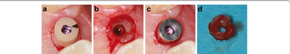
Each of the four gingival biopsies per patient (one control punch biopsy and three ring biopsies around the abutment materials) (Fig. 2d) are equally divided into quarters for the immunohistochemical analyses. The biopsy specimens are fixed immediately in 4 % neutral buffered formalin at room temperature and left there for 24 hours before embedding them in paraffin. Three micrometer thick sections are pre-pared prior to deparaffinization. A citrate buffer (pH 6.1; Target Retrieval Solution, Dako, Carpinteria, CA, USA) is applied. A steamer is used for antigen retrieval (20 minutes) and the slides cool off for 20 minutes. A serum blocking solution (Histostain-Plus Bulk Kit, Invitrogen®, Camarillo, CA, USA; 20 minutes) is applied. The specimens are incu-bated with the primary antibodies Anti-Neutrophil Elastase (over night; 4 °C; 1:500), Anti-MMP 8 (over night; room temperature; 4 μg/ml), Anti-IL 1β (over night; room temperature; 1:12.5) or Anti-MRP8 antibody (over night; 4 °C; 1:750), respectively (Abcam®, Cambridge, Great Britain), targeting the same molecules which are an-alyzed by the biomolecular analysis. Hydrogen peroxide 0.3 % is applied to block endogenous peroxidase (10 mi-nutes). A secondary biotinylated antibody (Histostain-Plus Bulk Kit, Invitrogen®, Camarillo, CA, USA) is used (10 minutes). Streptavidin conjugated with horseradish peroxidase (Histostain-Plus Bulk Kit, Invitrogen®, Camarillo, CA, USA) is applied (15 minutes). An AEC chromogen (AEC Single Solution, Invitrogen®, Camarillo, CA, USA) is applied (15 minutes) for final staining. The specimens are
[image:4.595.56.292.497.715.2]then counterstained with hematoxylin and mounted with Aquatex (Merck, Darmstadt, Germany). Negative controls are obtained by treating the sections similarly but, instead of the primary antibodies, Tris-buffered saline (TBS) is used. The staining is performed in serial sections from each specimen (e.g., lingual quarter of the control punch biopsy).
A digital image analysis system, which consists of a light microscope Axiophot (Carl Zeiss Jena, Oberkochen, Germany) and a digital color camera ProgRes C12 plus (Jenoptik, Jena, Germany) connected to a computer equipped with PeogRes-Software, is used to obtain im-ages of the microscopic samples (magnification 100× to 400×). Morphometric analyses are performed using the software ImageJ (National Institutes of Health, Bethesda, MD, USA) with the plugins Grid and CellCounter as pre-viously described [41].
Outcomes
The primary outcome measure is the biomolecular detec-tion of specific markers (MMP-8, IL-1β, PMN-elastase, MRP8/14) in the PICF samples. Secondary outcome mea-sures include immunohistochemical analyses and clinical parameters (PI, GI, PD, BOP).
Sample size
The study is biometrically categorized as a pilot study be-cause of the sample size of 24 patients. During establish-ment of the study design, there were no clinical results on which a biometrical sample size estimation could be based. The sample size has, therefore, been set at 24 patients. The weekly PICF sampling both buccally and lingually results in 24 PICF samples from each patient. Four histological specimens (one control punch biopsy and three ring biop-sies) are to be collected from each patient.
Randomization
Randomization is performed with a threefold crossover design. The sequence of the three abutment materials is randomly assigned, resulting in six groups (ZLT, ZTL, LZT, LTZ, TZL, TLZ) with four samples for each. The study is stratified into the two gender groups.
Randomization concealment is ensured because a clinic staff member not involved in the clinical trial performs the allocation according to a randomization list.
Any eligible patient is recorded in a screening list main-tained by the clinical investigators. Once a patient has given informed consent, a patient number and randomization are requested from the staff member performing the allocation according to the randomization list. The clinical investiga-tor enters the patient number into the patient list, which also includes the appointments of each patient. The patient number and sequence of the abutment materials are trans-ferred into the Case Report File of the baseline visit (sec-ond-stage surgery).
Blinding (masking)
Masking is not possible for the dentist or the patient be-cause of the visually distinguishable abutment materials. All analyses, however, are to be done masked because of the different personnel and spatial separation.
Statistical methods
An explorative data analysis will be performed. The data will be analyzed descriptively by means of absolute and relative frequency and medians and means, respectively, as well as measures of dispersion. The analysis of the pri-mary and secondary outcome measures will be performed using adequate statistical tests according to the distribu-tion patterns. Power analyses and sample size estimadistribu-tion will be done for similar clinical studies in the future.
Discussion
Against the background of a high prevalence of bio-logical peri-implant complications [3], the mechanisms of the interface between peri-implant soft tissue and transmucosal abutments are of interest. The aim of the present clinical study is to obtain a deeper understanding of the interaction mechanisms via PICF marker analyses and immunohistochemical analyses.
To date, only one recently published cross-sectional clin-ical study [39] has investigated the interaction of abutment materials (titanium versus zirconia) with peri-implant
Fig. 2Tissue sampling.aClinical situation after applying the stamping press around the zirconia abutment.bAfter removing the gingival ring
[image:5.595.59.538.89.179.2]tissue by means of PICF marker analyses. No significant differences were found between the biomarkers’ quan-tity (except for leptin). The present study has only the biomarker IL-1βin common with the other clinical study. Clinical parameters were not recorded (except for the presence or absence of plaque) at the time of PICF sam-pling. The authors recognize the benefits of a prospect-ive, randomized clinical trial including the collection of clinical indexes for correlation with biomarker expres-sion [39]. Our study design met those requirements. Moreover, we included a third material, lithium disili-cate ceramic, which is not as well- established as titan-ium and zirconia for use as abutment material. It has been shown, however, to be an increasingly relevant ma-terial for abutments [19, 20]. Titanium serves as “gold standard.”
PICF marker analyses have been established to detect peri-implant inflammation [21, 22]. For the PICF marker analyses, we use three well-established biomarkers (MMP-8, IL-1ß, PMN-elastase) and MRP8/14. To date, MRP8/14 has been used only rarely in PICF marker evaluations [33]. The combination of well-established biomarkers with a ra-ther new one allows us to evaluate its suitability for PICF marker analyses.
The role of plaque formation must be taken into account in terms of dental materials’interaction with peri-implant soft tissue. Plaque is associated with peri-implant mu-cositis [42, 43] and is correlated with elevated inflam-matory markers in PICF [28, 44]. In our study, we clinically determine the PI for correlation analyses with the number of biomarkers in PICF, which has not been sufficiently investigated because of the wide range of biomarkers. The formation of biofilm on dental mate-rials has been especially well-examined for titanium and zirconia [6–9, 45]. The results are inconsistent, however, as zirconia shows less bacterial adhesion and colonization in some studies [7, 45, 46] and no differ-ences in others [6, 8]. In the context of plaque forma-tion, the role of surface roughness also must be discussed. Excessive surface roughness of dental materials favors bio-film formation [11, 12] with more complex microbiota [10]. A threshold for the Ra value of abutment surfaces has been found (0.2μm), however, below which no further significant changes occur in regard to plaque accumula-tion [9]. The interacaccumula-tion between the three abutment ma-terials with the peri-implant soft tissue should not be biased by different surface roughness in the present study. Therefore, we adjusted the Ra values of all abutments for uniformity, as indicated in previous studies [8, 45], and well below the above-mentioned threshold.
Wound healing also influences the results of PICF marker analyses [47]. This aspect will not bias our study as the study design guarantees analogue wound healing condi-tions for all abutment materials. Each abutment is inserted
immediately after a circular gingival biopsy is performed with a stamping press. Thus, each abutment material is circularly in contact with open wound edges, and the 1-month PICF sampling takes place during the early wound healing period for each of the three abutments. This is a clinically relevant procedure especially for the
“one abutment–one time” concept, where the defini-tive abutment is inserted directly after implantation (direct loading) or directly after second-stage surgery (delayed loading) without removing it again [48–50].
The original biopsy material (punch biopsy from second-stage surgery) serves as histological control specimen as it has not been in contact with an abutment material so far. It will also be stained immunohistochemically to detect the four biomarkers.
Washout periods between the different abutment ma-terials may help to exclude an influence of the preceding abutment material on the tissues. Washout periods may be performed either by wearing a“neutral”abutment for a specific time between the tested abutments or by per-mitting tissues to heal without an abutment and punch biopsy again before inserting the next abutment. Follow-ing discussion with biometricians, the additional punch biopsies and time on top of an already prolonged proced-ure were deemed ethically unjustifiable. In addition, the newly formed cells and tissue in direct contact with each abutment are most likely to be affected and are circularly removed by the stamping press. Thus, we renounced washout periods in the trial. However, an influence of the preceding abutment material on the biomolecular and immunohistochemical findings of the next abutment material cannot be fully excluded. To the author’s know-ledge, there is no study which has tested this aspect yet. If our biometricians detect clear evidence of previous ment influence (worst-case scenario) only the first abut-ment material (n= 8 for each abutment material) shall be used for analysis.
The study combines biomolecular and immunohisto-chemical analyses–both aiming to detect the same bio-markers – and clinical analyses. This design allows a site-specific correlation analysis between those two tech-niques for biomarker detection as well as clinical indices to monitor gingival health.
Trial status
The screening and recruiting for the study began in August 2013 and is still ongoing. To date, the first eight patients have received a single-tooth implant. Five of the eight have finished the study.
Abbreviations
BOP:bleeding on probing; CONSORT: Consolidated Standards of Reporting Trials; ELISA: enzyme-linked immunosorbent assay; GI: Gingival Index; IL-1β: interleukin-1β; L: lithium disilicate; MMP-8: matrix metalloproteinase 8; MRP: myeloid-related protein; PD: probing depth; PI: Plaque Index; PICF: peri-implant crevicular fluid; PMN-elastase: polymorphonuclear elastase; T: titanium; TBS: Tris-buffered saline; Z: zirconia.
Competing interests
The authors declare that they have no competing interests.
Authors’contributions
KK made contributions to the conception and design and made substantial contributions to the coordination of the study, acquisition of data, and drafting the manuscript. HR made substantial contributions to the acquisition of funding, conception, design, and coordination of the study and was involved in drafting the manuscript. MG made contributions to the conception and design of the study, and acquisition of data. MM made contributions to the acquisition of data, especially for the biomolecular analyses. SZ made contributions to the conception and design of the study and biomolecular analyses, and revised the manuscript critically concerning the biomolecular analyses and in the analyses themselves. MU made contributions to the conception and design of the immunohistochemical analyses and revised the manuscript critically concerning the immunohistochemical analyses. AB made contributions to the conception and design of the immunohistochemical analyses and revised the manuscript critically concerning the immunohistochemical analyses and in the analyses themselves. RG made substantial contributions to the acquisition of funding, conception, design, and coordination of the study and was involved in drafting the manuscript. All authors read and approved the final manuscript.
Acknowledgments
This paper presents independent research funded to a major extent by Ivoclar Vivadent AG (Schaan, Liechtenstein).
Author details
1Department of Prosthetic Dentistry, Center of Dentistry, Ulm University, Ulm,
Germany.2Department of Clinical Chemistry, Ulm University, Ulm, Germany.
3Institut für Lasertechnologien in der Medizin und Messtechnik, Ulm
University, Ulm, Germany.
Received: 4 February 2015 Accepted: 28 September 2015
References
1. Donos N, Laurell L, Mardas N. Hierarchical decisions on teeth vs. implants in the periodontitis-susceptible patient: the modern dilemma. Periodontol 2000. 2012;59(1):89–110.
2. Lindhe J, Meyle J. Peri-implant diseases: Consensus Report of the Sixth European Workshop on Periodontology; Group D of European Workshop on Periodontology. J Clin Periodontol. 2008;35 Suppl 8:282–5. 3. Zitzmann NU, Berglundh T. Definition and prevalence of peri-implant
diseases. J Clin Periodontol. 2008;35 Suppl 8:286–91.
4. Abrahamsson I, Berglundh T, Glantz PO, Lindhe J. The mucosal attachment at different abutments. An experimental study in dogs. J Clin Periodontol. 1998;25(9):721–7.
5. Welander M, Abrahamsson I, Berglundh T. The mucosal barrier at implant abutments of different materials. Clin Oral Implants Res. 2008;19(7):635–41. 6. de Oliveira GR, Pozzer L, Cavalieri-Pereira L, de Moraes PH, Olate S, de Albergaria Barbosa JR. Bacterial adhesion and colonization differences between zirconia and titanium implant abutments: an in vivo human study. J Periodontal Implant Sci. 2012;42(6):217–23.
7. Rimondini L, Cerroni L, Carrassi A, Torricelli P. Bacterial colonization of zirconia ceramic surfaces: an in vitro and in vivo study. Int J Oral Maxillofac Implants. 2002;17(6):793–8.
8. Yamane K, Ayukawa Y, Takeshita T, Furuhashi A, Yamashita Y, Koyano K. Bacterial adhesion affinities of various implant abutment materials. Clin Oral Implants Res. 2013;24(12):1310–5.
9. Bollen CM, Papaioanno W, Van Eldere J, Schepers E, Quirynen M, van Steenberghe D. The influence of abutment surface roughness on plaque accumulation and peri-implant mucositis. Clin Oral Implants Res. 1996;7(3):201–11.
10. Quirynen M, van der Mei HC, Bollen CM, Schotte A, Marechal M, Doornbusch GI, et al. An in vivo study of the influence of the surface roughness of implants on the microbiology of supra- and subgingival plaque. J Dent Res. 1993;72(9):1304–9.
11. Subramani K, Jung RE, Molenberg A, Hammerle CH. Biofilm on dental implants: a review of the literature. Int J Oral Maxillofac Implants. 2009;24(4):616–26. 12. Teughels W, Van Assche N, Sliepen I, Quirynen M. Effect of material
characteristics and/or surface topography on biofilm development. Clin Oral Implants Res. 2006;17 Suppl 2:68–81.
13. Linkevicius T, Apse P. Influence of abutment material on stability of peri-implant tissues: a systematic review. Int J Oral Maxillofac Implants. 2008;23(3):449–56. 14. Rompen E. The impact of the type and configuration of abutments and their (repeated) removal on the attachment level and marginal bone. Eur J Oral Implantol. 2012;5(Suppl):83–90.
15. Degidi M, Artese L, Scarano A, Perrotti V, Gehrke P, Piattelli A. Inflammatory infiltrate, microvessel density, nitric oxide synthase expression, vascular endothelial growth factor expression, and proliferative activity in peri-implant soft tissues around titanium and zirconium oxide healing caps. J Periodontol. 2006;77(1):73–80.
16. Andersson B, Glauser R, Maglione M, Taylor A. Ceramic implant abutments for short-span FPDs: a prospective 5-year multicenter study. Int J Prosthodont. 2003;16(6):640–6.
17. Andersson B, Taylor A, Lang BR, Scheller H, Scharer P, Sorensen JA, et al. Alumina ceramic implant abutments used for single-tooth replacement: a prospective 1- to 3-year multicenter study. Int J Prosthodont. 2001;14(5):432–8. 18. Vigolo P, Givani A, Majzoub Z, Cordioli G. A 4-year prospective study to assess
peri-implant hard and soft tissues adjacent to titanium versus gold-alloy abutments in cemented single implant crowns. J Prosthodont. 2006;15(4):250–6. 19. Kurbad A, Kurbad S. CAD/CAM-based implant abutments. Int J Comput
Dent. 2013;16(2):125–41.
20. Roberts M. Strategies for integrating new restorative materials with digital technology and sound restorative principles. Compend Contin Educ Dent. 2013;34(1):52–7. 59.
21. Javed F, Al-Hezaimi K, Salameh Z, Almas K, Romanos GE. Proinflammatory cytokines in the crevicular fluid of patients with peri-implantitis. Cytokine. 2011;53(1):8–12.
22. Li JY, Wang HL. Biomarkers associated with periimplant diseases. Implant Dent. 2014;23(5):607–11.
23. Ataoglu H, Alptekin NO, Haliloglu S, Gursel M, Ataoglu T, Serpek B, et al. Interleukin-1beta, tumor necrosis factor-alpha levels and neutrophil elastase activity in peri-implant crevicular fluid. Clin Oral Implants Res. 2002;13(5):470–6. 24. Basegmez C, Yalcin S, Yalcin F, Ersanli S, Mijiritsky E. Evaluation of
periimplant crevicular fluid prostaglandin E2 and matrix metalloproteinase-8 levels from health to periimplant disease status: a prospective study. Implant Dent. 2012;21(4):306–10.
25. Boutros SM, Michalowicz BS, Smith QT, Aeppli DM. Crevicular fluid enzymes from endosseous dental implants and natural teeth. Int J Oral Maxillofac Implants. 1996;11(3):322–30.
26. Casado PL, Canullo L, de Almeida FA, Granjeiro JM, Barboza EP, Leite Duarte ME. Interleukins 1beta and 10 expressions in the periimplant crevicular fluid from patients with untreated periimplant disease. Implant Dent. 2013;22(2):143–50. 27. Petkovic AB, Matic SM, Stamatovic NV, Vojvodic DV, Todorovic TM, Lazic ZR,
et al. Proinflammatory cytokines (IL-1beta and TNF-alpha) and chemokines (IL-8 and MIP-1alpha) as markers of peri-implant tissue condition. Int J Oral Maxillofac Surg. 2010;39(5):478–85.
28. Yaghobee S, Khorsand A, Paknejad M. Comparison of interleukin-1beta levels in gingival crevicular fluid and peri-implant crevicular fluid and its relationship with clinical indexes. J Dent (Tehran). 2013;10(1):1–9. 29. Angelov N, Moutsopoulos N, Jeong MJ, Nares S, Ashcroft G, Wahl SM.
Aberrant mucosal wound repair in the absence of secretory leukocyte protease inhibitor. Thromb Haemost. 2004;92(2):288–97.
31. Khoury SB, Thomas L, Walters JD, Sheridan JF, Leblebicioglu B. Early wound healing following one-stage dental implant placement with and without antibiotic prophylaxis: a pilot study. J Periodontol. 2008;79(10):1904–12. 32. Korpi JT, Astrom P, Lehtonen N, Tjaderhane L, Kallio-Pulkkinen S, Siponen M,
et al. Healing of extraction sockets in collagenase-2 (matrix metalloproteinase-8)-deficient mice. Eur J Oral Sci. 2009;117(3):248–54. 33. Friedmann A, Friedrichs M, Kaner D, Kleber BM, Bernimoulin JP. Calprotectin
and cross-linked N-terminal telopeptides in peri-implant and gingival crevicular fluid. Clin Oral Implants Res. 2006;17(5):527–32.
34. Kido J, Kido R. Suryono, Kataoka M, Fagerhol MK, Nagata T. Calprotectin release from human neutrophils is induced by Porphyromonas gingivalis lipopolysaccharide via the CD-14-Toll-like receptor-nuclear factor kappaB pathway. J Periodontal Res. 2003;38(6):557–63.
35. Kido J, Nakamura T, Kido R, Ohishi K, Yamauchi N, Kataoka M, et al. Calprotectin in gingival crevicular fluid correlates with clinical and biochemical markers of periodontal disease. J Clin Periodontol. 1999;26(10):653–7.
36. Lundy FT, Chalk R, Lamey PJ, Shaw C, Linden GJ. Quantitative analysis of MRP-8 in gingival crevicular fluid in periodontal health and disease using microbore HPLC. J Clin Periodontol. 2001;28(12):1172–7.
37. Nakamura T, Kido J, Kido R, Ohishi K, Yamauchi N, Kataoka M, et al. The association of calprotectin level in gingival crevicular fluid with gingival index and the activities of collagenase and aspartate aminotransferase in adult periodontitis patients. J Periodontol. 2000;71(3):361–7.
38. Kido S. J, Hayashi N, Kataoka M, Nagata T. Effect of Porphyromonas gingivalis lipopolysaccharide, tumor necrosis factor-alpha, and interleukin-1beta on calprotectin release in human monocytes. J Periodontol. 2003;74(12):1719–24. 39. Barwacz CA, Brogden KA, Stanford CM, Dawson DV, Recker EN, Blanchette
D. Comparison of pro-inflammatory cytokines and bone metabolism mediators around titanium and zirconia dental implant abutments following a minimum of 6 months of clinical function. Clin Oral Implants Res. 2014. ahead of print.
40. Schulz KF, Altman DG, Moher D, Group C. CONSORT 2010 Statement: updated guidelines for reporting parallel group randomised trials. Trials. 2010;11:32.
41. Walschus U, Hoene A, Kochanowski A, Neukirch B, Patrzyk M, Wilhelm L, et al. Quantitative immunohistochemical examination of the local cellular reactions following implantation of biomaterials. J Microsc. 2011;242(1):94–9. 42. Mombelli A, Lang NP. The diagnosis and treatment of peri-implantitis.
Periodontol 2000. 1998;17:63–76.
43. Pontoriero R, Tonelli MP, Carnevale G, Mombelli A, Nyman SR, Lang NP. Experimentally induced peri-implant mucositis. A clinical study in humans. Clin Oral Implants Res. 1994;5(4):254–9.
44. Rakic M, Lekovic V, Nikolic-Jakoba N, Vojvodic D, Petkovic-Curcin A, Sanz M. Bone loss biomarkers associated with peri-implantitis. A cross-sectional study. Clin Oral Implants Res. 2013;24(10):1110–6.
45. Scarano A, Piattelli M, Caputi S, Favero GA, Piattelli A. Bacterial adhesion on commercially pure titanium and zirconium oxide disks: an in vivo human study. J Periodontol. 2004;75(2):292–6.
46. Tete S, Mastrangelo F, Bianchi A, Zizzari V, Scarano A. Collagen fiber orientation around machined titanium and zirconia dental implant necks: an animal study. Int J Oral Maxillofac Implants. 2009;24(1):52–8.
47. Emecen-Huja P, Eubank TD, Shapiro V, Yildiz V, Tatakis DN, Leblebicioglu B. Peri-implant versus periodontal wound healing. J Clin Periodontol. 2013;40(8):816–24.
48. Canullo L, Bignozzi I, Cocchetto R, Cristalli MP, Iannello G. Immediate positioning of a definitive abutment versus repeated abutment
replacements in post-extractive implants: 3-year follow-up of a randomised multicentre clinical trial. Eur J Oral Implantol. 2010;3(4):285–96.
49. Canullo L, Penarrocha D, Clementini M, Iannello G, Micarelli C. Impact of plasma of argon cleaning treatment on implant abutments in patients with a history of periodontal disease and thin biotype: radiographic results at 24-month follow-up of a RCT. Clin Oral Implants Res. 2015;26(1):8–14.
50. Grandi T, Guazzi P, Samarani R, Maghaireh H, Grandi G. One abutment-one time versus a provisional abutment in immediately loaded post-extractive single implants: a 1-year follow-up of a multicentre randomised controlled trial. Eur J Oral Implantol. 2014;7(2):141–9.
Submit your next manuscript to BioMed Central and take full advantage of:
• Convenient online submission
• Thorough peer review
• No space constraints or color figure charges
• Immediate publication on acceptance
• Inclusion in PubMed, CAS, Scopus and Google Scholar
• Research which is freely available for redistribution