ISSN Online: 2157-9415 ISSN Print: 2157-9407
Laparoscopic Choledochoduodenostomy
for Biliary Alleviation
Hirotaka Okamoto1,2, Suguru Maruyama1,2, Kazunori Takahashi1,2, Kenji Kawashima1, Toshio Fukasawa1, Hideki Fujii2
1Department of Surgery, Tsuru Municipal Hospital, Tsuru, Japan
2Gastrointestinal & Breast, Endocrine Surgery, Faculty of Medicine, University of Yamanashi, Yamanashi, Japan
Abstract
Choledochoduodenostomy (CDD) was very useful alternatives for treatment for patients with common bile duct (CBD) stones, especially recurrent stones, giant stones with choledochal dilatation, and difficult or failed cases by en-doscopic treatment. Furthermore, CDD was also applied to biliary bypass by malignant obstruction. In these days, minimal invasive laparoscopic approach is adapted in these disorders. We have conducted to perform a laparoscopic CDD for biliary alleviation for patients with endoscopic management of dif-ficult CBD stone with choledochal dilatation. A side-to-side CDD was created intra-corporeally using water-tight running absorbable suture by handmade 4-0 monofilament with double side needles, starting from the right side of choledochus, and continued along posterior wall until the left side of the cho-ledochus followed by anterior-wall anastomosis as the same manner. Five pa-tients were treated successfully by this laparoscopic procedure and remained well without bile leakage and reflux cholangitis for the short-term follow-up. The median operative time and intracorporeally anastomosis time were 182 (167 - 209) min and 33 (30 - 38) min, respectively. Median blood loss was 32 (little-90) ml, median hospital stay was 7 (5 - 14) days, and median follow-up time was 18 months. Although this series was relatively small, this laparos-copic technique is feasible and safe for biliary alleviation, especially for en-doscopic management of difficult or failed CBD stones, and would also poten-tially adapt to biliary bypass by malignant obstruction.
Keywords
Laparoscopic Choledochoduodenostomy, Biliary Alleviation, Endoscopic Management Difficult Choledocholithiasis
1. Introduction
Gallstone disease is one of the most common and popular digestive diseases [1]. How to cite this paper: Okamoto, H.,
Maruyama, S., Takahashi, K., Kawashima, K., Fukasawa, T. and Fujii, H. (2017) Lapa-roscopic Choledochoduodenostomy for Bi-liary Alleviation. Surgical Science, 8, 65-72. https://doi.org/10.4236/ss.2017.82008
Received: December 15, 2016 Accepted: January 19, 2017 Published: January 22, 2017
Copyright © 2017 by authors and Scientific Research Publishing Inc. This work is licensed under the Creative Commons Attribution International License (CC BY 4.0).
Approximately 10% to 18% patients who have cholecystectomy for gallbladder stones also have common bile duct (CBD) stones [2] [3]. The real incidence of primary CBD stone is controversial and varies widely from 4% to 56%, accord-ing to the different methods of identification or definition [4] [5]. Biliary infec-tion and biliary stasis have been implicated as important factors in formainfec-tion of primary duct stones [6].
Nowadays, endoscopic sphincterotomy (EST) is widely accepted as the treat-ment of choice for patients with CBD stones [7] [8], although the stone recur-rence rate was about 10% and EST difficult cases such as peri-papilla diverticu-lum existed to some extent [9] [10]. Laparoscopic common bile duct exploration (LCBDE) has been a partly accepted operative procedure for CBD stone with the technical and instrumental developments [11] [12]. On the other hand, tradi-tionally choledochoduodenostomy (CDD), choledochojejunostomy, and trans-duodenal sphincteroplasty are known to be the three major surgical alternatives. Of them, CDD was the simplest procedure to release the biliary stasis. Therefore, we attempted the CDD by laparoscopic procedure for the treatment of the en-doscopic management of difficult or failed CBD stones with choledochal dilata-tion. This study describes the advantages of laparoscopic CDD including a sim-ple, useful, effective laparoscopic minimal invasive technique.
2. Patients and Methods
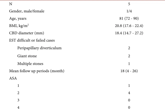
[image:2.595.212.534.480.696.2]Patients; Between June 2014 and April 2016, patients with choledocholithiasis which was difficult or failed to be treated by EST were eligible for the present study. Patients’ demographics were shown in Table 1. All cases had a choledo-chal dilatation, excluding the cases of CBD diameter less than 10 mm. Median
Table 1. Patients’ demographics.
N 5
Gender, male/female 1/4
Age, years 81 (72 - 90)
BMI, kg/m2 20.8 (17.6 - 22.4)
CBD diameter (mm) 18.4 (14.7 - 27.2)
EST difficult or failed cases
Peripapillary diverticulum 2
Giant stone 2
Multiple stones 1
Mean follow up periods (month) 18 (4 - 26) ASA
1 1
2 4
3 0
4 0
common bile duct diameter was 18.4 (14.7 - 27.2) mm. All five patients were EST difficult or failed cases such as peripapilla diverticulum, giant or multiple stones.
Surgical procedure; Laparoscopic operation was performed under general anesthesia with epidural analgesia. All procedures were done by one surgeon of authors. The patient was placed in the supine position with legs astride and arms opened. Traditional laparoscopic approach was performed for four ports access. In brief, initial 12-mm trocar (XCEL, Ethicon Endo-Surgery Inc, OH, USA) through 1.5-cm longitudinal incision was inserted via the umbilicus following CO2 pneumoperitoneum at 10 mmHg pressure. A 10-mm port was placed at the subxiphoidal region. Additional two trocars (5-mm) were also positioned right subcostal area at mid-clavicle and anterior axilla lines. A 10-mm flexible steera-ble laparoscope (OLYMPUS, Tokyo, Japan) was used. An additional needle tro-car (Microgrip, AMCO, Tokyo, Japan) was placed for retraction of the liver lat-eral segment.
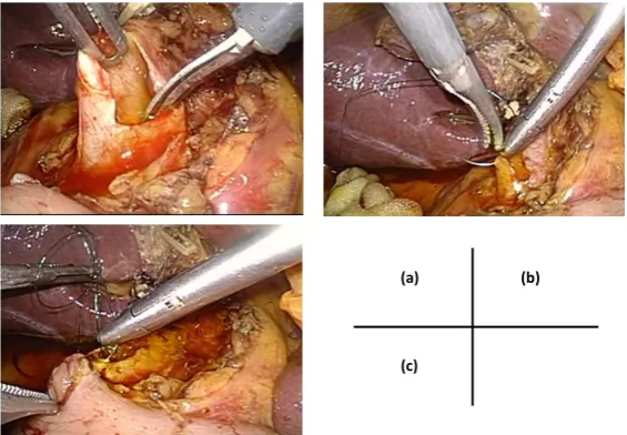
[image:3.595.232.515.482.678.2]Firstly, laparoscopic cholecystectomy was performed in a normograde fashion after the ligation and cut off the cystic-artery and cystic-duct. The gallbladder was not completely dissected from liver bed until forthcoming liver retraction by forceps. Laparoscopic choledochotomy was performed at anterior portion of the lower two-third of bile duct longitudinally and sharply with the endo-scissors approximately 2.0-cm in length (Figure 1(a)). Choledocholithotomy was done using by choledochoscope with bidirectional flex (OLYMPUS, CDS, Type-20, Tokyo, Japan). Stones can be removed by directional mechanical extraction, or stone basket for stone extraction under the direct vision of the choledochoscope (Cook TM Atlas Wire Stone Extractor). A longitudinal incision was made ap-proximately same length as that of the CBD in the duodenum with Harmonic scalpel (Ethicon Endo-Surgery Inc, OH, USA), after the ascertainment of the
Figure 1. Laparoscopic operative findings. (a) Longitudinal choledochotomy about 2.0 cm at the center of the common bile duct (CBD). Laparoscopic intracoporal suturing; (b) initial suture at the right side of central portion of choledochotomy; (c) posterior wall running suture.
(a) (b)
exact location of pylorus ring. A side-to-side CDD was created intracorporeally using running absorbable suture by 4-0 Maxon (COVIDIEN, MN, USA) ad-justed length about 15 cm with handmade double side needles, starting from the right to the left side of the choledochus with handling a needle from inside to outside of the bile duct and outside to inside of the duodenal wall (Figure 1(b)
and Figure 1(c)). The suture continued along posterior wall until the left side of the choledochus followed by anterior-wall suture. The anterior anastomosis was started from the right side of duodenum with handling a needle from inside to outside of the duodenal wall and outside to inside of the choledochus. Anterior side anastomosis was carried as the same manner of posterior side and finished at the left side by intracorporeal ligation of posterior and anterior threads. Fig-ure 2 illustrated how the intracorporeal choledochoduodenostomy was per-formed. Interlock sutures were added between posterior and anterior running sutures to prevent the anastomosis stenosis, indicating in Figure 2(b) and Fig-ure 2(d) (#).
After making sure that a tension-free anastomosis without biliary leakage, a standard Jacson-Pratt draine was placed at the lateral side of the anastomosis and brought out through the right-side port trocar.
This study was conducted and approved in accordance with the ethical prin-ciples outlined in the Declaration of Helsinki. Informed consent or its equivalent was obtained from a patient for this study.
3. Results
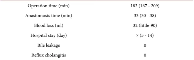
[image:4.595.234.515.479.678.2]In our case, median operation time was 182 min including 33 min of laparos-copic anastomosis time, and estimated median blood loss was 32 ml. Post oper-ative recovery was uneventful and discharged our hospital median 7 days later;
Figure 2. Illustration of the laparoscopic choledochoduodenostomy. (a) Initial suture of posterior wall anastomosis; (b) Final suture of posterior anastomosis; (c) Initial suture of anterior wall anastomosis; (d) Final suture of anterior anastomosis. # indicated the point sutured in interlock fashion.
(a)
Common Bile Duct
Duodenum
(b)
(c)
(d)
# #
the patient had no complications of the abdominal pain or high fever. Short- term outcome for up to 26 months showed also no complications such as bile leakage, reflux cholangitis, stone recurrence, and sump syndrome (Table 2). However, further long-term follow-up should be needed for an accurate estima-tion of stone recurrence or sump syndrome. Learning curve of intracorporeal anastomosis and operation time were showed in Figure 3. Completion time of intracorporeal anastomosis was gradually shortening, resulting in approximately 30 minutes. An operator became gradually skilled in intracorporeal suturing technique. Operation times were various and mainly depended on the preoper-ative inflammatory grade of cholecystitis or cholangitis and on fibrous adhesion of previous abdominal operation.
4. Discussions
The present study demonstrated that feasibility of laparoscopic CDD for patients especially with endoscopic management difficult or failed CBD stone with cho-ledochal dilatation. Furthermore, short-term outcomes of this laparoscopic pro-cedure were acceptable as a minimal invasive technique. Surgical results showed little blood loss, short hospital stay, no bile leakage, reflux cholangitis, and no mortality.
A first open side-to-side CDD was performed by Riedel in 1888, followed by successful CDD by Sprengel in 1891 [13]. Side-to-side CDD for the relief of dis-tal duct obstruction has several advantages. Its procedure is technically and phy-siologically simpler than sphincteroplasty or choledochojejunostomy and useful of the prophylaxis or treatment of residual, EST treatment difficult CBD stones. However, some surgeon avoided CDD due to the fear of potential complication of sump syndrome.
[image:5.595.209.540.614.717.2]Sump syndrome after CDD is characterized by upper abdominal pain or dis-comfort, rigors, pyrexia, jaundice and pancreatitis associated with elevated he-patobiliary enzymes. This syndrome thought to cause by stone, sludge or debris lodged in pool of the common bile duct distal to anastomosis, resulting in cho-langitis or hepatic abscess [14]. However, true incidence and resultant morbidity of sump syndrome are not well defined and has not been well examined. Recent studies indicate the rarity of the occurrence of reflux cholangitis and sump syn-drome [15] [16]. Some studies suggested a dilated common bile duct and con- Table 2. Short-term surgical outcomes of laparoscopic CDD.
Operation time (min) 182 (167 - 209)
Anastomosis time (min) 33 (30 - 38)
Blood loss (ml) 32 (little-90)
Hospital stay (day) 7 (5 - 14)
Bile leakage 0
Reflux cholangitis 0
struction of large or wide stoma would prevent these complications for the pro-cedure of CDD [17] [18].
The first laparoscopic CDD for the treatment of recurrent bile duct obstruc-tion was reported by Franklin et al. [19], in which the feasibility was also dem-onstrated. However, a rapid widespread of this procedure was not accomplished maybe due to requirement of significant advanced laparoscopic skill like handle of laparoscopic instrument and suturing intracorporeally technique. Conse-quently, published data of laparoscopic CDD are limited [20] [21] [22]. To overcome these problems, we used a dry box suturing training system and im-proved the technique. Furthermore, surgical practice shortens the completion time of intracorporeal CDD and improved the quality (Figure 3).
We confirmed the duodenal movability and tension prior to the beginning of anastomosis to decide whether Kocher’s maneuver was performed or not. In this series, Kocher’s maneuver was not carried out. In our procedure, the interlock sutures were added between the right and the left sides of posterior/anterior running sutures. The addition of the interlock suture was our original technique to prevent the anastomosis stenosis. Indeed, the stenosis was not observed for short-term follow-up period. Additionally, the stay suture of both sides between the common bile duct and duodenum was not made, because the procedure was becoming more complex by increasing number of handling threads.
[image:6.595.220.528.479.703.2]Our result in this report was acceptable even compared to other studies [20] [21] [22] [23]. Concerning about the development of sump syndrome, this com-plication was not observed for long-term follow-up in the small series of lapa-roscopic CDD [23] [24]. In laparoscopic CDD as well as open surgery, to pre-vent these complications a dilated common bile duct and construction of large or wide anastomosis should be needed. In addition, this laparoscopic technique
Figure 3. Completion times of operation and intracorporeal anastomosis of CDD by each case.
CASE
M
INUT
E
S
0 50 100 150 200 250 300
1 2 3 4 5
Operation time
might be adapted to biliary bypass operation by malignant disorder such as bile duct or pancreatic head cancers.
5. Conclusion
In conclusion, laparoscopic CDD is one of most reasonable operative methods for releasing biliary stasis, and also a feasible and safe technique in patients with CBD stone, especially EST of difficult or failed stone. In the case of endoscopic management of difficult stone, laparoscopic CDD should be a next therapeutic candidate. Further accumulating studies and long-term follow-up studies are required to evaluate this laparoscopic technique.
Acknowledgements
The authors have no conflicts of interest or financial disclose.
References
[1] Sandler, R.S., Everhart, J.E., Donowitz, M., Adams, E., Cronin, K., Goodman, C., et al. (2002) The Burden of Selected Digestive Diseases in United States. Gastroenter-ology, 122, 1500-1511. https://doi.org/10.1053/gast.2002.32978
[2] Lezoche, E., Paganini, Am, Carlei, F., Feliciotti, F., Lomanto, D. and Guerrieri, M. (1996) Laparoscopic Treatment of Gallbladder and Common Bile Duct Stones: A Prospective Study. World Journal of Surgery, 20, 535-541.
https://doi.org/10.1007/s002689900083
[3] Soltan, H.M., Kow, L. and Toouli, J. (2001) A Simple Scoring System for Predicting Bile Duct Stones in Patients with Cholelithiasis. Journal of Gastrointestinal Surgery, 5, 434- 437. https://doi.org/10.1016/S1091-255X(01)80073-1
[4] Saharia, P.C., Zuidema, G. and Cameron, J.L. (1977) Primary Common Duct Stones. Annals of Surgery, 185, 598-602.
https://doi.org/10.1097/00000658-197705000-00013
[5] Madden, J.L. (1973) Common Duct Stones; Their Origin and Surgical Management. Surgical Clinics of North America, 53, 1095-1113.
https://doi.org/10.1016/S0039-6109(16)40139-8
[6] Kaufman, H.S., Magnuson, T.H., Lillemoe, K.D., Frasca, P. and Pitt, H.A. (1989) The Role of Bacteria in Gallbladder and Common Duct Stone Formation.Annals of Surgery, 209, 584-591. https://doi.org/10.1097/00000658-198905000-00011
[7] Hill, J., Martin, D.F. and Tweedle, D.E. (1992) Risk of Leaving the Gallbladder in Situ after Endoscopic Sphincterotomy for Bile Duct Stones. British Journal of Sur-gery, 78, 554-557. https://doi.org/10.1002/bjs.1800780512
[8] Cotton, P.B., Geenen, J.E., Sherman, S., Cunningham, J.T., Howell, D.A., Carr- Locke, D.L., et al. (1998) Endoscopic Sphincterotomy for Stones by Experts Is Safe, Even in Younger Patients with Normal Ducts. Annals of Surgery, 227, 201-204.
https://doi.org/10.1097/00000658-199802000-00008
[9] Prat, F., Malak, N.A., Pelletier, G., Buffet, C., Fritsch, J., Choury, A.D., et al. (1996) Biliary Symptoms and Complications More than 8 Years after Endoscopic Sphinc-terotomy for Choledocholithiasis. Gastroenterology, 110, 894-899.
https://doi.org/10.1053/gast.1996.v110.pm8608900
https://doi.org/10.1016/S0016-5107(98)70086-0
[11] Memon, M.A., Hassaballa, H. and Memon, M.L. (2001) Laparoscopic Common Bile Duct Exploration: The Past, the Present, and the Future. The American Journal of Surgery,179, 309-315. https://doi.org/10.1016/S0002-9610(00)00346-9
[12] Tokumura, H., Umezawa, A., Cao, H., Sakamoto, N., Imaoka, Y., Ouchi, A., et al. (2002) Laparoscopic Management of Common Bile Duct Stones: Transcystic Ap-proach and Choledochotomy. Journal of Hepato-Biliary-Pancreatic Surgery, 9, 206-212. https://doi.org/10.1007/s005340200020
[13] Hess, W. (1997) Textbook of Bilio-Pancreatic Disease. Piccia Nuova Libraria, Pa-dova, 2828.
[14] Jan, A.U., Ahmad, I., Muneer, M. and Bukhari, H. (2001) Sump Syndrome—A Rar-ity after Choledochoduodenostomy. Journal of Postgraduate Medical Institute, 15, 183-189.
[15] Leppard, W.M., Shary, T.M., Adams, D.B. and Morqan, K.A. (2011) Choledocho-duodenostomy: Is It Really So Bad? Journal of Gastrointestinal Surgery,15, 754-757.
https://doi.org/10.1007/s11605-011-1465-2
[16] Okamoto, H., Miura, K., Wakana, H., Itakura, J. and Fujii, H. (2014) Current As-sessment of Choedochoduodenostomy for Bile Duct Stone. Japan Society for Sur-gical Infection, 11, 187-194.
[17] Madden, J.L., Chun, J.Y., Kandalaft, S. and Parekh, M. (1970) Choledochoduode-nostomy. An Unjustly Maligned Procedure? The American Journal of Surgery,119, 45-54. https://doi.org/10.1016/0002-9610(70)90010-3
[18] Akiyama, H., Ikezawa, H., Kameya, S., Iwasaki, M., Kuroda, Y. and Takeshita, T. (1980) Unexpected Problems of External Choledochoduodenostomy. The American Journal of Surgery,140, 660-665. https://doi.org/10.1016/0002-9610(80)90053-7
[19] Franklin, M.E. and Balli, J.E. (1997) Laparoscopic Common Bile Duct Bypass Pro-cedure. Seminars in Laparoscopic Surgery,4, 48-53.
https://doi.org/10.1177/155335069700400109
[20] Jeyapalan, M., Almeida, J.A., Michaelson, R.L. and Franklin, M.E.J. (2002) Lapa-roscopic Choledochoduodenostomy: Review of a 4-Year Experience with an Un-common Problem. Surgical Laparoscopy Endoscopy & Percutaneous Techniques, 12, 148-153. https://doi.org/10.1097/00129689-200206000-00002
[21] Gurbus, A.T., Watson, D. and Fenoglio, M.E. (1999) Laparopscopic Choledocho-duodenostomy. The American Surgeon,65, 212-214.
[22] Tinoco, R., El-Kadre, L. and Tinoco, A. (1999) Laparoscopic Choledochoduode-nostomy. Journal of Laparoendoscopic & Advanced Surgical Techniques, 9, 123- 126. https://doi.org/10.1089/lap.1999.9.123
[23] Khajanchee, Y.S., Cassera, M.A., Hammill, C.W., Swanström, L.L. and Hansen, P.D. (2012) Outcomes Following Laparoscopic Choledochoduodenostomy in the Man-agement of Benign Biliary Obstruction. Journal of Gastrointestinal Surgery,16, 801- 805. https://doi.org/10.1007/s11605-011-1768-3
[24] Tang, C.N., Siu, W.T., Ha, J.P. and Li, M.K. (2003) Laparoscopic Choledochoduo-denostomy. An Effective Drainage Procedure for Recurrent Pyogenic Cholangitis. Surgical Endoscopy and Other Interventional Techniques, 17, 1590-1594.
Submit or recommend next manuscript to SCIRP and we will provide best service for you:
Accepting pre-submission inquiries through Email, Facebook, LinkedIn, Twitter, etc. A wide selection of journals (inclusive of 9 subjects, more than 200 journals)
Providing 24-hour high-quality service User-friendly online submission system Fair and swift peer-review system
Efficient typesetting and proofreading procedure
Display of the result of downloads and visits, as well as the number of cited articles Maximum dissemination of your research work
Submit your manuscript at: http://papersubmission.scirp.org/