http://www.scirp.org/journal/ojemd ISSN Online: 2165-7432
ISSN Print: 2165-7424
Dysregulated miRNA Associated with
Transcription Factors of Insulin Gene
Expression in Chronic Pancreatitis
K. Murali Manohar
1, M. Sasikala
1*, P. Pavan Kumar
1, G. V. Rao
2, D. Nageshwar Reddy
21Institute of Basic Science and Translational Research, Asian Healthcare Foundation, Hyderabad, India 2Asian Institute of Gastroenterology, Somajiguda, Hyderabad, India
Abstract
Background/Aim: MicroRNAs with regulatory functions in gene expression are implicated in different diseases. The present study investigated differen-tially expressed miRNAs that possibly influence transcription factors involved in insulin gene expression in Chronic Pancreatitis (CP) employing bioinfor-matics approaches. Methods: Pancreatic tissues were collected from CP pa-tients undergoing partial pancreatectomy (n = 16) and controls (n = 15) un-dergoing resections for non-pancreatic malignancies. MiRNA profiles ob-tained using microarrays were validated by qRT-PCR. Target search involving miRWalk and TarBase as well as functional annotation employing KEGG (Kyoto encyclopedia of genes and genomes) and DAVID (Database for An-notation) databases were performed. Ingenuity pathway analysis (IPA) was used to construct networks relating miRNAs to their target genes. mRNA and proteins related to insulin gene transcription factors and hormones were eva-luated by qRT-PCR and western blotting followed by confirmation upon im-munofluorescent staining. Results:Microarray data revealed 10 up-regulated and 15 down-regulated miRNAs in CP as compared to controls (Log2 FC > 2). Bioinformatic analysis showed 8399 target genes and KEGG pathway analysis suggested a role for the dysregulated miRNAs in modulating cytokine signaling, fibrosis, JAK-STAT signaling and insulin synthesis. IPA analysis suggested a simplified network attributing dysregulated miRNAs to NFκB- dependent cytokine signaling. Further, associations could be noted between miRNA 200b with Maf A, 138-1 with Neuro D and 27b with FoxO1. Decreas-es in mRNA levels of Pdx1, Neuro D and increasDecreas-es of Maf A and FoxO1 tran-scription factors could be noted (P < 0.01) in CP. These results were con-firmed by western blotting and immunofluorescence staining. Conclusion:
How to cite this paper: Murali Manohar, K., Sasikala, M., Pavan Kumar, P., Rao, G.V. and Nageshwar Reddy, D. (2016) Dysregu-lated miRNA Associated with Transcription Factors of Insulin Gene Expression in Chronic Pancreatitis. Open Journal of Endocrine and Metabolic Diseases, 6, 205-227.
http://dx.doi.org/10.4236/ojemd.2016.610026
Received: August 20, 2016 Accepted: October 18, 2016 Published: October 21, 2016
Copyright © 2016 by authors and Scientific Research Publishing Inc. This work is licensed under the Creative Commons Attribution International License (CC BY 4.0).
http://creativecommons.org/licenses/by/4.0/
Our results identified dysregulation of miRNAs 138-1, 27b and 200b which were found to be associated with insulin gene transcription factors Neuro D, FoxO1 and Maf A respectively.
Keywords
MicroRNAs, Transcription Factors, Networks, β-Cell Dysfunction
1. Introduction
Chronic pancreatitis (CP) is a progressive inflammatory disorder ultimately culminating in exocrine and endocrine insufficiency contributing to associated maldigestion, abdominal pain and clinical diabetes. Endocrine insufficiency and clinical diabetes are popularly believed to occur as a result of fibrosis and β-cell destruction (apoptosis) in CP [1]. Several investigators demonstrated insulin se-cretory defects in patients with long standing CP [2]. It is held that insulin se-cretory defects arise initially as a result of β-cell dysfunction and overt clinical diabetes manifests from β-cell destruction much later in the course of the dis-ease. Such findings suggested a need to study the mechanism of β-cell dysfunc-tion under condidysfunc-tions of chronic inflammadysfunc-tion in CP. Diabetes mellitus, sec-ondary to chronic pancreatitis, is now categorized as Type 3C DM [3]. The complex pathophysiology of CP involving secondary diabetes presents distinct clinical and laboratory features, in contrast to autoimmunity and insulin resis-tance associated respectively with Type 1 and Type 2 DM. Currently it is re-ported that 5% - 10% in western population and 15% - 20% of diabetics in South East Asia have Type 3C DM [4]. Despite vast knowledge on Type 1 and Type 2 DM, Type 3C diabetes is relatively the less studied form of diabetes. Interesting-ly, our earlier reports demonstrated reduced islet response to glucose stimulation even in non-diabetic CP patients [5].
MicroRNAs are small, endogenously expressed noncoding RNAs (~21 - 25 nucleotides) known to influence basic cellular functions by regulating gene ex-pression. miRNAs bind to the 3’-untranslated region of mRNAs and lead to ei-ther transcriptional repression or mRNA degradation [6] [7]. Altered miRNA profiles have been implicated in different diseases such as diabetes, inflammato-ry diseases and various malignancies [8][9][10]. A few studies have also estab-lished basic functional role of miRNAs in islets (hormone secreting cell clusters) including miRNA375 in islet development, miRNA9 in regulating insulin secre-tory response, miRNA124 in regulating intracellular signaling and miRNA30d in induction of insulin production by targeting MAP4K4 [11][12][13][14]. Fur-ther, it was shown that miRNA21, 34a and 146 act as negative regulators of insu-lin signainsu-ling [15]. Increased expression of miRNA29 family in pre-diabetic NOD mice was shown to be associated with cytokine mediated β-cell dysfunction [16].
regula-tion between synthesis and secreregula-tion of pancreatic hormones. Expression of in-sulin gene in response to secretagogues is known to be controlled by various transcription factors such as Pdx1, neuro D, Maf A that bind to enhancer ele-ments in the promoter region of insulin gene [17]. Even though several investi-gators have reported altered miRNA profiles in CP and pancreatic cancer [18], relatively much less is known about the influence of aberrant miRNA(s) on β-cell function in CP; putative interactions between miRNAs and mRNAs of transcription factors of insulin gene expression have also not been elucidated under inflammatory conditions of CP. The established role of miRNAs in in-flammation [9] as well as the reported role of miRNAs in β-cell dysfunction in Type 2 DM [19] and in prediabetic NOD mice [16] have led us to hypothesize that aberrant miRNA expression in CP may also have an effect on insulin gene transcription, which may contribute to β-cell dysfunction.
The present study was thus conducted to (a) evaluate altered pattern of miR-NAs in CP and identify their putative targets including genes coding for islet hormones and associated transcription factors; (b) identify association of aber-rant miRNAs with signaling pathways involved in insulin gene expression and establish possible network(s) between miRNAs and target genes in CP using bioinformatics approaches.
2. Patients and Methods
Patients: Pancreatic tissues were obtained either from CP patients (n = 16) who had undergone partial pancreatectomy/lateral pancreaticojejunostomy for pain relief or from patients who had undergone pancreaticoduodenectomy for am-pullary/periampullary pathology without any history of CP (controls; n = 15), Histological confirmation of CP involved H&E staining of the resected speci-mens by a single pathologist experienced in pancreatic pathology who was blinded to the patient groups. Patients with CP having pancreatic malignancies, endocrine tumors, acute on CP were excluded. The study was initiated after the protocols were approved by the Institutional Ethics Committee of Asian Insti-tute of Gastroenterology and all the patients had given informed consent.
MicroRNA profiling and validation of dysregulated miRNAs
Target gene prediction and pathway analysis of dysregulated miRNAs Putative target genes for the differentially expressed miRNAs were identified and retrieved from miRWalk 2.0 database [21], which compiles and compares the predictions of other databases. Target genes that were experimentally vali-dated for dysregulated miRNAs were retrieved from Diana Lab Tarbase v.6 [22]. The putative and validated targets of dysregulated miRNAs were subjected to KEGG pathway annotation using DAVID functional annotation tool [23]. A two sided Fisher’s exact test and Chi-square test were used to classify the enrichment (Re) of pathway and false discovery rate (FDR) was calculated. A p value of <0.01 and an FDR of <0.05 were chosen for identification of enriched pathways.
IPA analysis to deduce network of altered miRNAs and target genes Differentially expressed miRNAs and the respective target genes were up-loaded into QIAGEN’s Ingenuity® Pathway Analysis (IPA®, QIAGEN Redwood City), which uses pooled genes as queries and then retrieves all other gene ob-jects related to query genes/miRNAs stored in knowledge base. Based on the re-lated genes a set of networks were generated with different scores. Networks that are considered to be best fit (implied by the score derived from the p-value) in-dicate the maximum likelihood of users’ list of genes in a network being found.
Evaluation of the network of transcription factors of insulin gene in CP One μg of total RNA was subjected to cDNA synthesis using oligodT, and M-MuLV Reverse transcriptase enzyme (Invitrogen). qRT-PCR for islet hor-mones (insulin, glucagon, somatostatin, polypeptide) and transcription factors (Pdx1, NeuroD, MafA, FoxO1) was performed using SYBR Green PCR Master Mix employing Step One Real Time PCR (Applied Bio systems). Primers were designed spanning multiple exons using IDT (Integrated DNA Technologies, Singapore) software available online. All RT-PCRs included no template controls and RT minus controls. For the relative quantification, 10 μL of master mix was prepared with 0.5 μL RT product, 0.2 μL volume each of appropriate forward and reverse primers, 5 μL SYBR green PCR master mix and 4.1 μL RNase free water. Samples were run in duplicates and data were normalized to human GAPDH. mRNA levels were relatively quantified by using ΔΔCT method (List of used Primers is given in Supplementary Table S1).
Western blot analysis and immunofluorescent staining for expression of Pdx1, Neuro D and MafA
Proteins were isolated from minced pancreatic tissue using 500 μl of RIPA ly-sis buffer with protease cocktail inhibitor (Sigma Aldrich, St Louis, USA) and probing the western blots either with rabbit anti-Pdx1 or rabbit anti-NeuroD or rabbit anti-MafA (Invitrogen, Shanghai, China) or anti β-actin (Santa cruz Bio-technology, USA) as per standard protocols [24].
antibo-dies for hormones; insulin and glucagon (guinea pig anti-insulin and mouse anti-glucagon at 1:200; Sigma Aldrich), somatostatin, polypeptide: (Invitrogen, Shanghai, China) and transcription factors (pancreatic duodenal homeobox; PDX-1, (rabbit anti PDX-1, rabbit anti MafA and Mouse Neuro D at 1:100 dilu-tion) followed by overnight incubation at 4˚C. After repeated washings with PBS, sections were treated with secondary antibodies tagged with fluorescent dyes guinea pig Alexa 488 for insulin and goat anti mouse Alexa 546 for gluca-gon at 1:200 dilutions; Invitrogen, Shanghai, China (goat anti rabbit Alexa 546 for PDX-1 and goat anti guinea pig Alexa 488 for insulin; Invitrogen, China, donkey anti goat Alexa 546 for Neuro D).Fluorescence images were captured using a Bioimager (CARV II, BD BioSciences, CA, USA).
Statistical analysis: The data were analyzed using statistical package for social sciences (SPSS version 20.0, Chicago, USA). A two tailed students P value of < 0.05 was considered statistically significant.
3. Results
Clinical characteristics of patients
A total of 16 patients with CP (male n = 12, mean age 34.44 ± 15.85 years), as confirmed by clinical, endoscopic and imaging (CT, MRI, EUS and ERCP) in-vestigations were recruited for the study. All of the CP patients had abdominal pain for 36 ± 21 months with radiological findings revealing the presence of intraductal calculi and main duct dilatation. Histopathological examination con-firmed intra and interlobular acinar atrophy and fibrosis in these patients. The control group (n = 15; male = 10, mean age 50.53 ± 9.53 years) were ascertained to be without CP.
Microarray experiments demonstrated differential expression profiles of miRNA in CP
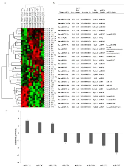
Significance analysis of Microarray (SAM) identified 52 differentially ex-pressed miRNAs in CP tissues as compared to control tissues (Supplementary
Table S2). Hierarchical clustering of the normalized expression data showed two
distinct clusters of miRNAs (Figure 1(a)). Further refinement with t-test and Bonferroni adjustment revealed a total of 25 differentially expressed miRNAs that includes 10 up-regulated and 15 down-regulated miRNAs with high signi-ficance (P < 0.05). The differentially expressed miRNAs detected by highest lo-garithmic fold changes between CP and adjacent control tissues are shown in
Figure 1(a). Among the up-regulated ones, miR-138-1 (3.12 fold change) and
among the down regulated miRNA-217, miR-216a (−4.85, −4.21) were highly significant (Figure 1(b)).
qRT-PCR analysis corroborated microarray miRNA expression profiles in CP
miR-376b) and five down-regulated miRNAs (miR-217, miR-200b, miR-27b, miR-29c and miR-375) miRNAs was validated by qRT-PCR. The results were in accordance with the microarray data indicating differential expression of these miRNAs in CP (P < 0.05). Of the seven miRNAs, differential expression of miR- 138, miR-767, miR-375 and miR-217 was significant (P < 0.001). The relative expression of differentially expressed miRNAs (log2 folds) is shown in Figure
1(c).
Target gene prediction of aberrant miRNA revealed fibrosis and insulin signaling pathways in CP
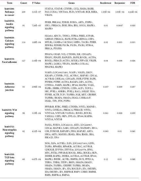
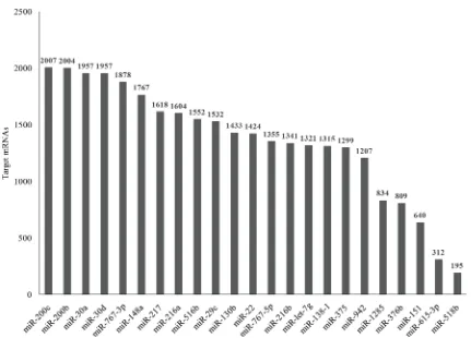
In silico analysis was performed to predict target mRNAs of the 25 differen-tially expressed miRNAs. The number of mRNA targets predicted for each of the differentially expressed miRNAs varied substantially. A three-way intersection of the three algorithms (Targetscan, miR Walk and miRNaDa) revealed 9577 target transcripts corresponding to 8399 unique official gene symbols. Functional an-notations of target genes using DAVID identified 12/53 significant pathways of which TGFβ, T cell receptor and cytokine-cytokine receptor interaction path-ways were related to inflammation and fibrosis. Similarly, pathpath-ways related to insulin synthesis, calcium signaling and Type2DM could be related to β cell spe-cific pathways as shown in the Table 1.
KEGG, ingenuity pathway analysis and putative networks in CP
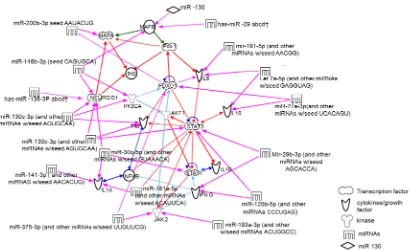
Fibrosis, inflammation, insulin signaling pathways and malignancy were found to be enriched on pathway analysis utilizing KEGG database. IPA analysis of 25 dysregulated miRNAs and respective target genes (predicted /validated) generated ten functional networks. Four of these networks that are relevant to CP include those involved in (i) inflammatory disease, fibrosis and cancer (ii) cytokine signaling, insulin synthesis and β cell function (iii) inflammatory re-sponse and cell morphology and (iv) immunological disease and cell cycle. Since CP is an inflammatory disease wherein islets have to function in an inflammato-ry milieu, we conducted further analysis relating miRNA expression to cytokine signaling and insulin gene transcription. Consequently, the final network gener-ated involved cytokines, transcription factors and miRNA. It depicts insulin gene transcription to be coordinately regulated by the three crucial transcription factors (Pdx1, NeuroD, MafA) and FoxO1 which are the confirmed targets of mir19, 130b, 30 family for Neuro D and miR 27 family for FoxO1 respectively. None of the altered miRNA targeted Pdx1 directly. This network also identifies a putative link between miR 200 family and MafA. Inflammatory cytokines (IL1 family, IL10, IFNγ, IL6) were found to be targets of differentially expressed miRNAs 191, 27, 29 and 191 respectively as reflected in the network (Figure 2). Validated targets of networks are shown in Supplementary Table S4.
Glucose responsive MafA expression increased and Pdx1, Neuro D ex-pression decreased in CP
Table 1. Putative targets and pathways that are associated with dysregulated microRNAs in chronic pancreatitis.
Term Count P Value Genes Bonferroni Benjamini FDR
hsa05200: Pathways
in cancer 209 5.31E−07
STAT5A, STAT5B, CTNNB1, CUL2, RARA, RARB, FAS, CCNA1, WNT10A, PLD1, WNT10B, BCR, RXRA,
VEGFA 1.05E−04 1.05E−04 6.64E−04
hsa04910: Insulin signaling pathway
91 7.26E−05 PHKB, PRKAG2, PDE3B, FOXO1, AKT1, PDPK1, SHC1, PRKACA, INSR, IRS4, IRS2, SOCS2, MAPK3,
MAPK9 0.01 0.0047 0.020
hsa04020: Calcium signaling pathway
114 1.08E−04
SLC8A3, ADCY1, TNNC1, ITPKA, PRKX, ATP2B1, GRIN2D, PRKACA, EGFR, HTR4, GRIN2A, CHP2, MYLK2, CAMK4, CACNA1I, GRIN1, TACR3, PHKB, EDNRA, EDNRB, PLCB4, PLCB1, PLCB2, HTR5A, PRKCA, PTGER3
0.02 0.005 0.013
hsa04510:
Focal adhesion 127 2.10E−04
PDGFA, CHAD, VCL, CTNNB1, ILK, CDC42P2, PDGFC, PDGFD, RAPGEF1, EGFR, ROCK1, ACTN4, ROCK2, PIK3CD, ACTN1, MYLK2, PPP1CB, VEGFB, MAPK1, LAMC3, VEGFA, MAPK3, COL1A2, PDGFRA, MAPK9
0.04 0.008 0.026
hsa04520: Adherens
junction 55 2.98E−04
WASF3, LOC100271831, WASF1, WASF2, LMO7, IQGAP1, CTNNB1, VCL, ACVR1C, MAP3K7, CDC42, ACVR1B, CSNK2A1, CDC42P2, INSR, PTPRJ, EGFR, PTPRM, PTPRF, ACTN4, BAIAP2, LEF1, ACTN1, CTNNA1, FARP2, MAPK1, EP300, MAPK3, WASL, FGFR1, ERBB2, CTNND1, CDH1, ACP1, TCF7L1, SRC, PVRL1, SORBS1, PVRL2, RAC1, SSX2IP, YES1, PTPRB, ACTB, TCF7, TGFBR1, NLK, MET, CREBBP, TGFBR2, SMAD4, SMAD2, SNAI2, CSNK2A1P, SNAI1, TJP1, FYN, PTPN1
0.057 0.009 0.037
hsa04310: Wnt signaling
pathway 98 3.19E−04
PPP2R5B, BTRC, PRKX, CTNNB1, WNT2, MAP3K7, WNT4, CSNK2A1, PRKACA, PRKACB, WNT6, WNT10A, WNT10B, VANGL1, ROCK1, ROCK2, VANGL2, CHP2, SKP1, DVL1L1, EP300, MAPK9, WNT5A, WNT5B
0.061 0.008 0.039
hsa04722: Neurotrophin
signaling pathway
82 4.52E−04
FASLG, FOXO3, LOC442113, AKT1, LOC440917, CDC42, MAP3K5, GAB1, CDC42P2, NGFRAP1, SHC1, CSK, FOXO3B, RAPGEF1, FRS2, MAP2K7, AKT3, SHC4, AKT2, MAP2K5, IRAK2, IRS4, IRAK1, IRS2, PIK3CD, TP53
0.085 0.01 0.045
hsa04350: TGF-beta signaling pathway
60 6.47E−04
NOG, E2F4, ACVRL1, E2F5, LOC100271831, GDF5, TGFB3, RPS6KB2, RPS6KB1, ACVR1C, ACVR1B, CDKN2B, ZFYVE9, ZFYVE16, LOC643778, IFNG, MYC, PITX2, PPP2R1B, ROCK1, RBL2, ROCK2, SKP1, INHBB, MAPK1, INHBA, ACVR2A, ACVR2B, EP300, MAPK3, INHBC, ACVR1, BMPR2, DCN, PPP2CA, THBS1, THBS2, TFDP1, BMP2, SMAD9, SMAD7, SMAD6, TGFBR1, CREBBP, TGFBR2, SMAD4, SMAD2, SMAD1, SP1, ID2, ROCK1P1, ID1, SMURF2, ID4, SMURF1, ID3, BMPR1B, BMP7, CHRD, BMP8B, BMP6, BMPR1A, BMP8A
Continued
hsa04010: MAPK signaling
pathway 158 0.002587266
MEF2C, FGF19, PDGFB, PDGFA, GNA12, FGF11, TGFB3, PRKX, PRKACG, MAP3K7, MAP3K6, MAP3K5, LOC100132771, MAP3K8, CDC42P2, PRKACA, FAS, PRKACB, RAPGEF2, MAP2K7, IL1A, MAP2K6, MAP2K5, EGFR, CHP2, PTPRR, FGF23, STK4, DDIT3, STK3, MAP4K3, MAP4K4, MAPK1, ARRB1
0.40 0.02 0.031
hsa04930: Type II diabetes
mellitus 34 0.004333089
HK2, PDX1, KCNJ11, LOC652797, HK2P1, SLC2A4, SLC2A2, HK3, PIK3R5, IRS4, IRS2, SOCS2, SOCS3, SOCS1, PIK3CD, LOC100131098, SOCS4, MAPK10, PRKCE, ADIPOQ, IRS1, MAPK1, GCK, PKM2, PKLR, MAPK3, CACNA1G, MAPK9, CACNA1E, MafA, IKBKB, CACNA1C, CACNA1D
0.57 0.036 0.045
hsa04660: T cell receptor
signaling pathway
68 0.008734447
CD8A, CD8B, PDCD1, IL10, MAP3K7, AKT1, PAK6, CDC42, FOS, IFNG, CDC42P2, PAK1, AKT3, AKT2, BCL10, CHP2, CDK4, CARD11, MAPK1, PRKCQ, NCK2, NCK1, MAPK3, NFKBIE, GRB2, KRAS, RASGRP1, SOS1, ICOS, ZAP70, NFAT5, CD4
0.82 0.067 0.039
hsa04060: Cytokine-cytoki
ne receptor interaction
151 0.01225817
PDGFB, IL6ST, PDGFA, IL18, GDF5, TNFSF15, TGFB3, IL15, CXCL11, CXCL12, TNFSF18, IL10, CXCL10, IL11, ZFP91, IFNG, CCR10, IL15RA, CSF3R, PDGFC, FAS, IFNK, IL1A, EGFR, LIFR, IL25, IL26, TNFRSF14
[image:9.595.89.500.395.646.2]0.913023416 0.086 0.030
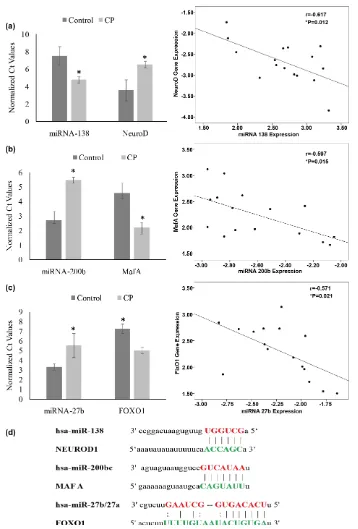
tered in CP. While the expression of Pdx1 and neuro D decreased by 5.02, 2.96 fold, that of MafA increased by more than 2.41 folds in CP. On the other hand FoxO1, a transcription factor known to be increased under conditions of oxida-tive stress, showed two fold increase in CP in comparison to controls. Similarly expression of genes coding for islet hormones namely insulin, glucagon, soma-tostatin, and pancreatic polypeptide (PP-1) indicated 4.3, 2.8, 4.4 and 1.9 log2 fold decrease respectively in CP patients as compared to control tissues (Figure 3(a)). Results obtained with qRT-PCR were akin to those obtained with western blot analysis, indicating increased MafA (~3 times) and decreased NeuroD and Pdx1 in CP as compared to controls (Figure 3(b)). These observations were va-lidated upon immunofluorescence staining of the paraffin embedded sections for insulin, glucagon, somatostatin, NeuroD and Pdx1 (Figure 3(c)).
Differentially expressed miRNA negatively correlated with NeuroD, MafA and FoxO1 expression in CP
The IPA network indicated three putative links between differentially ex-pressed miRNAs and β-cell specific transcription factors: miR-200b (−2.75) and MafA (2.41); miR-138 (2.76) and Neuro D (−2.965); and miR-27b (−2.24) and FoxO1 (2.21). miRNA targets of the transcription factors were confirmed by matching the seed sequences of miRNAs and genes coding for β cell specific transcription factors as shown below. Pearson correlation analysis revealed a significant (P < 0.05) negative correlation between mRNA levels with miRNA levels; NeuroD and miR-138 (r = −0.617, P = 0.012), MafA and miR-200b (r = −0.597, P = 0.015), FoxO1 and miR-27b (r = −0.571, P = 0.021) (Figure 4).
4. Discussion
Micro RNAs are known to play a pivotal role in controlling basic cellular func-tions by regulating gene expressions. We conducted this study to explore whether there is any dysregulation of pancreatic miRNAs under the inflamma-tory conditions prevalent in CP and to identify putative links between differen-tially expressed miRNAs and transcription factors that coordinately regulate in-sulin gene expression contributing to secondary diabetes occurring in the dis-ease. Our results demonstrate a network of three miRNAs (miR200b, 138 and 27b) regulating insulin gene transcription factors along with cytokines in CP.
Figure 3. Islet Hormones and transcription factors in CP. (a) Altered expression patterns of islet hormones and transcription fac-tors in CP tissues identified by qRT-PCR. The mRNA transcript abundance was normalized to that of GAPDH. Relative expres-sion was analyzed by 2^-ΔΔCT method; (b) Protein expression of β-cell specific transcription factors (NeuroD, PDX1 and MafA) in
between these two studies in miRNA expressions may be ascribed to differences in etiopathogenesis of CP (largely alcoholic in Western countries and idiopathic in tropical countries). Although these studies focused on miRNA expression patterns during progression of CP to pancreatic cancer, no study has been con-ducted to find an association between dysregulated miRNA and transcription factors involved in regulating insulin gene expression in CP. Such an identifica-tion of networks relating aberrant pancreatic miRNA expression to insulin gene transcription is of relevance in unraveling the mechanism of endocrine dysfunc-tion in CP.
and aberrant β-cell functions associated with CP.
Although comprehensive search was conducted for target gene prediction, networks were generated restricting the analysis to target genes that are asso-ciated with oxidative stress, cytokine signaling, Inflammation and insulin sig-naling since they are highly relevant to the progression of CP and manifestation type 3CDM. An attempt was also made to understand the intricate relations be-tween the dysregulated miRNAs, target genes, and β-cell dysfunction in CP em-ploying Ingenuity Pathway Analysis. A curated network was generated involving miRNAs, target genes of cytokine signaling, oxidative stress, insulin synthesis, β cell function. Interestingly, the resultant network revealed a web consisting of various proinflammatory cytokines (IL6, IFNγ, IL1β, and TNFα), JAK2, NFκB signaling pathways, statins (STAT1 and STAT3), effectors of β-cell function (MafA, NeuroD and Pdx1), β-cell development (MafB), β cell oxidative stress protective transcription factor (FoxO1) and insulin. Earlier results from our la-boratory and that of others have shown increased expression of proinflammato-ry cytokines [41] in CP and a role for NFκB [42] and oxidative stress was impli-cated in the inflammation in CP [43]. The network established in the present study has also identified links between cytokines and miRNAs (IL6 -miR191; IL10-miR27; IFNγ miR125b and 29C; IL1 miR141; TNFα miR 130 and miR19b) involving putative links between miR27-IL10 [44], miR29-IFNγ [45], TNFα- miR130 [46], that were previously established. In the network, NFκB and STAT proteins have network links with IFNγ, IL1A, IL1B TNFα, IL6 and IL10), while transcription factors and miRNAs (NeuroD-miR138, 19b, 130a, 30c, 148b; Ma-fA-200b; MafB-miR130b, miR29; FoxO1-miR27a; STAT3-miR29b, 130a; STAT1- miR30c) were associated. Interestingly, none of the differentially expressed miRNA targeted Pdx1 (crucial β cell transcription factor). Results of this study established possible links between miRNAs, cytokines, oxidative stress and tran-scription factors of insulin gene and β-cell functions in CP.
binding site in the Pdx1 promoter [47]. FoxO1 and Pdx1 have also been reported to show mutually exclusive nuclear localization [48][49] [50]. Increased oxida-tive stress, arising from inflammation and decreased miR 27b could result in raised FoxO1 and diminished Pdx1 expression in CP as observed in this study could be due to raised FoxO1. Transgenic mice with FoxO1 gain of function in the pancreas exhibited glucose intolerance [51]. Conversely, FoxO1 ablation in β-cells resulted in increased insulin secretion [52]. Further, in vitro studies showed suppression of miRNA-27 resulted in increased FoxO1 protein levels and a consequent decrease in cell number [53]. Hence we believe that increased FoxO1 resulting from increased oxidative stress leads to dysregulation in β cell functions.
Insulin gene transcription is known to be coordinately regulated by transcrip-tional activators such as Neuro D, Maf A and Pdx1 [54] [55][56][57]. To our knowledge, this is the first report to show increased expression of Maf A in CP both at mRNA and protein levels. We had earlier shown that reduced expression of Pdx1 is associated with decreased β-cell function [5]. It is well established that Pdx1, Neuro D and Maf A bind to the insulin promoter to initiate insulin gene transcription. Even though FoxO1 is known to activate gene expression (Maf A) in response to oxidative stress via acetylation [58], decrease in Pdx1 and Neuro D in CP might result in loss of coordinated transcription of insulin gene result-ing in decreased β cell function. Despite acetylated FoxO1 acting as a protective factor to β cells in response to oxidative stress under acute metabolic change, it fails to preserve β cell function in chronic conditions [58].
5. Conclusion
In summary, present study identified dysregulated miRNAs and indicated the pathways they target in CP. Dysregulated miRNA target genes are significantly enriched in pathways related to fibrosis and EMT (miR-29, 200, 217 miRNA clusters), inflammation/cytokines (miR-27 family) and insulin signaling path-ways (miR-30 family and miR-375). Putative links were also identified upon bioinformatic analysis between miRNAs (miR-138, 200b, 27a/27b) and tran-scription factors of insulin gene (Neuro D, Maf A, and FoxO1) suggesting that miRNAs, such as miR138-1, 27b and 200b, might be important regulators of crucial transcription factors involved in insulin gene expression in CP.
Acknowledgements
The authors are thankful to Prof C Subramanyam for editing the manuscript.
References
[1] Braganza, J.M., Lee, S.H. and Mc Cloy, R.F. (2011) Chronic Pancreatitis. Lancet, 377, 1184-1197. https://doi.org/10.1016/S0140-6736(10)61852-1
Beta-Cell Area in Patients with Chronic Pancreatitis. Gastroenterology, 136, 513- 522. https://doi.org/10.1053/j.gastro.2008.10.083
[3] American Diabetes Association (2009) Diagnosis and Classification of Diabetes Mellitus. Diabetes Care, 32, S62-S67. https://doi.org/10.2337/dc09-s062
[4] Ewald, N. and Bretzel, R.G. (2013) Diabetes Mellitus Secondary to Pancreatic Dis-eases (Type 3c)—Are We Neglecting an Important Disease? European Journal of Internal Medicine, 24, 203-206. https://doi.org/10.1016/j.ejim.2012.12.017
[5] Mitnala, S., Pondugala, P.K. and Guduru, V.R. (2010) Reduced Expression of Pdx-1 Is Associated with Decreased Beta Cell Function in Chronic Pancreatitis. Pancreas, 39, 856-862. https://doi.org/10.1097/MPA.0b013e3181d6bc69
[6] Bartel, D.P. (2009) MicroRNAs: Target Recognition and Regulatory Functions. Cell, 136, 215-233. https://doi.org/10.1016/j.cell.2009.01.002
[7] Valencia-Sanchez, M.A., Liu, J., Hannon, G.J. and Parker, R. (2006) Control of Translation and Mrna Degradation by Mirnas and Sirnas. Genes & Development, 20, 515-524. https://doi.org/10.1101/gad.1399806
[8] Tang, X., Tang, G. and Ozcan, S. (2008) Role of Micrornas in Diabetes. Biochimica et Biophysica Acta, 1779, 697-701. https://doi.org/10.1016/j.bbagrm.2008.06.010
[9] Contreras, J. and Rao, D.S. (2012) Micrornas in Inflammation and Immune Res-ponses. Leukemia, 26, 404-413. https://doi.org/10.1038/leu.2011.356
[10] Schultz, N.A., Dehlendorff, C. and Jensen, B.V. (2014) MicroRNA Biomarkers in Whole Blood for Detection of Pancreatic Cancer. JAMA, 311, 392-404.
https://doi.org/10.1001/jama.2013.284664
[11] Kloosterman, W.P., Lagendijk, A.K. and Ketting, R.F. (2007) Targeted Inhibition of Mirna Maturation with Morpholinos Reveals a Role for Mir-375 in Pancreatic Islet Development. PLOS Biology,5, e203. https://doi.org/10.1371/journal.pbio.0050203
[12] Plaisance, V., Abderrahmani, A. and Perret-Menoud, V. (2006) Microrna-9 Con-trols the Expression of Granuphilin/Slp4 and the Secretory Response of Insulin- Producing Cells. Journal of Biological Chemistry, 281, 26932-26942.
https://doi.org/10.1074/jbc.M601225200
[13] Baroukh, N., Ravier, M.A. and Loder, M.K. (2007) Microrna-124a Regulates Foxa2 Expression and Intracellular Signaling in Pancreatic Beta-Cell Lines. Journal of Bi-ological Chemistry, 282, 19575-19588. https://doi.org/10.1074/jbc.M611841200
[14] Zhao, X., Mohan, R., Ozcan, S. and Tang, X. (2012) Microrna-30d Induces Insulin Transcription Factor Mafa and Insulin Production by Targeting Mitogen-Activated Protein 4 Kinase 4 (Map4k4) in Pancreatic Beta-Cells. Journal of Biological Chemi-stry, 290, 31155-31164. https://doi.org/10.1074/jbc.M112.362632
[15] Roggli, E., Britan, A. and Gattesco, S. (2010) Involvement of Micrornas in the Cy-totoxic Effects Exerted by Proinflammatory Cytokines on Pancreatic Beta-Cells.
Diabetes, 59, 978-986. https://doi.org/10.2337/db09-0881
[16] Roggli, E., Gattesco, S. and Caille, D. (2012) Changes in Microrna Expression Con-tribute to Pancreatic Beta-Cell Dysfunction in Prediabetic Nod Mice. Diabetes, 61, 1742-1751. https://doi.org/10.2337/db11-1086
[17] Aramata, S., Han, S.I., Yasuda, K. and Kataoka, K. (2005) Synergistic Activation of the Insulin Gene Promoter by the Beta-Cell Enriched Transcription Factors Mafa, Beta2, and Pdx1. Biochimica et Biophysica Acta, 1730, 41-46.
https://doi.org/10.1016/j.bbaexp.2005.05.009
to Differentiate Pancreatic Adenocarcinoma from Normal Pancreas and Chronic Pancreatitis. JAMA, 297, 1901-1908. https://doi.org/10.1001/jama.297.17.1901
[19] Lovis, P., Roggli, E. and Laybutt, D.R. (2008) Alterations in Microrna Expression Contribute to Fatty Acid-Induced Pancreatic Beta-Cell Dysfunction. Diabetes, 57, 2728-2736. https://doi.org/10.2337/db07-1252
[20] Kalluri Sai Shiva, U.M., Kuruva, M.M. and Mitnala, S. (2014) MicroRNA Profiling in Periampullary Carcinoma. Pancreatology, 14, 36-47.
https://doi.org/10.1016/j.pan.2013.10.003
[21] Dweep, H., Sticht, C., Pandey, P. and Gretz, N. (2011) MiRWalk—Database: Predic-tion of Possible Mirna Binding Sites by “Walking” the Genes of Three Genomes.
Journal of Biomedical Informatics, 44, 839-847.
https://doi.org/10.1016/j.jbi.2011.05.002
[22] Sethupathy, P., Corda, B. and Hatzigeorgiou, A.G. (2006) Tarbase: A Comprehen-sive Database of Experimentally Supported Animal Microrna Targets. RNA, 12, 192-197. https://doi.org/10.1261/rna.2239606
[23] Huang, W., Sherman, B.T. and Lempicki, R.A. (2009) Systematic and Integrative Analysis of Large Gene Lists Using David Bioinformatics Resources. Nature Proto-cols, 4, 44-57. https://doi.org/10.1038/nprot.2008.211
[24] Dong, H., Morgan, K., Adams, D. and Wang, H. (2012) Prevention of Beta Cell Death in Chronic Pancreatitis. Advances in Bioscience and Biotechnology, 3, 782- 787. https://doi.org/10.4236/abb.2012.326098
[25] Bauer, A.S., Keller, A. and Costello, E. (2012) Diagnosis of Pancreatic Ductal Ade-nocarcinoma and Chronic Pancreatitis by Measurement of Microrna Abundance in Blood and Tissue. PLoS ONE, 7, e34151.
https://doi.org/10.1371/journal.pone.0034151
[26] Mogilyansky, E. and Rigoutsos, I. (2013) The Mir-17/92 Cluster: A Comprehensive Update on Its Genomics, Genetics, Functions and Increasingly Important and Nu-merous Roles in Health and Disease. Cell Death & Differentiation, 20, 1603-1614.
https://doi.org/10.1038/cdd.2013.125
[27] Chun, A.C., Dong, Y. and Yang, W. (2013) Smad7 Suppresses Renal Fibrosis via Altering Expression of Tgf-Beta/Smad3-Regulated Micrornas. Molecular Therapy, 21, 388-398. https://doi.org/10.1038/mt.2012.251
[28] Xiao, J., Meng, X.M. and Huang, X.R. (2012) Mir-29 Inhibits Bleomycin-Induced Pulmonary Fibrosis in Mice. Molecular Therapy, 20, 1251-1260.
https://doi.org/10.1038/mt.2012.36
[29] Van Rooij, E., Sutherland, L.B., Thatcher, J.E., DiMaio, J.M., Naseem, R.H., Mar-shall, W.S., Hill, J.A. and Olson, E.N. (2008) Dysregulation of Micrornas after Myocardial Infarction Reveals a Role of Mir-29 in Cardiac Fibrosis. Proceedings of the National Academy of Sciences of the United States of America, 105, 13027- 13032. https://doi.org/10.1073/pnas.0805038105
[30] Shek, F.W., Benyon, R.C. and Walker, F.M. (2002) Expression of Transforming Growth Factor-Beta 1 by Pancreatic Stellate Cells and Its Implications for Matrix Secretion and Turnover in Chronic Pancreatitis. American Journal of Pathology, 160, 1787-1798. https://doi.org/10.1016/S0002-9440(10)61125-X
[31] Wang, B., Koh, P. and Winbanks, C. (2011) Mir-200a Prevents Renal Fibrogenesis through Repression of Tgf-Beta2 Expression. Diabetes, 60, 280-287.
[32] Oba, S., Kumano, S. and Suzuki, E. (2010) Mir-200b Precursor Can Ameliorate Renal Tubulointerstitial Fibrosis. PLoS ONE,5, e13614.
https://doi.org/10.1371/journal.pone.0013614
[33] Hu, L.H., Ji, J.T. and Li, Z.S. (2015) Potential Application of Mirnas as Diagnostic and Therapeutic Tools in Chronic Pancreatitis. Journal of Cellular and Molecular Medicine, 19, 2049-2057. https://doi.org/10.1111/jcmm.12603
[34] Kato, M., Putta, S. and Wang, M. (2009) Tgf-Beta Activates Akt Kinase through a Microrna-Dependent Amplifying Circuit Targeting Pten. Nature Cell Biology, 11, 881-889. https://doi.org/10.1038/ncb1897
[35] Yan, M.X., Ren, H.B. and Kou, Y. (2012) Involvement of Nuclear Factor Kappa B in High-Fat Diet-Related Pancreatic Fibrosis in Rats. Gut Liver, 6, 381-387.
https://doi.org/10.5009/gnl.2012.6.3.381
[36] Stevens, T., Conwell, D.L. and Zuccaro, G. (2004) Pathogenesis of Chronic Pancrea-titis: An Evidence-Based Review of Past Theories and Recent Developments. Amer-ican Journal of Gastroenterology, 99, 2256-2270.
https://doi.org/10.1111/j.1572-0241.2004.40694.x
[37] Joglekar, M.V., Joglekar, V.M. and Hardikar, A.A. (2009) Expression of Islet-Spe- cific microRNAs during Human Pancreatic Development. Gene Expression Pat-terns, 9, 109-113. https://doi.org/10.1016/j.gep.2008.10.001
[38] Hennessy, E. and O’Driscoll, L. (2008) Molecular Medicine of Micrornas: Structure, Function and Implications for Diabetes. Expert Reviews in Molecular Medicine, 10, e24. https://doi.org/10.1017/s1462399408000781
[39] Poy, M.N., Eliasson, L. and Krutzfeldt, J. (2004) A Pancreatic Islet-Specific Micro-rna Regulates Insulin Secretion. Nature, 432, 226-230.
https://doi.org/10.1038/nature03076
[40] Van de Bunt, M., Gaulton, K.J. and Parts, L. (2013) The Mirna Profile of Human Pancreatic Islets and Beta-Cells and Relationship to Type 2 Diabetes Pathogenesis.
PLoS ONE, 8, e55272. https://doi.org/10.1371/journal.pone.0055272
[41] Pavan Kumar, P., Radhika, G. and Rao, G.V. (2012) Interferon Gamma and Gly-cemic Status in Diabetes Associated with Chronic Pancreatitis. Pancreatology, 12, 65-70. https://doi.org/10.1016/j.pan.2011.12.005
[42] Sah, R.P., Dudeja, V., Dawra, R.K. and Saluja, A.K. (2013) Cerulein-Induced Chronic Pancreatitis Does Not Require Intra-Acinar Activation of Trypsinogen in Mice. Gastroenterology, 144, 1076-1085.
https://doi.org/10.1053/j.gastro.2013.01.041
[43] Schoenberg, M.H., Birk, D. and Beger, H.G. (1995) Oxidative Stress in Acute and Chronic Pancreatitis. American Journal of Clinical Nutrition, 62, 1306S-1314S. [44] Xie, N., Cui, H. and Banerjee, S. (2014) Mir-27a Regulates Inflammatory Response
of Macrophages by Targeting Il-10. Journal of Immunology, 193, 327-934.
https://doi.org/10.4049/jimmunol.1400203
[45] Ma, F., Xu, S. and Liu, X. (2011) The Microrna Mir-29 Controls Innate and Adap-tive Immune Responses to Intracellular Bacterial Infection by Targeting Interfe-ron-Gamma. Nature Immunology, 12, 861-869. https://doi.org/10.1038/ni.2073
[46] Kim, C., Lee, H. and Cho, Y.M. (2013) Tnfalpha-Induced Mir-130 Resulted in Adi-pocyte Dysfunction during Obesity-Related Inflammation. FEBS Letters,587, 3853- 3858. https://doi.org/10.1016/j.febslet.2013.10.018
FoxO1 Links Insulin Signaling to Pdx1 Regulation of Pancreatic Beta Cell Growth.
Journal of Clinical Investigation, 110, 1839-1847.
https://doi.org/10.1172/JCI200216857
[48] Hashimoto, N., Kido, Y. and Uchida, T. (2006) Ablation of Pdk1 in Pancreatic Beta Cells Induces Diabetes as a Result of Loss of Beta Cell Mass. Nature Genetics, 38, 589-593. https://doi.org/10.1038/ng1774
[49] Kawamori, D., Kaneto, H. and Nakatani, Y. (2006) The Forkhead Transcription Factor FoxO1 Bridges the Jnk Pathway and the Transcription Factor Pdx-1 through Its Intracellular Translocation. Journal of Biological Chemistry, 281, 1091-1098.
https://doi.org/10.1074/jbc.M508510200
[50] Okada, T., Liew, C.W. and Hu, J. (2007) Insulin Receptors in Beta-Cells Are Critical for Islet Compensatory Growth Response to Insulin Resistance. Proceedings of the National Academy of Sciences of the United States of America, 104, 8977-8982.
https://doi.org/10.1073/pnas.0608703104
[51] Kikuchi, O., Kobayashi, M. and Amano, K. (2012) FoxO1 Gain of Function in the Pancreas Causes Glucose Intolerance, Polycystic Pancreas, and Islet Hypervascula-rization. PLoS ONE, 7, e32249. https://doi.org/10.1371/journal.pone.0032249
[52] Miyazaki, S., Minamida, R. and Furuyama, T. (2012) Analysis of FoxO1-Regulated Genes Using FoxO1-Deficient Pancreatic Beta Cells. Genes Cells, 17, 758-767.
https://doi.org/10.1111/j.1365-2443.2012.01625.x
[53] Guttilla, I.K. and White, B.A. (2009) Coordinate Regulation of FoxO1 by Mir-27a, Mir-96, and Mir-182 in Breast Cancer Cells. Journal of Biological Chemistry, 284, 23204-23216. https://doi.org/10.1074/jbc.M109.031427
[54] Naya, F.J., Stellrecht, C.M. and Tsai, M.J. (1995) Tissue-Specific Regulation of the Insulin Gene by a Novel Basic Helix-Loop-Helix Transcription Factor. Genes & Development, 9, 1009-1019. https://doi.org/10.1101/gad.9.8.1009
[55] Zhao, L., Guo, M. and Matsuoka, T.A. (2005) The Islet Beta Cell-Enriched Mafa Ac-tivator Is a Key Regulator of Insulin Gene Transcription. Journal of Biological Chemistry, 280, 11887-11894. https://doi.org/10.1074/jbc.M409475200
[56] Kaneto, H., Miyatsuka, T. and Kawamori, D. (2008) Pdx-1 and Mafa Play a Crucial Role in Pancreatic Beta-Cell Differentiation and Maintenance of Mature Beta-Cell Function. Endocrine Journal, 55, 235-252.
https://doi.org/10.1507/endocrj.K07E-041
[57] Petersen, H.V., Serup, P. and Leonard, J. (1994) Transcriptional Regulation of the Human Insulin Gene Is Dependent on the Homeodomain Protein Stf1/Ipf1 Acting through the Ct Boxes. Proceedings of the National Academy of Sciences of the United States of America, 91, 10465-1049. https://doi.org/10.1073/pnas.91.22.10465
Supplementary Tables and Figures
Table S1. Primers list.
Gene Forward primer Reverse primer Product Size Annealing Temp
Insulin CTAGTGTGCGGGGAACG CACGCTTCTGCAGGGAC 148 bp 58
Somatostatin ACTCCGTCAGTTTCTGCAG CTGGGACAGATCTTCAGGTTC 136 bp 58.5
Glucagon CATTGCTTGGCTGGTGAAAG GCGGCAAGATTATCAAGAATGG 135 bp 58
Polypeptide GTATGCAGCTGATCTCCGTAG GGCATTATAAGTCCAGCGGG 149 bp 57
MafA GAGAAGTGCCAACTCCAGAG GCCAGCTTCTCGTATTTCTCC 101 bp 60
Pdx-1 TGAAGTCTACCAAAGCTCACG GGAACTCCTTCTCCAGCTCTA 132 bp 58
NeuroD CCAGGGTTATGAGACTATCACTG TCCTGAGAACTGAGACACTCG 149 bp 60
FoxO1 GCAATGGCTATGGCAGAATG AGTGTAACCTGCTCACTAACC 240 bp 57.5
GAPDH AATCCCATCACCATCTTCCAG AAATGAGCCCCAGCCTTC 122 bp 60
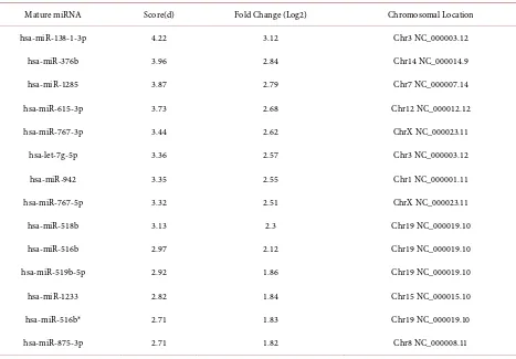
Table S2. Differentially expressed miRNAs identified by SAM (52). (a) Up regulated (14); (b) Down regulated (38). (a)
Mature miRNA Score(d) Fold Change (Log2) Chromosomal Location
hsa-miR-138-1-3p 4.22 3.12 Chr3 NC_000003.12
hsa-miR-376b 3.96 2.84 Chr14 NC_000014.9
hsa-miR-1285 3.87 2.79 Chr7 NC_000007.14
hsa-miR-615-3p 3.73 2.68 Chr12 NC_000012.12
hsa-miR-767-3p 3.44 2.62 ChrX NC_000023.11
hsa-let-7g-5p 3.36 2.57 Chr3 NC_000003.12
hsa-miR-942 3.35 2.55 Chr1 NC_000001.11
hsa-miR-767-5p 3.32 2.51 ChrX NC_000023.11
hsa-miR-518b 3.13 2.3 Chr19 NC_000019.10
hsa-miR-516b 2.97 2.12 Chr19 NC_000019.10
hsa-miR-519b-5p 2.92 1.86 Chr19 NC_000019.10
hsa-miR-1233 2.82 1.84 Chr15 NC_000015.10
hsa-miR-516b* 2.71 1.83 Chr19 NC_000019.10
[image:20.595.71.538.377.702.2](b)
Mature miRNA Score(d) Log2 Fold Change Chromosomal Location
hsa-miR-217 −3.35 −4.64 2p16.1
hsa-miR-216a −3.32 −3.84 2p16.1
hsa-miR-375 −3.48 −3.64 2q35
hsa-miR-216b −2.92 −3.32 2p16.1
hsa-miR-148a −3.36 −3.18 7p15.2
hsa-miR-22-3p −3.87 −2.74 17p13.3
hsa-miR-30a-5p −3.47 −2.56 6q13
hsa-miR-200c-3p −2.36 −2.47 12p13.31
hsa-miR-30d-5p −4.57 −2.40 8q24.22
hsa-miR-200b-3p −2.16 −2.32 1p36.33
hsa-miR-151-3p −3.96 −2.25 8q24.3
hsa-miR-130b-3p −2.67 −2.25 22q11.21
hsa-miR-29c-5p −3.44 −2.18 1q32.2
hsa-miR-29a-3p −2.82 −2.06 7q32.3
hsa-miR-27b-3p −3.3 −2.06 9q22.32
hsa-miR-19b-3p −2.12 −1.79 13q31.3
hsa-miR-191-5p −2.56 −1.74 3p21.31
hsa-miR-152 −2.97 −1.56 17q21.32
hsa-miR-339-3p −3.73 −1.51 7p22.3
hsa-miR-181a-5p −3.66 −1.47 9q33.3
hsa-miR-99a-5p −2.29 −1.47 21q21.1
hsa-miR-125b-5p −2.19 −1.43 11q24.1
hsa-miR-141-3p −2.26 −1.40 12p13.31
hsa-miR-30a-3p −3.99 −1.36 6q13
hsa-miR-193b-3p −4.48 −1.32 16p13.2
hsa-miR-34a-5p −2.16 −1.32 1p36.33
hsa-miR-28-3p −2.71 −1.22 3q28
hsa-miR-127-3p −2.4 −1.22 14q32.2
hsa-miR-29b-3p −2.44 −1.09 7q32.3
hsa-miR-1184 −2.6 −1.06 Xq28
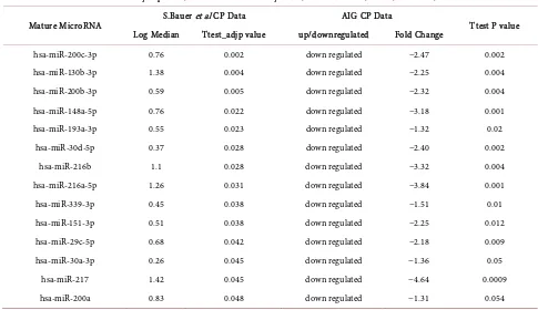
Table S3. Comparison of dysregulated miRNA in CP with other studies.
Differentially expressed microRNAs commonly found in S.Bauer CP data and AIG CP data
Mature MicroRNA S.Bauer et al CP Data AIG CP Data Ttest P value Log Median Ttest_adjp value up/downregulated Fold Change
hsa-miR-200c-3p 0.76 0.002 down regulated −2.47 0.002 hsa-miR-130b-3p 1.38 0.004 down regulated −2.25 0.004 hsa-miR-200b-3p 0.59 0.005 down regulated −2.32 0.004 hsa-miR-148a-5p 0.76 0.022 down regulated −3.18 0.001 hsa-miR-193a-3p 0.55 0.023 down regulated −1.32 0.02
hsa-miR-30d-5p 0.37 0.028 down regulated −2.40 0.002 hsa-miR-216b 1.1 0.028 down regulated −3.32 0.004 hsa-miR-216a-5p 1.26 0.031 down regulated −3.84 0.001 hsa-miR-339-3p 0.45 0.038 down regulated −1.51 0.01 hsa-miR-151-3p 0.51 0.038 down regulated −2.25 0.012 hsa-miR-29c-5p 0.68 0.042 down regulated −2.18 0.009 hsa-miR-30a-3p 0.26 0.045 down regulated −1.36 0.05
hsa-miR-217 1.42 0.045 down regulated −4.64 0.0009 hsa-miR-200a 0.83 0.048 down regulated −1.31 0.054
Table S4. Interactions of dysregulated miRNAs and target genes: relevance to CP.
[image:22.595.59.534.415.712.2]IPA Network (Figure)
miRNA miRNA target Regulatory feedback
loop Confirmed biological functions Proposed role in CP Ref. nos. Gene FC mRNA FC
Figure 2 miR130b decrease MafB decrease miR-130b —MafB MicroRNA fingerprints during human megakaryocytopoiesis. factors may be involved in Maf family transfription
beta cell dysfunction 23863625
Figure 2 miR-27b decrease FoxO1 increase miR-27b —FoxO1 by miR-27a, miR-96, and miR-182 Coordinate regulation of FOXO1 in breast cancer cells.
FoxO1 gain of function in pancreas causes glucose
intolerance, polycystic pancreas, and islet hyper
vascularization.
19574223, 22384192
Figure 2 miR-29 family decrease IFN G increase —IFN g miR-29
miR29 controls innate and adaptive immune responses to intracellular bacterial infection
bytargeting interferon-γ.
Interferon γ and glycemic status in diabetes associated with chronic pancreatitis and
its role in fibrogenesis
21785411, 22487478, 20971881
Figure 2 miR375 decrease JAK2 known Not miR375 —JAK2
miR-375 may function as a tumor suppressor to regulate gastric cancer cell proliferation potentially
by targeting the JAK2 oncogene, implicating a role of miR-375 in the pathogenesis of gastric cancer
JAK2 mediated pathways were shown to stimulate replication
and survival of β-cells
Continued
Figure 2 miR-217 decrease SIRT1 increase —SIRT1 miR217
—EMT
Chronic pancreatitis and pancreatic cancer demonstrate active epithelial-mesenchymal transition profile, regulated by
miR-217-SIRT1 pathway
miR217 is highly pancreas specific and it is shown to involve in progression of CP
to pancreatic cancer
25172416
Figure 2, miR-191 decrease IL1 increase miR191 —IL1
hsa-miR-191 Is a Candidate Oncogene Target for Hepatocellular Carcinoma
Therapy
No proposed role of miR191, however elevation of proinflammatory cytokines (IL1a,
IL1b) were reported in CP
20924108, 22487478
Figure 2 miR-181 decrease SMAD increase miR-181 —TGFB
—SMAD
miR-181a can affect the phosphorylation of Smad2 through
activation of TGF-β signaling
Inhibition of transforming growth factor beta decreases
pancreatic fibrosis and protects the pancreas against
chronic injury in mice.
22714950, 15502860
Figure 2 miR-200b decrease ZEB1 increase miR-200b —ZEB1 Participation of miR-200 in pulmonary fibrosis implicated in progression of miR-200 family were fibrosis
[image:23.595.84.515.347.657.2]25172416, 22189082
Table shows the miRNAs and corresponding target genes that are part of the network represented in Figure 2. These interactions of miRNAs and target genes that are relevant to CP have been validated in previous studies. Ref Nos column shows the pubmed IDs of the validated interaction that were pub-lished.
Submit or recommend next manuscript to SCIRP and we will provide best service for you:
Accepting pre-submission inquiries through Email, Facebook, LinkedIn, Twitter, etc. A wide selection of journals (inclusive of 9 subjects, more than 200 journals)
Providing 24-hour high-quality service User-friendly online submission system Fair and swift peer-review system
Efficient typesetting and proofreading procedure
Display of the result of downloads and visits, as well as the number of cited articles Maximum dissemination of your research work
Submit your manuscript at: http://papersubmission.scirp.org/