Development and Validation of a Quantitative, One-Step, Multiplex,
Real-Time Reverse Transcriptase PCR Assay for Detection of Dengue
and Chikungunya Viruses
Monika Simmons,aTodd Myers,aCarolina Guevara,bDonald Jungkind,cMaya Williams,aHuo-Shu Houngd
Viral and Rickettsial Diseases Department, Naval Medical Research Center, Silver Spring, Maryland, USAa
; Virology and Emerging Infections Department, U.S. Naval Medical Research Unit No. 6, Lima, Perub
; St. George’s University School of Medicine, St. George, Grenadac
; Division of Viral Diseases, Walter Reed Army Institute of Research, Silver Spring, Maryland, USAd
Dengue virus (DENV) and chikungunya virus (CHIKV) are important human pathogens with common transmission vectors
and similar clinical presentations. Patient care may be impacted by the misdiagnosis of DENV and CHIKV in areas where both
viruses cocirculate. In this study, we have developed and validated a one-step multiplex reverse transcriptase PCR (RT-PCR) to
simultaneously detect, quantify, and differentiate between four DENV serotypes (pan-DENV) and chikungunya virus. The assay
uses TaqMan technology, employing two forward primers, three reverse primers, and four fluorophore-labeled probes in a
sin-gle-reaction format. Coextracted and coamplified RNA was used as an internal control (IC), and
in vitro-transcribed DENV and
CHIKV RNAs were used to generate standard curves for absolute quantification. The diagnostic 95% limits of detection (LOD)
within the linear range were 50 and 60 RNA copies/reaction for DENV (serotypes 1 to 4) and CHIKV, respectively. Our assay was
able to detect 53 different strains of DENV, representing four serotypes, and six strains of CHIKV. No cross-reactivity was
ob-served with related flaviviruses and alphaviruses, To evaluate diagnostic sensitivity and specificity, 89 clinical samples positive or
negative for DENV (serotypes 1 to 4) and CHIKV by the standard virus isolation method were tested in our assay. The multiplex
RT-PCR assay showed 95% sensitivity and 100% specificity for DENV and 100% sensitivity and specificity for CHIKV. With an
assay turnaround time of less than 2 h, including extraction of RNA, the multiplex quantitative RT-PCR assay provides rapid
diagnosis for the differential detection of two clinically indistinguishable diseases, whose geographical occurrence is increasingly
overlapping.
D
engue is a mosquito-borne viral infection in humans that is of
global importance. An estimated 390 million infections occur
each year, of which 96 million result in clinically apparent
infec-tions, and in some epidemics the mortality rate may reach 5% (
1
,
2
). Dengue viruses (DENVs) are members of the family
Flaviviri-dae
and consist of four antigenically distinct serotypes which
ex-hibit 65% to 70% sequence homology (
3
). Disease manifestations
range from mild undifferentiated acute dengue fever (DF) to
se-vere dengue hemorrhagic fever (DHF) and dengue shock
syn-drome (DSS) (
4
,
5
). After an incubation period of 2 to 10 days, a
primary infection presents with the acute onset of high fever
(
ⱖ
40°C), accompanied by headache, retroorbital pain,
general-ized myalgias, arthralgias, and malaise. The early phase of DHF or
DSS has similar clinical features; however, after defervescence,
new abdominal pain, nausea, and vomiting, followed by
throm-bocytopenia and a rise in hematocrit, may progress to shock
lead-ing to multiorgan dysfunction, life-threatenlead-ing hemorrhage, and
death. DF symptoms may be clinically indistinguishable from
acute febrile illness due to other infectious diseases such as
influ-enza, malaria, measles, and chikungunya virus (CHIKV)
infec-tions. CHIKVs are alphaviruses belonging to the family
Togaviri-dae
. CHIKV causes an acute illness including fever, rash, severe
incapacitating arthralgia, headache, fatigue, vomiting, and
con-junctivitis (
6
). Although many patients infected with CHIKV
re-cover within a few weeks of onset, neurologic disease and fatalities
have been reported (
7
,
8
). CHIKV was first isolated during the
1952-1953 epidemic of arthralgic disease in Tanzania and has been
reported to have affected millions of people in Africa and
South-east and Central Asia (
9–14
). Since December 2013, CHIKV has
been reported throughout the Americas, and local transmission
has been observed on almost every island in the Caribbean, with
more than 1,400,000 cases as of May 2015 (
15–18
). It is difficult to
predict how long the outbreak in the Americas will last. Both
DENV and CHIKV are transmitted to humans by
Aedes aegypti
and
Aedes albopictus
mosquitoes, which cocirculate in many of the
tropical and subtropical areas of the world. As with dengue,
chi-kungunya could become an endemic disease in this region. Since
dengue is endemic throughout the Americas where there are
on-going chikungunya outbreaks and their illnesses are clinically
sim-ilar, both DENV and CHIKV should be included in the differential
diagnosis of people with acute febrile illness.
Accurate and rapid differential diagnosis of DENV and CHIKV
infections is crucial for proper clinical management of patients
presenting with an acute febrile illness. To distinguish dengue
fever from chikungunya fever is critical in patients with DHF,
which has the potential for a life-threatening outcome. The
quan-Received9 February 2016Returned for modification26 February 2016
Accepted12 April 2016
Accepted manuscript posted online20 April 2016
CitationSimmons M, Myers T, Guevara C, Jungkind D, Williams M, Houng H-S.
2016. Development and validation of a quantitative, one-step, multiplex, real-time reverse transcriptase PCR assay for detection of dengue and chikungunya viruses. J Clin Microbiol 54:1766 –1773.doi:10.1128/JCM.00299-16.
Editor:Y.-W. Tang, Memorial Sloan-Kettering Cancer Center
Address correspondence to Monika Simmons, [email protected]. Copyright © 2016, American Society for Microbiology. All Rights Reserved.
on May 16, 2020 by guest
http://jcm.asm.org/
tification of virus in clinical samples can be used as a marker of the
degree of viral replication in patients at high risk of severe disease,
as well as for monitoring the response to treatment. CHIKV has
demonstrated the capacity to emerge and spread quickly, and
in-fected people develop high viremia, which makes heightened
sur-veillance a priority.
There are three main types of laboratory tests currently used
for the diagnosis of DENV and CHIKV: virus propagation and
isolation in susceptible cell lines, reverse transcriptase PCR
(RT-PCR), and serology (immunoglobulin M [IgM] and IgG
enzyme-linked immunosorbent assays [ELISAs]) (
19
). Serological
meth-ods are not appropriate in the first 5 to 6 days after the clinical
onset. In early-stage infection, while the patient is viremic, nucleic
acid amplification by PCR is the most rapid diagnostic tool. The
use of molecular methods has increased, and several PCR-based
assays have been developed for the diagnosis of DENV and
CHIKV with human samples (
20–25
). The objective of this study
was to develop a multiplex assay for the rapid, differential,
quan-titative diagnosis of DENV and CHIKV in a single-reaction
for-mat. This assay targets the 3
=
untranslated region (UTRs) of
CHIKV and DENV serotypes 1 through 4. The assay uses TaqMan
technology employing two forward primers, three reverse
prim-ers, and four fluorophore-labeled probes in a single-reaction
for-mat. A real-time (closed) system was chosen because of increased
speed, increased sensitivity, and lower risk of contamination (
26
).
We used an exogenous internal control (IC) to increase the
reli-ability of the RT-PCR results.
In vitro
-transcribed RNA was used
for the standard curve for both DENV and CHIKV. The assay has
been validated on material from spiked human sera and positive
and negative field samples, and the results showed that the method
is highly sensitive and specific for the early differential diagnosis of
suspected DENV and CHIKV cases.
MATERIALS AND METHODS
DENV sequences and assay design.The forward primer for the pan-DENV TaqMan assay was chosen from the last 200 nucleotides of the 3=
noncoding sequence of dengue virus (DENV) types 1 to 4 (DENV-1, GenBank accession numberM87512.1; DENV-2,M20558.1; DENV-3, M93130.1; DENV-4,M14931) using Primer Express software (Applied Biosystems, Foster City, CA). Fluorogenic probes specific for DENV-2, DENV-3, and DENV-4 as well as two reverse primers detecting all four DENV serotypes have been described previously (27). A DENV-1-specific probe was designed and added to the multiplex assay. The CHIKV primers and probe were also designed based on its 3=-end genomic sequence (GenBank accession numberAF369024.2, S27, African prototype) using Geneious software (Biomatters, Auckland, New Zealand). The DENV
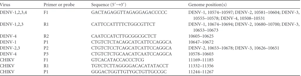
fluorogenic nucleotide probes were labeled with 5=6-carboxyfluorescein (FAM) and 3=Black Hole Quencher (BHQ) dyes. The CHIKV nucleotide probe was labeled with 5=cyanine 5 (Cy5) and 3=BHQ (Integrated DNA Technologies, Coralville, IA). Sequences and genome positions for all oligonucleotide primers and fluorogenic probes are shown inTable 1.
Viruses.Cell culture supernatant harvested from Vero 81 (WHO) cells infected with DENV-1 (West Pac 74), DENV-2 (S16803), DENV-3 (CH53489), DENV-4 (341750), and CHIKV (181/25) (seeTable 2) was used as virus stock for spiking negative human sera. The concentration of viral RNA orin vitro-transcribed RNA was measured as genome equiva-lents (GE)/gene copies. Genomic RNA preparations from 53 dengue vi-ruses (including 10 strains of DENV-1, 24 strains of DENV-2, 10 strains of DENV-3, and 9 strains of DENV-4), 6 strains of CHIKV, Japanese en-cephalitis (JE) virus (SA 14-14-2, attenuated vaccine strain), yellow fever (YF) virus (17D, attenuated vaccine strain), Bebaru virus (BEBV) (ATCC VR-600), Ross River virus (RRV) (ATCC VR-373), and Sindbis virus (SBV) (ATCC VR-1248) (seeTables 2,4, and5) were obtained from BEI Resources-ATCC (Manassas, VA). DENV laboratory strains and JE and YF virus vaccine strains were received from the Walter Reed Army Insti-tute of Research (WRAIR) (Silver Spring, MD), CHIKV R-91142, CHIKV S-27, CHIKV SL-15649, and all other DENV strains were received from BEI Resources-ATCC, the CHIKV proficiency panel was obtained from CDC (Fort Collins, CO), and CHIKV 181-25 (attenuated vaccine strain) was received from Shuenn-Jue Wu (NMRC, MD).
RNA standards. The target regions of DENV-3 (CH53489) and CHIKV (181/25) were amplified by RT-PCR (SuperScript III Platinum one-step qRT-PCR kit with ROX) (Invitrogen, Grand Island, NY) from supernatant of infected cells, generating a 119-bp fragment and a 188-bp fragment, respectively. The forward (DENV F and CHIKV F) and reverse (DENV R1 and CHIKV R1) primers are listed inTable 1. Amplicons were detected by 2% agarose gel electrophoresis, and 2l of the PCR product was used to clone amplicons into TOPO TA cloning kit with PCR 2.1 TOPO (Invitrogen). The presence of the cloned inserts was confirmed by restriction digestion (SpeI and NotI) followed by gel electrophoresis. Re-striction digest plasmids were purified using the QIAprep spin miniprep kit (Qiagen Inc., Valencia, CA) and were CsCl purified for quantification by spectrophotometry. The copy number or genome equivalents (GE) of the plasmids were calculated based on their concentration and molecular weight. Purified linearized plasmids were used as templates forin vitro
[image:2.585.41.546.77.205.2]transcription (IVT) with the MEGAscript kit (Ambion, Thermo Fisher, Waltham, MA) according to the manufacturer’s instructions. The IVT products were then treated with Turbo DNase for 15 min at 37°C, fol-lowed by inactivation of DNase at 70°C for 15 min. Tenfold dilution series of plasmids and IVT were prepared in duplicate to generate standard curves. The GE of the IVT preparations were calibrated using the calcu-lated plasmid concentrations. For an exogenous internal control (IC) to be used as extraction and amplification control, DENV-1 Armored RNA was purchased from Asuragen Inc. (Austin, TX).
TABLE 1Oligonucleotide primers and fluorogenic probes used in the quantitative multiplex RT-PCR assay
Virus Primer or probe Sequence (5=¡3=) Genome position(s)
DENV-1,2,3,4 F1 GACTAGAGGTTAGAGGAGACCCCC DENV-1, 10574–10597; DENV-2, 10581–10604; DENV-3, 10555–10578; DENV-4, 10508–10531
DENV-1,2,3 R1 CATTCCATTTTCTGGCGTTCT DENV-1, 10674–10694; DENV-2, 10680–10700; DENV-3, 10653–10673
DENV-4 R2 CAATCCATCTTGCGGCGCTCT 10605–10625 DENV-1 P1 CTGTCTCTACAGCATCATTCCAGGCA 10647–10672
DENV-2,3 P2 CTGTCTCCTCAGCATCATTCCAGGCA DENV-2, 10653–10678; DENV-3, 10626–10651 DENV-4 P3 CTGTCTCTGCAACATCAATCCAGGCA 10578–10603
CHIKV F1 GTCACATACCACCCTCG 11169–11185 CHIKV R1 TGYCTCTTAGGGGACACATATACCT 11332–11356 CHIKV P1 GGGACTGGTTGTYGCTGTTGCCGC 11244–11267
on May 16, 2020 by guest
http://jcm.asm.org/
One-step multiplex real-time RT-PCR assay.A real-time, one-step, multiplex RT-PCR assay was developed for the detection and quantitation of DENV and CHIKV RNAs in cell culture supernatants or human serum. Real-time PCR was performed under College of American Pathologists (CAP) restricted conditions in a CAP-accredited laboratory. Care was taken to perform RNA extraction, master mix preparation, template ad-dition, and thermal cycling in different areas to avoid contamination. A total volume of 25l of reaction mixture consisted of 5l of extracted RNA, 12.5l of 2⫻RT buffer (Superscript III Platinum qRT-PCR kit with ROX), 0.5l of SuperScript III RT/PlatinumTaqmix, 0.2l of RNase inhibitor (40 units/l) (RNaseout; Invitrogen), 170 nM DENV.F1, 110 nM DENV.R1 and DENV.R2, 60 nM DENV1.P1, DENV.P2, and DENV.P3, and 150 nM CHIKV.F, CHIKV.R, and CHIKV.P. For the DENV probes, the 5=and 3=ends were labeled with 6-carboxyfluorescein (FAM) and Black Hole Quencher (BHQ), respectively, whereas cyanine 5 (Cy5) and BHQ were used for the CHIKV probe. The ABI 7500 DX gene detection system (Applied Biosystems, Carlsbad, CA) was used for PCR cycling, real-time data collection, and analysis. The amplification condi-tions were 50°C for 30 min, 95°C for 2 min, and then 40 cycles of 95°C for 15 s and 60°C for 40 s. The total run time for this protocol was 1 h 10 min. All reactions were carried out in MicroAmp Fast 96-well reaction plates (ABI). For clinical sample testing, all samples and controls were run in duplicate, and each plate contained a six-dilution standard curve of both DENV and CHIKV IVT product, an H2O nontemplate control (NTC), an
H2O extraction control, an H2O extraction control containing an
exoge-nous positive internal control (IC) (DENV-1 Armored RNA), extracted clinical sample, and extracted clinical sample containing the IC. The ac-ceptance criteria for a successful run were a slope between⫺3.1 and⫺3.6 for both standard curves and correlation coefficient (r) values greater than 0.98. Samples were considered negative if the IC was positive but the patient sample without IC was negative.
Analysis of linearity, diagnostic LOD, and precision.In vitr o-tran-scribed RNA was used to establish standard curves for the quantitation of viral copy numbers and analysis of analytical sensitivity. Linearity studies were performed on serial 10-fold dilutions of both DENV and CHIKV IVT RNAs. Dilutions of IVT RNA ranged from 5⫻107GE to 5 GE in
triplicate. The IVT stock solutions for standard curves were diluted in nuclease-free water, prepared as 10-l one-time-use aliquots, and stored at⫺80°C for future use. The linear range was established with 10-fold dilutions of IVT RNA and by fitting a best-fit line to the data by regression analysis, where ther2value for this line was⬎0.98. Acceptance criteria for
a successful run were established as a slope for the standard curve between
⫺3.1 and⫺3.6 with an efficiency of⬎90%.
To establish the 95% limit of detection (LOD), the lowest concentra-tions of RNA from the linear-range study were used as the starting points for another five 2-fold dilutions. The LOD was defined as the lowest con-centration of viral RNA that can be detected inⱖ95% of 24 replicates with the amplification curves above the threshold of 0.04. The precision of the multiplex RT-PCR assay was evaluated using six 10-fold dilutions of IVT RNAs to generate standard curves, with one run per day in quadruplicate over 3 days by two different operators (seeTable 2). Fresh dilutions were made for each run from one-time-use aliquots.
Specificity and sensitivity.The specificity and sensitivity of the mul-tiplex RT-PCR were determined using extracted RNA preparations from 53 dengue viruses, including 10 strains of DENV-1, 24 strains of DENV-2, 10 strains of DENV-3, and 9 strains of DENV-4, and 6 strains of CHIKV. To determine cross-reactivity of the dengue virus primers and probes, two related flaviviruses, JE virus and YF virus, at concentrations of 2⫻104
GE/reaction were evaluated in the assay. To determine the specificity of the CHIKV primers and probe, we included BEBV, RRV, and SBV at 100 ng/reaction (seeTable 4). For the initial determination of sensitivity and cross-reactivity between DENV serotypes in the multiplex assay, serum from a healthy individual was spiked with 50, 500, or 5,000 GE of DENV-1 to -4 (seeTable 3). A total of 12 spiked samples were run in duplicate in two separate runs.
Clinical samples.A total of 89 human serum samples were used to evaluate the multiplex RT-PCR for its diagnostic potential. Fifty clinical samples provided by St. George’s University (St. George, Grenada) were leftover specimen from serum samples collected in 2014 for clinically required testing for suspected dengue. Deidentified residual samples were shipped to the Naval Infectious Diseases Diagnostic Laboratory (NIDDL) (Silver Spring, MD) for use in the validation of our assay. The use of these deidentified coded samples for research purposes was covered under an Institutional Review Board (IRB) protocol (PJT-12-13, Collection of Hu-man Samples for Use as Controls in Diagnostic Assays) allowing use of leftover samples collected for diagnostic purposes from febrile persons suspected of a possible arbovirus infection. Nineteen DENV isolation-confirmed and 20 location- and date-matched DENV-negative clinical samples from Honduras, Venezuela, and Peru were collected between 2010 and 2014 under study protocol NMRCD.2010.0010. This protocol was approved by the NAMRU-6 IRB in compliance with all U.S. federal regulations governing the protection of human subjects. The chikungu-nya virus RT-PCR proficiency panel was obtained from the CDC at Fort Collins, CO, and consisted of three samples representing chikungunya virus collected in India in 2006.
Isolation of viral RNA.Viral RNA was extracted from 200l of serum samples or supernatant of infected cells using the EZ1 DSP virus kit (Qiagen) according to the protocol suggested by the manufacturer, using the EZ1 Advanced XL (Qiagen) automated extraction system. The RNA was eluted with 60l of elution buffer, aliquoted, and immediately stored at⫺80°C until further use.
Virus growth and isolation.Vero cells were grown in Eagle’s mini-mum essential medium (EMEM) (Sigma-Aldrich, St. Louis, MO) supple-mented with 10% fetal bovine serum (FBS) (Sigma), 1% penicillin-strep-tomycin (Mediatech Inc., Manassas, VA), and 1% glutamine (Sigma, St. Louis, MO) at 37°C and 5% CO2. Isolation of viruses from acute-phase
patient samples was carried out by adsorption of 0.3 ml of sample in 0.3 ml of EMEM–2% FBS to cells in a 12-cm2flask for 1 h at 37°C. After
adsorp-tion, the cells were replenished with 7 ml of EMEM–2% FBS and moni-tored for cytopathic effect. Cells were harvested on appearance of cyto-pathic effect (compared to uninfected cells) or were kept for 14 days, with a medium change on day 7. The identification of the virus isolates ob-tained from the clinical samples was carried out by immunofluorescence using virus-specific antibodies.
Statistics.Basic statistical analysis, including determination of means and standard deviations, was performed using Excel software (Microsoft, Bellevue, WA).
RESULTS
Primer design.
This assay targets the distinctive 3
=
untranslated
regions (UTRs) of CHIKV and four serotypes of DENV. The
DENV/CHIK TaqMan assay consists of two forward primers,
three reverse primers, and four fluorophore-labeled probes in a
single-reaction format. The resultant real-time RT-PCR system
can be used to differentially detect CHIKV as well as dengue virus
serotypes 1 through 4. The oligonucleotide sequences for the
re-verse primers targeting DENV-1 through -4 and
fluorescence-labeled probes specific to DENV-2, -3, and -4 have been
previ-ously described (
27
,
28
). A universal forward primer was designed
from a homologous region, and an additional DENV-1 probe was
included in the multiplex assay to ensure the detection of all four
DENV serotypes. The oligonucleotide sequences and genome
po-sitions of all primers and probes for the multiplex assay are listed
in
Table 1
.
Assay performance and analytical sensitivity.
The multiplex
assay was optimized as a single-well method incorporating a
DENV group-specific forward primer, two DENV reverse primers
which also serve at RT primers, a CHIKV primer pair, and four
TaqMan probes for the detection of DENV serotypes 1 through 4
on May 16, 2020 by guest
http://jcm.asm.org/
and CHIKV. The detection limits for both the DENV complex and
CHIKV were determined through 10-fold serial dilutions of
in
vitro
-transcribed RNA copies. The samples at each concentration
were tested in quadruplicate in one run per day over 3 days by two
different operators for a total of 24 runs per dilution (
Table 2
). The
linear dynamic range for the multiplex RT-PCR assay was between
50 and 5
⫻
10
6GE per reaction for DENV and between 60 and 6
⫻
10
6GE for CHIKV (
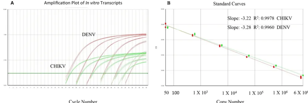
Fig. 1
). The total imprecision (percent
coef-ficient of variation [CV]) for the threshold cycle (
C
T) values for
the six concentrations ranged from 1.4% to 2.1% for DENV and
from 1.3% to 3.2% for CHIKV. The accepted amplification
effi-ciency for each run was set at a slope of
⫺
3.1 and
⫺
3.6 with the
correlation coefficient greater than 0.98. The 95% limit of
detec-tion (LOD) and limit of quantitadetec-tion (LOQ) in which the signal
was linear for the DENV complex (serotypes 1 to 4) and CHIKV
RT-PCRs were 50 (3
⫻
10
3GE/ml) and 60 (3.6
⫻
10
3GE/ml) gene
copies per reaction, respectively (
Table 2
). To ensure that the assay
is not negatively impacted by components in human serum and to
confirm the sensitivity and LOD for all DENV serotypes, dengue
virus-negative human serum was spiked with three different
dilu-tions of previously titrated virus stocks. Each dilution was
ex-tracted separately and assayed in two separate runs (
Table 3
). The
results indicate equal sensitivity to all four DENV serotypes, and
the assay is not impacted by components in human serum. The
cutoff for positivity was defined by a
C
Tvalue of
ⱕ
37, based on 50
independent runs where 100% of all replicates were below this
value (data not shown).
Analytical specificity.
Analytical specificity distinguishes
be-tween the target analyte and other components in the sample and
was expressed as inclusivity and exclusivity (
29
). Extracted RNAs
from 53 dengue viruses, representing all four serotypes of DENV
and six strains of CHIKV, were tested in duplicate to determine
inclusivity of the multiplex RT-PCR. Two related flaviviruses (JE
virus and YF virus) and three related alphaviruses (BEBV, RRV,
and SBV) were tested to confirm exclusivity of the assay. The
mul-tiplex assay detected all DENV and CHIKV samples tested (
Table
4
). The five related flaviviruses and alphaviruses were not
de-tected.
Diagnostic sensitivity and specificity.
To establish estimates
of diagnostic sensitivity and specificity, clinical samples which had
been defined positive or negative by virus isolation in Vero cells
were used. Nucleic acid extracted from a total of 89 clinical
sam-ples positive or negative for DENV-1 to -4 and CHIKV were used
for the calculation of diagnostic sensitivity and specificity (
Table
5
). Of the 19 samples positive for DENV by virus isolation, 18
samples (95%) tested positive by multiplex RT-PCR, and all 20
negative-control samples were negative in both assays. The one
sample negative in the PCR but positive by virus isolation showed
a
C
Tvalue of 38.7 after a repeated run, which is below the validated
LOD
C
Tof 37 and is therefore recorded as negative. For the 50
positive and negative CHIKV clinical samples, 40 (100%) samples
were positive by virus isolation and positive by RT-PCR. Ten
(100%) negative-control sera were negative in both assays.
DISCUSSION
There are several studies describing the development of real-time
PCR assays for the detection of DENV and CHIKV (
20–25
,
27
,
[image:4.585.39.287.88.249.2]28
). Two previous studies reported on the development of an
RT-PCR assay for the detection of dengue viruses using primers
TABLE 2Detection and precision analysis of the DENV/CHIKV multiplex RT-PCR
Virusa GE/assay
No. of replicatesb
CTvalue
Mean SD % CV
DENV-3 5⫻106 24 17.84 0.26 1.5 5⫻105 24 21.08 0.34 1.6 5⫻104 24 24.43 0.35 1.4 5⫻103 24 27.70 0.39 1.4 5⫻102 24 31.17 0.50 1.6 5⫻101 24 34.55 0.72 2.1
CHIKV 6⫻106 24 18.08 0.28 1.5 6⫻105 24 21.30 0.31 1.5 6⫻104 24 24.69 0.38 1.5 6⫻103 24 28.14 0.38 1.3 6⫻102 24 31.36 0.80 2.5 6⫻101 24 34.59 1.10 3.2
a
The multiplex RT-PCR was carried out on RNAs extracted from DENV and CHIKV
in vitrotranscripts.
b
Quadruplicate runs over 3 days were carried out by two different operators.
FIG 1(A) Amplification plot for DENV/CHIKV multiplex assay (10-fold dilutions, triplicate), (B) The linear dynamic range of the multiplex RT-PCR is shown as gene copy number for DENV (5⫻101to 5⫻106) and CHIKV (6⫻101to 6⫻106).
on May 16, 2020 by guest
http://jcm.asm.org/
[image:4.585.46.542.533.699.2]and probes designed in the 3
=
UTR region (
30
,
31
). These are
assays either not quantitative or use a two-step method. Other
real-time dengue virus RT-PCR assays use either a three- or
four-step cycling method, which increases assay time, or use a SYBR
green-based method, which is prone to nonspecific reaction
prod-ucts leading to increased background and false positives (
20
,
32–
34
). In 2010, the international external quality control assessment
for the molecular diagnosis of dengue infections published their
findings for 37 laboratories performing 46 tests and showed that
80% of these tests lacked sensitivity, specificity, or both (
35
).
Sim-ilarly, several groups have developed singleplex real-time RT-PCR
assays for the detection of CHIKV in clinical samples (
24
,
25
,
36–39
). Some of these assays are not quantitative or use plasmid
DNA as a standard for quantitation (
36
,
37
). Another study
re-cently described the development of a multiplex real-time
RT-PCR for the simultaneous detection of DENV and CHIKV with a
detection limit of 1 to 50 PFU and an assay cutoff value
C
Tof 32,
due to nonspecific signal observed in the nontemplate control
(
40
).
In this study, we describe the development and validation of a
more sensitive, rapid, single-reaction, one-step, multiplex
real-time RT-PCR for the detection, quantitation, and differentiation
of DENV-1 to -4 and CHIK in patient samples. This assay targets
the 3
=
untranslated regions (UTRs) of all four DENV serotypes for
species-specific detection but cannot be used for serotype
deter-mination. Serotype-specific determination is usually not required
and does not affect clinical care and management, especially
out-side areas where dengue is endemic. In areas where DENV and
CHIKV cocirculate, it is important to be able to differentiate
be-tween them due to different disease progression and outcome.
Real-time PCR has brought true quantitation into the diagnostic
laboratory. It allows monitoring of the progress of an infection
and may be necessary when there is a lack of sequential specimens
to indicate changes in virus levels or when viral load is used to
differentiate active versus persistent infection. Our assay contains
two differently labeled probes, and it uses external standard curves
for quantitation that consist of serial dilutions of identically
am-plified templates within the same experimental run. The linear
dynamic range in the assay covers six log
10copies of nucleic acid
template, where the lower limit was set at the LOD
C
Tof 37. To
control for plate-to-plate variation, we adopted an acceptance
cri-terion where the slope of the standard curves has to be within
⫺
3.1
to
⫺
3.6 with the correlation coefficient greater than 0.98. Due to
the broad dynamic range of the assay, template quantitation from
samples containing a large range of concentrations can be
accom-plished without the need to repeat the assay using a diluted
sam-ple. In addition to a nontemplate control and standard curves on
every plate serving as negative and positive controls, both external
and internal controls have been applied. Both patient sample and
H
2O controls were extracted with and without a spiked IC and
served as false-negative and contamination/amplification
con-trols. Inhibition of the IC amplification in the presence of high
DENV or CHIKV RNA levels has been observed, but this was not
detrimental since the IC is used to monitor false-negative results.
Using this format, up to nine samples, including all controls, can
be analyzed in about 2 h on a single plate, including RNA
extrac-tion. This assay fulfilled the validation criteria of specificity,
lin-earity, and precision.
The sensitivity of the multiplex assay was evaluated by testing
triplicate 10-fold serial dilutions of
in vitro
-transcribed RNAs for
both DENV and CHIKV. The linear dynamic range of the assay
was between 50 and 5
⫻
10
6GE per reaction for DENV and
be-tween 60 and 6
⫻
10
6GE for CHIKV. Sensitivity of 95% was
achieved, with
C
Tvalues in the range of 33 to 36, when samples
included 50 or 60 GE for both targets, and these were therefore
defined as the limit of detection (LOD). The limit of
quantifica-tion (LOQ) corresponds to the last diluquantifica-tion in which the signal is
linear and has an acceptable CV of
⬍
5%, which in this assay is
equal to the LOD.
In this study, we demonstrated that the one-step multiplex
RT-PCR assay was able to detect 53 different serotypes of DENV
and 6 different strains of CHIKV at concentrations ranging from
10
3to10
7GE per reaction. The assay was found to have high
[image:5.585.38.549.79.274.2]ana-lytical specificity, and no false-positive results were seen from
test-ing of relatively high concentrations of related flavivirus and
al-phavirus RNAs. This study assessed the reproducibility of the
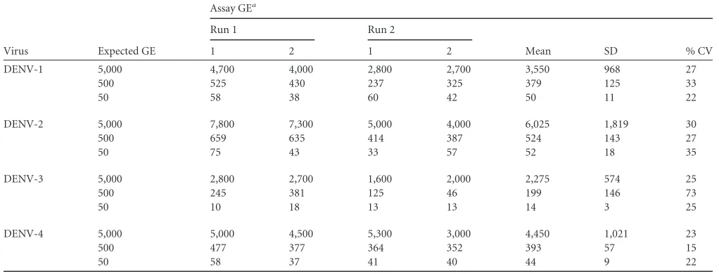
TABLE 3Detection limit of the multiplex RT-PCR assay for the four DENV serotypes in spiked human serum
Virus Expected GE
Assay GEa
Run 1 Run 2
Mean SD % CV
1 2 1 2
DENV-1 5,000 4,700 4,000 2,800 2,700 3,550 968 27
500 525 430 237 325 379 125 33
50 58 38 60 42 50 11 22
DENV-2 5,000 7,800 7,300 5,000 4,000 6,025 1,819 30
500 659 635 414 387 524 143 27
50 75 43 33 57 52 18 35
DENV-3 5,000 2,800 2,700 1,600 2,000 2,275 574 25
500 245 381 125 46 199 146 73
50 10 18 13 13 14 3 25
DENV-4 5,000 5,000 4,500 5,300 3,000 4,450 1,021 23
500 477 377 364 352 393 57 15
50 58 37 41 40 44 9 22
a
For each virus, a dilution series including the lowest LOD in two separate runs is shown.
on May 16, 2020 by guest
http://jcm.asm.org/
assay by testing a range of concentrations from
in vitro
-tran-scribed RNA. The assay was highly reproducible for detecting
vi-rus at concentrations ranging from 50 to 6
⫻
10
6gene copy
num-bers per reaction. Furthermore, low intra-assay and interassay
variabilities were shown in measuring
C
Tvalues over a wide range
of concentrations, with different operators on different days,
ranging from 1.4% to 3.2%. We have evaluated the diagnostic
application of the multiplex RT-PCR assay using 89 archived
se-rum samples representing all four serotypes of DENV and
CHIKV. For the detection of DENV, the multiplex RT-PCR had a
95% diagnostic sensitivity and 100% specificity, whereas for
CHIKV detection there was 100% positive agreement between the
multiplex RT-PCR assay and the gold standard of virus isolation
with both positive and negative samples tested.
In conclusion, the newly developed and validated one-step,
multiplex quantitative RT-PCR enables simultaneous, sensitive,
and specific detection and quantitation of DENV serotypes 1 to 4
or CHIKV in clinical specimens. The dynamic range of the new
assay encompasses the copy numbers expected in diagnostic
sam-ples. The analytical sensitivity of the assay is 50 and 60 GE/reaction
(or 10 and 12 copies/
l) for DENV-1 to -4 and CHIKV,
respec-tively. The assay includes an exogenous internal control for the
detection of false-negative results and two standard curves for
quantitation, is technically robust, and has high-throughput
po-tential. The screening assay described in the current study
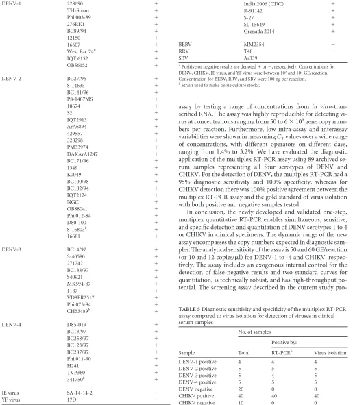
pro-TABLE 4Viruses used to determine the multiplex RT-PCR specificity/ inclusivity
Virus Strain Resulta
DENV-1 228690 ⫹
TH-Sman ⫹
Phi 003-89 ⫹
276RK1 ⫹
BC89/94 ⫹
12150 ⫹
16607 ⫹
West Pac 74b ⫹
IQT 6152 ⫹
OBS6152 ⫹
DENV-2 BC27/96 ⫹
S-14635 ⫹
BC141/96 ⫹
P8-1407MS ⫹
10674 ⫹
S2 ⫹
IQT2913 ⫹
ArA6894 ⫹
429557 ⫹
328298 ⫹
PM33974 ⫹
DAKArA1247 ⫹
BC171/96 ⫹
1349 ⫹
K0049 ⫹
BC100/98 ⫹
BC102/94 ⫹
IQT2124 ⫹
NGC ⫹
OBS8041 ⫹
Phi 012-84 ⫹
D80-100 ⫹
S-16803b ⫹
16681 ⫹
DENV-3 BC14/97 ⫹
S-40580 ⫹
271242 ⫹
BC188/97 ⫹
S40921 ⫹
MK594-87 ⫹
1187 ⫹
VD8PR2517 ⫹ Phi 075-84 ⫹ CH53489b ⫹
DENV-4 D85-019 ⫹
BC13/97 ⫹
BC258/97 ⫹
BC123/97 ⫹
BC287/97 ⫹
Phi 011-90 ⫹
H241 ⫹
TVP360 ⫹
341750b ⫹
JE virus SA-14-14-2 ⫺
[image:6.585.44.292.85.701.2] [image:6.585.38.553.102.693.2]YF virus 17D ⫺
TABLE 4(Continued)
Virus Strain Resulta
CHIKV 181/25b ⫹
India 2006 (CDC) ⫹
R-91142 ⫹
S-27 ⫹
SL-15649 ⫹
Grenada 2014 ⫹
BEBV MM2354 ⫺
RRV T48 ⫺
SBV Ar339 ⫺
a
Positive or negative results are denoted⫹or⫺, respectively. Concentrations for DENV, CHIKV, JE virus, and YF virus were between 103and 107GE/reaction.
Concentration for BEBV, RRV, and SBV were 100 ng per reaction.
[image:6.585.300.544.579.696.2]bStrain used to make tissue culture stocks.
TABLE 5Diagnostic sensitivity and specificity of the multiplex RT-PCR assay compared to virus isolation for detection of viruses in clinical serum samples
Sample
No. of samples
Total
Positive by:
RT-PCRa Virus isolation
DENV-1 positive 4 4 4 DENV-2 positive 5 5 5 DENV-3 positive 5 4 5 DENV-4 positive 5 5 5 DENV negative 20 0 0 CHIKV positive 40 40 40 CHIKV negative 10 0 0
aIn comparison with the results of the reference method (virus isolation), the multiplex
RT-PCR assay had 95% sensitivity and 100% specificity for DENV and 100% agreement for CHIKV.
on May 16, 2020 by guest
http://jcm.asm.org/
vides the means to rapidly detect and differentiate five distinct
viruses in the same sample in less than 2 h, including extraction of
RNA. The recent occurrence of both DENV and CHIKV in
ever-closer geographical areas highlights the need to rapidly detect and
differentiate these two diseases. This assay has been validated as an
in-house diagnostic test in the CAP-accredited NIDDL and is used
as a routine diagnostic tool for DENV and CHIKV infection.
ACKNOWLEDGMENTS
We thank Susana Widjaja for technical assistance and Kenneth Eckels and Shuenn-Jue Wu for providing virus stock. We thank Juan Perez, Instituto Nacional de Salud del Perú, DIRESA Loreto, Hospital La Merced (Junin), CS Pachitea (Piura), Stalin Vilcarromero, Crystyan Siles, Michel Valerio, and Victor Ocaña for their work in the field. We thank I. Lorenzana (Universidad Autonoma de Honduras) and G. Comach (LARDIDEV, Venezuela) for their help in collecting patient samples.
The views expressed in this article are those of the authors and do not necessarily reflect the official policy or position of the Department of the Navy, the Department of Defense, or the U.S. Government.
We report no competing interests.
FUNDING INFORMATION
This work was funded by a grant from the Armed Forces Health Surveil-lance Branch (AFHSB) Global Emerging Infections SurveilSurveil-lance and Re-sponse (GEIS) work unit number P0085-14-NM. The funders had no role in study design, data collection and interpretation, or the decision to submit the work for publication.
REFERENCES
1.Ranjit S, Kissoon N.2011. Dengue hemorrhagic fever and shock syn-dromes. Pediatr Crit Care Med 12:90 –100. http://dx.doi.org/10.1097 /PCC.0b013e3181e911a7.
2.Bhatt S, Gething PW, Brady OJ, Messina JP, Farlow AW, Moyes CL, Drake JM, Brownstein JS, Hoen AG, Sankoh O, Myers MF, George DB, Jaenisch T, Wint GR, Simmons CP, Scott TW, Farrar JJ, Hay SI.2013. The global distribution and burden of dengue. Nature496:504 –507.http: //dx.doi.org/10.1038/nature12060.
3.Rico-Hesse R.1990. Molecular evolution and distribution of dengue vi-ruses type 1 and 2 in nature. Virology174:479 – 493.http://dx.doi.org/10 .1016/0042-6822(90)90102-W.
4.Halstead SB.1988. Pathogenesis of dengue: challenges to molecular biol-ogy. Science239:476 – 481.http://dx.doi.org/10.1126/science.3277268. 5.Gubler DJ, Clark GG. 1995. Dengue/dengue hemorrhagic fever: the
emergence of a global health problem. Emerg Infect Dis1:55–57.http://dx .doi.org/10.3201/eid0102.952004.
6.Robinson M.1956. An epidemic of a dengue-like fever in the southern province of Tanganyika. Central Afr J Med2:394.
7.Gérardin P, Fianu A, Malvy D, Mussard C, Boussaïd K, Rollot O, Michault A, Gaüzere B-A, Bréart G, Favier F.2011. Perceived morbidity and community burden after a Chikungunya outbreak: the TELECHIK survey, a population-based cohort study. BMC Med9:5.http://dx.doi.org /10.1186/1741-7015-9-5.
8.Mavalankar D, Shastri P, Bandyopadhyay T, Parmar J, Ramani KV.
2008. Increased mortality rate associated with chikungunya epidemic, Ahmedabad, India. Emerg Infect Dis14:412.http://dx.doi.org/10.3201 /eid1403.070720.
9.Karabatsos N (ed). 1985. International catalogue of arthropod-borne viruses, 3rd ed. American Society for Tropical Medicine and Hygiene, San Antonio, TX.
10. Carey DE.1971. Chikungunya and dengue: a case of mistaken identity? J Hist Med Allied Sci26:243–262.
11. Rao T.1966. Recent epidemics caused by Chikungunya virus in India, 1963-1965. Sci Culture32:215.
12. Nimmannitya S, Halstead SB, Cohen SN, Margiotta MR.1969. Dengue and chikungunya virus infection in man in Thailand, 1962-1964. I. Ob-servations on hospitalized patients with hemorrhagic fever. Am J Trop Med Hyg18:954 –971.
13. Carey DE, Myers RM, DeRanitz C, Jadhav M, Reuben R.1969. The 1964
chikungunya epidemic at Vellore, South India, including observations on concurrent dengue. Trans R Soc Trop Med Hyg63:434 – 445.http://dx.doi .org/10.1016/0035-9203(69)90030-3.
14. Jupp P, McIntosh B.1988. Chikungunya virus disease, p 137–157.In
Monath TP (ed), The arboviruses: epidemiology and ecology, vol 2. CRC Press, Boca Raton, FL.
15. Powers AM.2015. Risks to the Americas associated with the continued expansion of chikungunya virus. J Gen Virol96:1–5.http://dx.doi.org/10 .1099/vir.0.070136-0.
16. Rodríguez-Morales AJ, Paniz-Mondolfi AE.2015. Venezuela: far from the path to dengue and chikungunya control. J Clin Virol66:60 – 61.http: //dx.doi.org/10.1016/j.jcv.2015.02.020.
17. Fischer M, Staples JE.2014. Notes from the field: chikungunya virus spreads in the Americas-Caribbean and South America, 2013-2014. MMWR Morb Mortal Wkly Rep63:500 –501.
18. Pan American Health Organization. 2015. Chikungunya: number of reported cases of chikungunya fever in the Americas. Pan American Health Organization, Washington, DC.
19. Blacksell SD, Jarman RG, Gibbons RV, Tanganuchitcharnchai A, Mam-men MP, Nisalak A, Kalayanarooj S, Bailey MS, Premaratna R, de Silva HJ.2012. Comparison of seven commercial antigen and antibody en-zyme-linked immunosorbent assays for detection of acute dengue infec-tion. Clin Vaccine Immunol19:804 – 810.http://dx.doi.org/10.1128/CVI .05717-11.
20. Waggoner JJ, Abeynayake J, Sahoo MK, Gresh L, Tellez Y, Gonzalez K, Ballesteros G, Balmaseda A, Karunaratne K, Harris E.2013. Develop-ment of an internally controlled real-time reverse transcriptase PCR assay for pan-dengue virus detection and comparison of four molecular dengue virus detection assays. J Clin Microbiol51:2172–2181.http://dx.doi.org /10.1128/JCM.00548-13.
21. Waggoner JJ, Abeynayake J, Sahoo MK, Gresh L, Tellez Y, Gonzalez K, Ballesteros G, Guo FP, Balmaseda A, Karunaratne K.2013. Comparison of the FDA-approved CDC DENV-1-4 real-time reverse transcription-PCR with a laboratory-developed assay for dengue virus detection and serotyping. J Clin Microbiol 51:3418 –3420. http://dx.doi.org/10.1128 /JCM.01359-13.
22. Ito M, Takasaki T, Yamada K, Nerome R, Tajima S, Kurane I.2004. Development and evaluation of fluorogenic TaqMan reverse transcriptase PCR assays for detection of dengue virus types 1 to 4. J Clin Microbiol
42:5935–5937.http://dx.doi.org/10.1128/JCM.42.12.5935-5937.2004. 23. Lanciotti RS, Calisher CH, Gubler DJ, Chang GJ, Vorndam AV.1992.
Rapid detection and typing of dengue viruses from clinical samples by using reverse transcriptase-polymerase chain reaction. J Clin Microbiol
30:545–551.
24. Pastorino B, Bessaud M, Grandadam M, Murri S, Tolou HJ, Peyrefitte CN.2005. Development of a TaqMan® RT-PCR assay without RNA ex-traction step for the detection and quantification of African Chikungunya viruses. J Virol Methods124:65–71.http://dx.doi.org/10.1016/j.jviromet .2004.11.002.
25. Laurent P, Le Roux K, Grivard P, Bertil G, Naze F, Picard M, Stai-kowsky F, Barau G, Schuffenecker I, Michault A.2007. Development of a sensitive real-time reverse transcriptase PCR assay with an internal con-trol to detect and quantify chikungunya virus. Clin Chem53:1408 –1414. http://dx.doi.org/10.1373/clinchem.2007.086595.
26. Mackay IM, Arden KE, Nitsche A.2002. Real-time PCR in virology. Nucleic Acids Res30:1292–1305.http://dx.doi.org/10.1093/nar/30.6.1292. 27. Houng HS, Chung-Ming Chen R, Vaughn DW, Kanesa-thasan, N.
2001. Development of a fluorogenic RT-PCR system for quantitative iden-tification of dengue virus serotypes 1-4 using conserved and serotype-specific 3=noncoding sequences. J Virol Methods95:19 –32.http://dx.doi .org/10.1016/S0166-0934(01)00280-4.
28. Houng HH, Hritz D, Kanesa-thasan, N.2000. Quantitative detection of dengue 2 virus using fluorogenic RT-PCR based on 3=-noncoding sequence. J Virol Methods86:1–11.http://dx.doi.org/10.1016/S0166 -0934(99)00166-4.
29. Christensen DR, Hartman LJ, Loveless BM, Frye MS, Shipley MA, Bridge DL, Richards MJ, Kaplan RS, Garrison J, Baldwin CD.2006. Detection of biological threat agents by real-time PCR: comparison of assay performance on the RAPID, the LightCycler, and the Smart Cycler platforms. Clin Chem 52:141–145. http://dx.doi.org/10.1373/clinchem .2005.052522.
30. Gurukumar KR, Priyadarshini D, Patil JA, Bhagat A, Singh A, Shah PS, Cecilia D.2009. Development of real time PCR for detection and
on May 16, 2020 by guest
http://jcm.asm.org/
titation of Dengue Viruses. Virol J6:10.http://dx.doi.org/10.1186/1743 -422X-6-10.
31. Alm E, Lesko B, Lindegren G, Ahlm C, Söderholm S, Falk KI, Lager-qvist N.2014. Universal single-probe RT-PCR assay for diagnosis of den-gue virus infections. PLoS Negl Trop Dis8:e3416.http://dx.doi.org/10 .1371/journal.pntd.0003416.
32. Dos Santos HW, Poloni TR, Souza KP, Muller VD, Tremeschin F, Nali LC, Fantinatti LR, Amarilla AA, Castro HL, Nunes MR, Casseb SM, Vasconcelos PF, Badra SJ, Figueiredo LT, Aquino VH.2008. A simple one-step real-time RT-PCR for diagnosis of dengue virus infection. J Med Virol80:1426 –1433.http://dx.doi.org/10.1002/jmv.21203.
33. Chien LJ, Liao TL, Shu PY, Huang JH, Gubler DJ, Chang GJ.2006. Development of real-time reverse transcriptase PCR assays to detect and serotype dengue viruses. J Clin Microbiol44:1295–1304.http://dx.doi.org /10.1128/JCM.44.4.1295-1304.2006.
34. Shu PY, Chang SF, Kuo YC, Yueh YY, Chien LJ, Sue CL, Lin TH, Huang JH.2003. Development of group- and serotype-specific one-step SYBR green I-based real-time reverse transcription-PCR assay for dengue virus. J Clin Microbiol41:2408 –2416.http://dx.doi.org/10.1128/JCM.41 .6.2408-2416.2003.
35. Domingo C, Niedrig M, Teichmann A, Kaiser M, Rumer L, Jarman RG, Donoso-Mantke O.2010. 2nd international external quality control
as-sessment for the molecular diagnosis of dengue infections. PLoS Negl Trop Dis4:e833.http://dx.doi.org/10.1371/journal.pntd.0000833. 36. Edwards CJ, Welch SR, Chamberlain J, Hewson R, Tolley H, Cane PA,
Lloyd G.2007. Molecular diagnosis and analysis of Chikungunya virus. J Clin Virol39:271–275.http://dx.doi.org/10.1016/j.jcv.2007.05.008. 37. Carletti F, Bordi L, Chiappini R, Ippolito G, Sciarrone MR,
Capo-bianchi MR, Di Caro A, Castilletti C. 2007. Rapid detection and quantification of Chikungunya virus by a one-step reverse transcrip-tion–polymerase chain reaction real-time assay. Am J Trop Med Hyg
77:521–524.
38. Grivard P, Le Roux K, Laurent P, Fianu A, Perrau J, Gigan J, Hoarau G, Grondin N, Staikowsky F, Favier F.2007. Molecular and serological diagnosis of Chikungunya virus infection. Pathol Biol (Paris)55:490 – 494. http://dx.doi.org/10.1016/j.patbio.2007.07.002.
39. Kumar C, Johnson AA, Gopal D.2007. Molecular characterization of chikungunya virus from Andhra Pradesh, India & phylogenetic relation-ship with Central African isolates. Indian J Med Res126:534.
40. Cecilia D, Kakade M, Alagarasu K, Patil J, Salunke A, Parashar D, Shah P.2015. Development of a multiplex real-time RT-PCR assay for simul-taneous detection of dengue and chikungunya viruses. Arch Virol160:
323–327.http://dx.doi.org/10.1007/s00705-014-2217-x.