Infectious Diseases: Research and Treatment 2013:6 39–50
doi: 10.4137/IDRT.S11263
This article is available from http://www.la-press.com.
© the author(s), publisher and licensee Libertas Academica Ltd.
This is an open access article published under the Creative Commons CC-BY-NC 3.0 license.
Open Access
Full open access to this and thousands of other papers at
http://www.la-press.com. R e v I e w
An Update on Global Tuberculosis (TB)
Balkis A. Talip1,#, Roy D. Sleator2,#, Colm J. Lowery1, James S.G. Dooley1 and william J. Snelling1 1Northern Ireland Centre for Food and Health (NICHe), School of Biomedical Sciences, University of Ulster, Coleraine. 2Department of Biological Sciences, Cork Institute of Technology, Cork, Ireland. #These co-authors contributed equally to this publication. Corresponding author email: snellingwilliam@hotmail.com
Abstract: Tuberculosis globally results in almost 2 million human deaths annually, with 1 in 4 deaths from tuberculosis being human
immunodeficiency virus/acquired immunodeficiency syndrome (HIV/AIDS)-related. Primarily a pathogen of the respiratory system,
aerobic Mycobacterium tuberculosis complex (MTBC) infects the lungs via the inhalation of infected aerosol droplets generated by people with pulmonary disease through coughing. This review focuses on M. tuberculosis transmission, epidemiology, detection meth-ods and technologies.
Introduction
Mycobacterium tuberculosis was first discovered in
1882 by Robert Koch and is one of almost 200 myco
-bacterial species which have been detected by
molec-ular techniques.1 The genus Actinobacteria (given its
own family, the Mycobacteriaceae) includes pathogens
known to cause serious diseases in mammals, includ
-ing tuberculosis (MTBC) and leprosy (M. leprae).1
Mycobacteria are grouped neither as Gram-positive nor Gram-negative bacteria, and have a relatively high average G+C content of 61 to 71 mol% for all
species, except M. leprae (57 mol%).2,3 The genome
size varies from species to species ranging from 4 to
7 million base pairs (bp).2,3 90% of the genome
repre-sents coding regions that potentially encode . 6,000
proteins.2,3 MTBC consists of M. tuberculosis,
M. bovis, M. bovis BCG (bacillus Calmette-Guérin),
M. africanum M. caprae, M. microti, M. canettii and M. pinnipedii, all of which share genetic homology,
with no significant variation between sequences (∼0.01 to 0.03%), although differences in phenotypes
are present.4 Some species such as M. smegmatis and
M. tuberculosis are able to grow in a limited oxygen environment. Cells in the genus have a typical rod, or slightly curved-shape, with dimensions of 0.2 to
0.6 µm by 1 to 10 µm.1
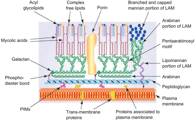
Mycobacterium tuberculosis has a waxy mycolic acid lipid complex coating on its cell surface, making
the cells impervious to Gram staining (Fig. 1). Thus, a common staining procedure used is Ziehl-Neelsen
(ZN) staining.1 The outer compartment of the cell wall
contains lipid-linked polysaccharides consisting of lipoarabinomannan (LAM), lipomannan, phthiocerol dimycocerosate, trehasole, and phosphatidylinosi-tol mannoside, with sulfolipid only being present in
M. tuberculosis (Fig. 1).5,6 The outer compartment of
the cell wall is soluble and has a role in interacting
with the host immune system. The insoluble inner compartment of the cell wall consists of peptidogly-can (PG), arabinogalactan (AG), and mycolic acid (MA). Mycobacteria have some unique qualities that are divergent from members of the Gram-positive group, such as the presence of mycolic acids in the
cell wall.5,7
Transmission
MTBC and M. leprae replication occurs in the
tis-sues of warm-blooded human hosts. This air-borne pathogen is transmitted from an active pulmonary
tuberculosis patient by coughing.7,8 Droplet nuclei,
approximately 1 to 5 µm in size “meander” in the
air and are transmitted to susceptible individuals by
inhalation. Mycobacteria are incapable of replicating
in or on inanimate objects. The risk of infection is dependent to the load of the bacillus that has been inhaled, level of infectiousness, person-to- person
Mycolic acids
Galactan
Phospho-diester bond
PIMs Trans-membrane proteins
Proteins associated to plasma membrane
Plasma membrane Peptidoglycan Arabinan Lipomannan portion of LAM Pentaarabinosyl motif
Arabinan portion of LAM Porin
Complex free lipids Acyl
[image:2.595.126.469.506.716.2]glycolipids Branched and cappedmannan portion of LAM
Figure 1. Schematic representation of Mycobacterium, showing the main components of the outer and inner layer of the cell wall.
notes: The presence of mycolic acid in the complex of covalent-linked MA-AG-PG is a unique character for identification of the genus Mycobacterium and
plays an important role as a permeability barrier. Reproduced with permission from elsevier.76
contact perimeter and the immune competency of potential hosts. Due to the size of the droplets inhaled into the lungs, the infection penetrates the defense systems of the bronchi and enters the terminal alve-oli. Invading bacteria are then engulfed by alveolar
macrophage and dendritic cells.7–9 The cell- mediated
immune response alleviates the multiplication of
M. tuberculosis and halts infection. Tuberculosis can also infect other vital organs of the human body such as the kidneys, spine and brain. Infected indi-viduals with strong immune systems are generally
able to combat the infection within 2 to 8 weeks
post-infection, when the active cell-mediated immune
response stops further multiplication of M.
tubercu-losis. With latent tuberculosis, the tubercle (infected macrophage or granuloma) bacilli are not completely eradicated from immune system, appearing
asymp-tomatic for a longer period of time.7–9 Although
tuberculosis is curable, available treatment regimens
merely slow the infection rate rather than eliminating
the infection from the population. However, the use
of first-line drugs to treat M. tuberculosis does have
an effect within 6 months in susceptible patients. For the unsusceptible patient, second-line drug
treat-ments are introduced that are used to treat the mul
-tidrug-resistant tuberculosis (MDR-TB).8 Latently
infected individuals are non-infectious, and the
infection can be detected by tuberculin skin tests
(TST) and interferon gamma release assays (IGRA).8
While latent tuberculosis has a low potential to cause recurrent infection (approximately 10%), it has been reported that reactivation of latent tuberculosis (active tuberculosis) may occur after years of post-infection. The risk of reactivation is greater in
HIV-infected patients (15%).8 Intracellular replication
is initiated before responses of the immune system occurs in the lymph nodes and other extra pulmo-nary sites, and thus avoids eradication by the
host-immune system.8,9
Natural division and growth of Mycobacterium
species differs based on slowly-growing to
rapidly-growing characteristics. Slowly-rapidly-growing
Mycobac-terium (SGM) require more than 7 days for visible
growth, as opposed to rapidly-growing
Mycobacte-rium (RGM) which requires , 7 days when grown
on Löwenstein-Jensen (L∼J) medium. However, the
RGM may require . 1 week if grown from clini
-cal specimens.10,11 In healthy adults, tuberculosis
progression is generally slow. Tuberculosis infection shows several significant clinical manifestations in
pulmonary and extra-pulmonary sites.12 Prolonged
coughing, severe weigh-lost, night sweats, low-grade fever, dyspnoea and chest pain are clinical symptoms indicated from pulmonary infections. The
extra-pulmonary manifestations of M. tuberculosis
infection include meningitis, pleuritis, pericarditis, synovitis, cervical lymphadenitis and infections of
the skin, joints, bones and internal organs.8,13 In
HIV-infected tuberculosis patients, both types of clinical manifestation are significant due to the rapid progres-sion of infection.
The RGM are grouped as non-tuberculous myco-bacteria (NTM) and are opportunistic pathogens that
can grow within 7 days in specific growth media.14,15
To date, more than 130 species of RGM have been
found.14 Infections due to the RGM are an emerging
health problem in the United States.15 The most com
-mon RGM to cause human diseases are M. abscessus,
M. chelonae, M. fortuitum, and M. massiliense and
these have been linked with health care-associated
pseudo-outbreaks.14,15 Chronic pulmonary disease
and skin/soft-tissue infections are the 2 most
com-mon disorders due to these organisms.15 Clinical
out-comes in the treatment of M. abscessus infections
are generally disappointing.15 Less common human
pathogenic RGM species include M. phocacium,
M. mucogenicum and M. smegmatis group (known
as sensu stricto).14,15 The clinical and microbiologic
features of 115 cases involving RGM isolated at the University of Texas from 2000–2005 were examined
using 16S ribosomal RNA gene sequencing analysis.14
At least 15 RGM species were included: M. abscessus
(43 strains [37.4%]), M. fortuitum complex (33 strains
[28.7%]), and M. mucogenicum (28 strains [24.3%])
were the most common, accounting for 90.4%.14 Most
M. abscessus (32/43) were isolated from respiratory
sources, whereas most M. mucogenicum (24/28) were
from blood cultures.14 Antimicrobial susceptibility
tests showed that M. abscessus was the most resistant
species; M. mucogenicum was most susceptible.14
From blood and catheter sources, 46 strains (40.0%) were isolated; 44 represented bacteremia or
catheter-related infections.14 These infections typically
mani-fested as a high fever (mean temperature, 38.9 °C),
with a high number of RGM colonies cultured.14
antibiotic therapy.14 Six strains (M. abscessus and
M. fortuitum only) were from skin, soft tissue, and
wound infections.14 There were 59 strains from
respi-ratory sources, and 28 of these represented
defini-tive to probable infections.14 Prior lung injuries and
co-isolation of other pathogenic organisms were
common.14 Overall, 78 RGM strains (67.8%) caused
true to probable infections without direct deaths.14
The majority of RGM have been found to be sensitive to amikacin, gatifloxacin, moxifloxacin,
ciprofloxacin and norfloxacin.16 Although the
major-ity of health care-associated pseudo-outbreak
dis-eases are treatable using antibiotics, the emergence
of multidrug-resistant M. abscessus strains worsen
the pseudo-outbreak infection scenario, due to the
most-virulent and chemotherapy-resistant strains.16,17
Existing diagnostic methods for MDR-TB are slower and take longer to perform in comparison to diag-nostics for MTBC common infections. Second-line drug treatment for MDR-TB is relatively expensive due to the long treatment regime required to ensure
effectiveness and prevent recurring infections.16,17
epidemiology
Although all members of MTBC cause infection in
humans, M. bovis, M. bovis BCG, M. Microti and M. pinnipedii commonly infect the warm-blooded animal as their primary host. It is likely that
trans-mission occurs from animal to human.18–20 Primarily,
animal to human TB transmission cases have been observed in people who have worked or resided in
a particular environment.20–23 Unlike M. tuberculosis,
non- tuberculous mycobacteria (NTM) are found in the environment, particularly moist habitats such
as lakes, rivers and damp-soil. M. avium complex
(MAC), M. genavense, M. kansasii, M. xenopi,
M. simiae, M. gordonae and some RGM have been
recovered from tap water.14,24,25 Some NTM play
important roles in nosocomial disease and pseudo-outbreaks and have been isolated from skin, upper respiratory tract, intestinal tract and genital tract of
non-symptomatic individuals.26
Mycobacterium bovis is a slow-growing (16 to 20 hour generation time) aerobic bacterium and is the causative agent of tuberculosis in cattle (known as
bovine TB). Mycobacterium bovis can also jump the
species barrier and cause tuberculosis in humans. The
European badger (Meles meles) has been identified as
a wildlife reservoir of bovine tuberculosis and a source
of transmission to cattle in Britain and Ireland.27 Both
behavioral ecology and statistical ecological model-ing have indicated the long-term persistence of the disease in some badger communities, and this is pos-tulated to account for the high incidence of bovine
tuberculosis in cattle across large tracts of England
and Wales.27 Since few of the badgers collected in
road traffic accidents between 1972 and 1990 had tuberculosis in counties such as Cheshire, where the
disease had until shortly before that been rife in the
cattle population, the role of badgers as reservoirs in spreading disease in similar counties outside the
south-west of England has to be questioned.27
Mycobacterium africanum and M. canetti tuber -culosis infection is mainly reported in tropical
Africa.28,29M. africanum can be subdivided into type I
(M. bovis-like) and II (M. tuberculosis-like) based on chromosomal deletion distribution and geographic
origin.29,30 Infected patients exhibit similar
pulmo-nary clinical features when infected with both strains.
However, patients infected with M. africanum type II
are more likely to present with major lung
complica-tions.31 Although the main reservoir of M. canetti is
unknown, patients show M. tuberculosis-related
pul-monary clinical features as well as lymphadenitis in tuberculosis-infected children.
Global Infection Trends
From 2009 to 2012, the majority of tuberculo -sis cases were reported from less-developed areas,
with the highest death rates being recorded in the
poorest regions of Africa, India, China and Southeast Asia (approximately 20 to 40 rate per 100,000
population).13,32 The higher prevalence of MTBC
in developing countries is known to involve several factors such as poverty and limited access to health-care systems, close-contact with infected tuberculosis patients, poor nutrition, and reactivation of latent
tuberculosis among HIV-infected individuals.1,12,33,34
The World Health Organization (WHO) Millennium Development Goal (MDG) target to halt and reverse
the TB epidemic by 2015 has already been achieved.35
New cases of TB have been falling for several years
and fell at a rate of 2.2% between 2010 and 2011.35
The TB mortality rate has decreased 41% since 1990 and the world is on track to achieve the global target
rates are also falling in all of WHO’s 6 regions and in most of the 22 high-burden countries that account for over 80% of the world’s TB cases. At the country level, Cambodia demonstrates what can be achieved in a low- income and high-burden country: new data
show a 45% decrease in TB prevalence since 2002.35
However, the global burden of TB remains enormous. In 2011, there were an estimated 8.7 million new cases of TB (13% co-infected with HIV) and 1.4 million people died from TB, including almost 1 million deaths among HIV-negative individuals and 430 000 among
people who were HIV-positive.35 Global progress also
conceals regional variations; the African and European regions are not on track to halve 1990 levels of
mor-tality by 2015.35 Between 1995 and 2011, 51 million
people were successfully treated for TB in countries that had adopted the WHO strategy, saving 20 million
lives.35 Progress in responding to multidrug-resistant
TB (MDR-TB) remains slow. While the number of cases of MDR-TB notified in the 27 high MDR-TB
burden countries is increasing and reached almost
60 000 worldwide in 2011, this is 1 in 5 (19%) of the
notified TB patients estimated to have MDR-TB.35
There has been further progress in implementing col-laborative TB/HIV activities (first recommended by WHO in 2004). These saved an estimated 1.3 million
lives between 2005 and the end of 2011.35
Overall, the reported number global mortality rates
from tuberculosis decreased in 2011 among 22 high
TB burden countries.35 However, the actual number
of deaths from HIV-related tuberculosis cases may still be significantly higher than previously recorded, due to poor diagnosis and the fact that death may not be recorded as being due to tuberculosis. The highest number of HIV-related tuberculosis cases peaked in Africa during the 1980s and increased steadily up to 2004, when extensive treatment and monitoring slowed
the epidemic.36 The rise of MDR-TB cases has become
a major concern for the WHO. There were an esti-mated 630,000 cases of MDR-TB among the world’s
12 million prevalent cases reported in 2011 (Table 1).35
This surveillance data is crucial to effectively monitor-ing the control and spread of global TB.
Diagnosis of
M. tuberculosis
from clinical specimens
The most frequent type of sample used to detect the
presence of M. tuberculosis is respiratory expectorate
or sputum. Samples are taken from potential patients after clinical manifestations are confirmed by chest X-rays, except for the HIV infected and elderly patients that do not show typical pulmonary
clinical features.10,11,37 Conventionally, 3 sputum
samples collected from persistent coughing patients are processed to ensure a sufficient number of
bacilli.37,38 The sample is suspended in sterile saline
(0.85%) or bovine albumin (0.2%) and centrifuged ($3000 × g, 15 minutes) prior to inoculation of the
sediment.37,38
Mycobacterium tuberculosis detection from pre-processed samples is performed by acid-fast staining to identify the presence of acid-fast bacillus (AFB), followed by culturing on solid media. Due to the
slowly-growing character of M. tuberculosis, at least
4 to 8 weeks are required for visible growth on solid
media.37,38 The Centers for Disease Control and
Pre-vention (CDC) recommends that positive results from AFB smears must be reported after 24 hours of receipt
of the specimen.12,38 The standard culture media used
in many diagnostic laboratories for identification of
M. tuberculosis isolated from sputum samples are L∼J, Kirchner solid/liquid media, and Middlebrook (7H9, 7H10 and 7H11) formulation agar and broth. Following confirmation of tuberculosis from stan-dard protocols, an AFB microscopy analysis must be performed to ensure the prescribed treatment is
successful.38,40
Microscopy Techniques
The most common microscopy technique used for the detection of mycobacteria is light microscopy com-bined with a ZN stain smear and is rapid and cheaper in comparison to other methods described below. The sensitivity detection of ZN staining microscopy
anal-ysis can be as low as 5 × 10³ AFBs/mL and detection
proficiency may be between 22% to 80% of culture
positive respiratory specimen.10,11 Previous studies
have shown that using . 5 mL of a sputum sample
increased detection sensitivity by 3-fold AFBs/mL, in contrast to processed/concentrated sputum
sam-ples (1 × 10³:1 × 10⁶ AFB/mL).39 Whilst the
speci-ficity of performing smears to detect mycobacteria is quite high, prolongation of decontamination and con-centrating processes from sputum samples, as well
as shortening culture incubation time, may result in
Table
1.
e
stimated burden of disease caused by
TB, 201
1. Numbers in thousands.
a population Mortality b prevalence Incidence HIV -positive
incident TB cases
Best c Low High Best Low High Best Low High Best Low High Afghanistan 32358 13 5.3 23 110 55 190 61 51 73 0.3 0.2 0.4 Bangladesh 150494 68 29 120 620 300 1100 340 280 400 0.6 0.3 1 Brazil 196655 5.6 4.6 6.8 91 36 170 83 69 97 16 13 19 Cambodia 14305 9.1 4.2 16 120 99 140 61 52 70 3.1 2.6 3.6 China 1347565 47 45 49 1400 1200 1600 1000 890 1100 13 8.6 17 DR_Congo 67758 36 16 65 350 180 570 220 190 250 34 27 41 e thiopia 84734 15 11 20 200 160 240 220 160 280 38 28 49 India d 1241492 300 190 430 3100 2100 4300 2200 2000 2500 94 72 120 Indonesia 242326 65 29 120 680 310 1200 450 380 540 15 11 20 Kenya 41610 9.2 4.7 15 120 63 200 120 110 120 47 45 49 Mozambique 23930 11 4 22 120 56 200 130 91 180 83 58 110 Myanmar 48337 23 11 40 240 190 310 180 160 210 18 15 22 Nigeria 162471 27 6.1 64 280 71 620 190 90 330 50 23 86 Pakistan 176745 59 26 110 620 280 1100 410 340 490 1.5 1 2.1 Philippines 94852 28 25 31 460 400 520 260 210 310 1.1 0.6 1.6 Russian Federation 142836 22 22 23 180 72 330 140 120 160 9.3 7.4 11 South Africa 50460 25 11 44 390 200 630 500 410 600 330 270 390 Thailand 69519 9.8 4.2 18 110 51 200 86 71 100 13 10 15 Uganda 34509 5 2.1 9 63 33 100 67 54 81 35 28 42 UR_T anzania 46218 6.4 3.3 11 82 43 130 78 73 83 30 28 32 vietnam 88792 30 12 55 290 130 500 180 140 220 14 11 18 Zimbabwe 12754 6 2.4 11 70 37 110 77 59 96 46 36 58 High-burden Countries e 4370719 820 680 980 9700 8300 11000 7100 6800 7500 890 810 970 AFR 857382 220 180 270 2500 2100 3000 2300 2100 2400 870 800 950 AMR 943019 21 18 24 330 250 420 260 240 280 37 34 40 e MR 608628 99 61 150 1000 660 1500 660 590 740 8.7 7.6 9.9 e UR 899500 45 44 46 500 370 650 380 350 400 23 20 25 S e AR 1830361 480 350 630 5000 3800 6300 3500 3200 3700 140 120 170 w PR 1808797 130 100 150 2500 2200 2800 1700 1500 1800 36 31 42 Global 6947687 990 840 1100 12000 10000 13000 8700 8300 9000 1100 1000 1200 n otes: aNumbers for mortality , prevalence and incidence shown to two significant fig ures. Totals (HBCs, regional and global) are computed prior to rounding; bmortality excludes deaths among HIV -positive TB cases. Deaths among HIV -positive TB cases are classified as HIV deaths according to ICD-10; cbest, low and high indic ate the point estimate and lower and upper bounds of the 95% uncertainty interval; destimates for India have not yet been officially approved by the Ministry of Health and Family W elfare, Government of India, and should therefore be considered provisi onal; eAFR: w HO African Region, AMR: w HO Region of the Americas, e MR: w HO e astern Mediterranean Region, e UR: w HO e uropean Region, S e AR: w HO South-e ast
Asia Region and WPR: WHO W
estern Pacific Region. Reproduced with permission from W
orld Health Organization (WHO).
Fluorescent microscopy with fluorochrome stains,
such as single auramine O or the combination of
auramine-rhodamine B, is quicker to perform and
more sensitive.38 The auramine O fluorescence is
enhanced on binding to DNA or RNA as opposed to
the carbol fuchsin dye that stains the cell membrane
in ZN staining. Despite being more sensitive and tak-ing less time to perform, fluorescent microscopy uses mercury vapor as the light source and so may pose
a health risk to the lab operative.40.41 Furthermore,
there is a requirement for a dark room, equipment and reagents are more expensive. As a result small laboratories may be cost-compromised. Despite quick results, microscopy analysis using either ZN or fluorochrome staining dyes may potentially give false-positive results due to cross contamination from many sources, such as the presence of Gram-
positive-like Nocardia sp.42 False-positive results may also be
due to the handling process, suitability of stain used
for NTM, the transfer of AFB from slides to slides through immersion oil and misinterpretation of results
by untrained lab personnel.43 In addition, light and
fluorescent microscopy is not adequately sensitive for the detection of MDR-TB and cannot differentiate
between MTBC and NTM.44,45
A peptide nucleic acids (PNAs) fluorescent tech-nique is another alternative in microscopy, which
can be used to distinguish between M.
tuberculo-sis and NTM directly in respiratory specimens.38,46
This technique uses a peptide-like structure to replace the sugar phosphate backbone and binds at specific sequences such as 16S rRNA. Due to the hydrophobicity of PNA, it can enter the
myco-bacterial cell wall and bind to intracellular nucleic
acid sequences and is visualized using fluorescent
microscopy.46
culture Techniques
Conventional methods for tuberculosis detection using AFB microscopic staining are insensitive due to the misinterpretation with other genus of AFB group (Gordonia, Norcadia, Rhodococcus and Tsukamurella). Sub-culturing concentrated respiratory specimens,
together with clinical indicators, remains the most reli -able method of mycobacterial identification. Culture on solid or broth media (selective and non- selective)
remains the gold standard method for detection of the
phenotypic Mycobacterium.47,48 The option of using
solid or liquid media depends on the routine practices and preferences of the laboratory; however, both must be optimized to ensure the rapid detection and reduc-tion of risks for cross contaminareduc-tion. The common media used to culture mycobacterial species is L∼J or Kirchner and various Middlebrook formulations (7H9,
7H10 and 7H11).38,49 Egg-based L-J media is most
commonly used to recover M. tuberculosis and can
poorly recover other species without modifications
such as additional of glycerol (M. ulcerans) or
sub-stitution of glycerol with pyruvate (M. bovis). Visible
colonies may be observed within 18 to 24 days.49,50
Visible growth on an agar-based media (Middlebrook formulation) may be observed in 10 to 12 days. Early growth of microscopic colonies can be observed at
10× and 100× magnifications due to media trans-parency after 11 days (7H11). Agar-based media can be used for drug susceptibility testing in com-parison to egg-based media as this is due to the inhibition of contaminant growth. Like L∼J media,
substitution and addition of certain chemical com -pounds facilitates the recovery of mycobacterial
species.48–50 Addition of 0.1% enzymatic
hydro-lysate of casein Middlebrook 7H11 improves the
recovery of isonazid-resistant M. tuberculosis. The
addition of antimicrobial agents enables the elimina -tion of contaminating organisms and plays a role of selective media. However, the use of selective media should be in conjunction with either egg- or
media-based media.48–50
Immunodiagnostic Tests
for Tuberculosis
The immuno-based tests are performed following clinical diagnosis. The classic serologic test for tuber-culosis is tuberculin-serological-testing. However, this method is incapable of distinguishing active tubercu-losis disease from past-sensitization of BCG, it has unknown predictive values and it allows
cross-reac-tion with NTM.51,52 In clinical application, serological
tests have not been used widely due to the insensitivity and low antigen specificity. 2 systems of whole-blood IGRA determination kits, T-SPOT.TB and QuantiF-ERON Gold In-Tube (QFNG-IT) are also available to
use with the TST.51–53 Both systems use the principles
of response of T-cell IFN-γ to M. tuberculosis
spe-cific antigens (ESAT-6, CFP-10 and TB 7.7) for .16
is based on an enzyme-linked immunospot assay methodology that requires approximately 250,000 peripheral blood mononuclear cells per test well. In contrast, the QFNG-IT is performed by employing
whole blood to the M. tuberculosis antigen pre-coated
tube which is simpler, quicker and reproducible.53
Studies have shown that several variables affect the diagnosis of pulmonary tuberculosis including age, country of origin, symptoms, AFB positivity and
dura-tion of exposure of patient to infecdura-tion.51–54 Detection
sensitivity is higher with IGRA testing kits
(QFNG-IT and T-SPOT.TB) in comparison with TST.55,56
Although several comparison studies of IGRA for M.
tuberculosis have been carried out, their clinical appli-cation is still limited, especially among at-risk
popula-tions and children.55–57
Nucleic Acid Amplification
(nAA) Assays
The limitations of AFB microscopy analysis and the length of time required for culture methods have led
to the development of NAA assays.58–60 Polymerase
chain reaction (PCR) provides a rapid and specific diagnostic technique for the simultaneous detec-tion and identificadetec-tion of MTBC or NTM species
from clinical samples.59–61 However, NAA assays
are relatively expensive, requiring a fully equipped laboratory and trained personnel. Several commer-cially-available kits based on classical PCR or iso-thermal amplification qualitative assays have been
developed. These include Amplified Mycobacterium
tuberculosis direct test (AMTD), the AMPLICOR
Mycobacterium tuberculosis Test (AMPLICOR M. tuberculosis), the COBAS AMPLICOR M. tubercu-losis test and the COBAS TaqMan M. tuberculosis
test.62,63
AMTD is the only commercial NAA assay kit approved by Food and Drug Administration (FDA) for smear-positive and smear-negative respiratory specimens due to the higher sensitivity and
speci-ficity compared to other NAA assays.37,59,61–63 The
AMTB or Gen-Probe assay uses the transcriptional mediated amplification (TMA) and hybridiza-tion protechybridiza-tion methods to amplify an RNA target
gene (16S rRNA) at constant temperature (42oC),
in ,4 hours.59,63 There are 2 types of AMTD assays,
Gen-Probe AMTD and AMTD2. The Gen-Probe
AMTD assay detects MTBC rRNA in a single tube, contrary to AMTD2, which enables detection of all
organisms within MTBC.61 Following clinical mani
-festations, smear-positive and smear negative respi-ratory samples are cultured for tuberculosis infection confirmation and requires up to 8 weeks to obtain
culture conversion prior to results validation.64
Therefore, detection using the NAA assay enables early confirmation, especially for culture-negative samples. NAA testing for specimens collected from patients treated with anti-tuberculosis drugs within 12 months is excluded to avoid false-positive results.
BD ProbeTec also uses isothermal amplification.65,66
The amplification method used is strand displace-ment amplification, which amplifies a specific
trans-posable element of M. tuberculosis (IS6110).65–67 The
line-probe assay involves amplification followed by reverse hybridization of amplicons to immobilized
and membrane-bound probes.68 The commercial first
line probe assay provides detection for
rifampin-resis-tant M. tuberculosis from culture and NTM including
M. kansasii, M. xenopi, M. avium, M. chelonae, M. scrofulaceum, M. gordonae and M. intracellulare.68
However, a second line-probe assay (GenoType
MTBDRplus) was developed to detect the mutation
of rpoB and inhA mutation (rifampin and isoniazid resistance) in MTBC, enabling the detection of NTM
isolated directly from clinical specimens.69
The simultaneous detection of multiple genetic
sequences of M. tuberculosis and mutation in drug
resistance can be performed using oligonucleotide
microarray technology.70,71 A DNA microarray for
simultaneous detection of various drug resistant
tuberculosis targeting different genetic markers (katG,
inhA, rpoB, rpsL and gyrA) is actively being
devel-oped.40,60 The use of microarray technology may lead
to the development of a new biomarker for detection of MDR- and XDR-TB. The urgent need for quali-fied tuberculosis biomarkers enables the prediction of reactivation and cure and also indicates
vaccine-induced protection.73–75
The Xpert MTB/RIF is a rapid molecular test that can diagnose TB and rifampicin resistance
within 100 minutes directly from sputum samples.35
countries; South Africa (37% of purchased tests) is
the leading adopter.35 A 41% price reduction (from
US$ 16.86 to US$ 9.98) in August 2012 should
accelerate uptake.35 An on-demand patient detection
system such as the GeneX-pert system by Cepheid
is one of the current detection assays that detects
the rifampin-resistance. This single-tube molecular beacon-based real-time PCR assay targets a muta-tion in the rifampin resistance-determining region
(RRDR) of the rpoB gene.72 Although this assay has
been approved by the WHO, it is unable to detect
isoniazid resistance.72
Discussion
Improved and affordable molecular-based detection technologies for MTBC, including highly sensitive, rapid and less-laborious techniques are needed espe-cially in high-risk populations with poorly-resourced laboratories. There are critical funding gaps for TB
care and control.35 Between 2013 and 2015, up to
US$ 8 billion per year is needed in low- and mid-dle-income countries, with an annual funding gap of
up to US$ 3 billion.35 International donor funding is
especially critical to sustain recent gains and make further progress in 35 low-income countries (25 in Africa), where donors provide more than 60% of
cur-rent funding.35
The modern concept of drug discovery utilizes the integrated knowledge of genomics,
proteom-ics, molecular biology and systems biology to
identify more specific targets. The development of new drugs and new vaccines is also progress-ing. New or re-purposed TB drugs and novel TB regimens to treat drug-sensitive or drug resistant TB are advancing in clinical trials and regulatory
review.35 11 vaccines to prevent TB are moving
through developmental stages.35 However, to be
significantly successful, these potential vaccine(s) must be made affordable and available to develop-ing countries.
Whole-genome sequencing of bacteria has recently emerged as a cost-effective and convenient approach for addressing many microbiological
questions.77 In developed countries, the
applica-tion of next- generaapplica-tion sequencing may soon be sufficiently fast, accurate and cheap to be used in routine clinical microbiology practice, where it
could replace many complex current techniques
with a single, more efficient workflow.77 During
the past 20 years, microbial detection methods that are genetically based, such as real-time PCR and peptide nucleic acid fluorescent hybridization, coexisted with traditional microbiological meth-ods and were typically based on the identification
of individual genetic targets.78 For these meth
-ods to be successful, a potential cause of infec-tion must be suspected. More recently, multiplex PCR and multiplex RT-PCR were used to enable more broad-range testing based on panels of
sus-pected pathogens.78 PCR-electrospray ionization
mass spectrometry (PCR-ESI/MS) has emerged as a technology that is capable of identifying nearly all known human pathogens either from
micro-bial isolates or directly from clinical specimens.78
Assay primers are strategically designed to target one or more of the broad pathogen categories:
bacterial, mycobacterial, fungal, or viral.78 With
broad-range amplification followed by detection of mixed amplicons, the method can identify genetic
evidence of known and unknown pathogens.78 This
unique approach supports a higher form of inquiry, asking the following question: What is the genetic evidence of known or unknown pathogens in the
patient sample?78 This approach has advantages
over traditional assays that commonly target the presence or absence of one or more pathogens
with known genetic composition.78 However, the
widespread application of these relatively new techniques in clinical laboratories will require significant retraining and method validation, and
without significant financial aid will remain in
more wealthy countries.
Acknowledgements
Acknowledge any contributions not in the nature of authorship here. RDS is coordinator of the FP7-PEO-PLE-2012-IAPP grant ClouDx-i.
Author contributions
WJS. All authors reviewed and approved of the final manuscript.
Funding
BAT was funded through a SLAB-KPT scholarship from the Ministry of Higher Education Malaysia and Universiti Tun Hussein Onn Malaysia.
competing Interests
Author(s) disclose no potential conflicts of interest.
Disclosures and ethics
As a requirement of publication the authors have pro-vided signed confirmation of their compliance with
ethical and legal obligations including but not lim -ited to compliance with ICMJE authorship and com-peting interests guidelines, that the article is neither under consideration for publication nor published elsewhere, of their compliance with legal and
ethi-cal guidelines concerning human and animal research
participants (if applicable), and that permission has been obtained for reproduction of any copyrighted material. This article was subject to blind, indepen-dent, expert peer review. The reviewers reported no competing interests. Provenance: the authors were invited to submit this paper.
References
1. Pfyffer GE, Palicova F. Mycobacterium: General Characteristics, Laboratory Detection, and Staining Procedures. In: Versalovic J, Carroll KC, Funke G, Jorgensen JH, Landry M, Warnock DW, editors. Manual of Clinical Micro-biology, Volume 1, 10th ed. 2011; Washington: ASM Press; pp. 472–502. 2. Cole ST, Brosch R, Parkhill J, et al. Deciphering the biology of Mycobacterium
tuberculosis from the complete genome sequence. Nature. 1998;393(6685):537–44. 3. Zakham F, Belayachi L, Ussery D, et al. Mycobacterial species as case-study of
comparative genome analysis. Cell Mol Biol. 2011;57(Suppl):OL1462–9. 4. Homolka S, Post E, Oberhauser B, et al. High genetic diversity among
Mycobacterium tuberculosis complex strains from Sierra Leone. BMC Microbiol. 2008;8(1):103.
5. Hett EC, Rubin EJ. Bacterial growth and cell division: a mycobacterial per-spective. Microbiol Mol Biol Rev. 2008;72(1):126–56.
6. Draper P. The outer parts of the mycobacterial envelope as permeability barriers. Front Biosci. 1998;3:D1253–61.
7. Brennan PJ. Structure, function and biogenesis of the cell wall of Mycobac-terium tuberculosis. Tuberculosis (Edinb). 2003;83(1–3):91–7.
8. Ahmad S. Pathogenesis, immunology and diagnosis of latent Mycobacte-rium tuberculosis infection. Clin Dev Immunol. 2011;2011:814943. 9. Obregon-Henao A, Duque-Correa MA, Rojas M. Stable extracellular RNA
frag-ments of Mycobacterium tuberculosis induce early apoptosis in human mono-cytes via a caspase-8 dependent mechanism. PloS ONE. 2012;7(1):e29970. 10. Brown-Elliott BA, Wallace RJJ. Mycobacterium: Clinical and laboratory
characteristics of rapidly growing mycobacteria. In: Versalovic J, Carroll KC, Funke G, Jorgensen JH, Landry M, Warnock DW, editors. Manual of Clinical Microbiology, Volume 1, 10th ed. 2011; Washington: ASM Press; pp. 525–38.
11. Richter E, Brown-Elliott BA, Wallace RJJ. Mycobacterium: Laboratory Characteristics of Slowly Growing Mycobacteria. In: Versalovic J, Carroll KC, Funke G, Jorgensen JH, Landry M, Warnock DW, editors. Manual of Clinical Microbiology, Volume 1, 10th ed. 2011; Washington: ASM Press; pp. 503–24.
12. Knechel NA. Tuberculosis: Pathophysiology, clinical features, and diagnosis. Crit Care Nurse. 2009;29(2):34–43.
13. Fitzgerald DW, Sterling TR, Haas DW. Mycobacterium tuberculosis. In: Mandel GL, Bennett JE, Dolin R, editors. Principles and Practice of Infec-tious Diseases, 7th ed. 2010; Philidelphia: Churchill Livingstone Elsevier; pp. 3129–63.
14. Han XY, De I, Jacobson KL. Rapidly growing mycobacteria: Clinical and microbiologic studies of 115 cases. Am J Clin Pathol. 2007;128(4):612–21. 15. Chan ED, Bai X, Kartalija M, Orme IM, Ordway DJ. Host immune response to rapidly growing mycobacteria, an emerging cause of chronic lung disease. Am J Respir Cell Mol Biol. 2010;43(4):387–93.
16. Gayathri R, Therese K, Deepa P, Mangai S, Madhavan H. Antibiotic susceptibility pattern of rapidly growing mycobacteria. J Postgrad Med. 2010;56(2):76–8.
17. Leao SC, Tortoli E, Vianna-Niero C, et al. Characterization of mycobacteria from a major Brazilian outbreak suggests that revision of the taxanomic sta-tus of members of Mycobacterium chelonae-M. abscessus group is needed. J Clin Microbiol. 2009;47(9):2691–8.
18. de la Rua-Domenech R. Human Mycobacterium bovis infection in the United Kingdom: Incidence, risks, control measures and review of the zoonotic aspects of bovine tuberculosis. Tuberculosis (Edinb). 2006;86(2): 77–109.
19. Leung A, Tran V, Wu Z, et al. Novel genome polymorphisms in BCG vac-cine strains and impact on efficacy. BMC Genomics. 2008;9:413.
20. Kiers A, Klarenbeek A, Mendelts B, van Soolingen D, Koëter G. Transmission of Mycobacterium pinnipedii to humans in a zoo with marine mammals. Int J Tuberc Lung Dis. 2008;12(12):1469–73.
21. Romero B, Aranaz A, Bezos J, et al. Drug susceptibility of Spanish Mycobacterium tuberculosis complex isolates from animals. Tuberculosis (Edinb). 2007;87(6):565–71.
22. Cisneros LF, Valdivia AG, Waldrup K, et al. Surveillance for Mycobacterium bovis transmission from domestic cattle to wild ruminants in a Mexican wildlife-livestock interface area. Am J Vet Res. 2012;73(10):1617–25. 23. Cvetnic Z, Katalinic-Jankovic V, Sostaric B, et al. Mycobacterium caprae
in cattle and humans in Croatia. Int J Tuberc Lung Dis. 2007;11(6): 652–8.
24. Esteban J, Martín-de-Hijas NZ, Fernandez AI, Fernandez-Roblas R, Gadea I. Epidemiology of infections due to nonpigmented rapidly growing mycobacteria diagnosed in an urban area. Eur J Clin Microbiol Infect Dis. 2008;27(10):951–7.
25. Stout JE, Gadkowski LB, Rath S, Alspaugh JA, Miller MB, Cox GM. Pedicure-associated rapidly growing mycobacterial infection: an endemic disease. Clin Infect Dis. 2011;53(8):787–92.
26. Lim JM, Kim JH, Yang HJ. Management of infections with rapidly grow-ing mycobacteria after unexpected complications of skin and subcutaneous surgical procedures. Arch Plast Surg. 2012;39(1):18–24.
27. Atkins PJ, Robinson PA. Bovine tuberculosis and badgers in Britain: relevance of the past. Epidemiol Infect. 2013;25:1–8.
28. Miltgen J, Morillon M, Koeck JL, et al. Two cases of pulmonary tuberculo-sis caused by Mycobacterium tuberculotuberculo-sis subsp canetti. Emerg Infect Dis. 2002;8(11):1350–2.
29. de Jong BC, Antonio M, Gagneux S. Mycobacterium africanum—Review of an important cause of human tuberculosis in West Africa. PLoS Negl Trop Dis. 2010;4(9):e744.
30. Mostowy S, Onipede A, Gagneux S, et al. Genomic analysis distinguishes Mycobacterium africanum. J Clin Microbiol. 2004;42(8):3594–9. 31. de Jong BC, Hill PC, Aiken A, et al. Clinical presentation and outcome of
tuberculosis patients infected by M. africanum versus M. tuberculosis. Int J Tuberc Lung Dis. 2007;11(4):450–6.
33. Balakrishnan S, Vijayan S, Nair S, et al. High diabetes prevalence among tuberculosis cases in Kerala, India. PLoS One. 2012;7(10):e46502. 34. El Khechine A, Henry M, Raoult D, Drancourt M. Detection of
Mycobacte-rium tuberculosis complex organisms in the stools of patients with pulmo-nary tuberculosis. Microbiology. 2009;155(7):2384–9.
35. World Health Organization (WHO). Global Tuberculosis Report. 2012; Geneva: WHO Press; ISBN 978 92 4 156450 2.
36. World Health Organization (WHO). Global Tuberculosis Control: A short
update to the 2009 report. 2009; Geneva: WHO Press; ISBN 978 92 4
159886 6.
37. Perkins MD, Cunningham J. Facing the crisis: improving the diagnosis of tuberculosis in the HIV era. J Infect Dis. 2007;196(Suppl 1):S15–27. 38. Watterson SA, Drobniewski FA. Modern laboratory diagnosis of
mycobac-terial infections. J Clin Pathol. 2000;53(10):727–32.
39. Steingart KR, Ng V, Henry M, et al. Sputum processing methods to improve the sensitivity of smear microscopy for tuberculosis: A systematic review. Lancet Infect Dis. 2006;6(10):664–74.
40. Wilson ML. Recent advances in the laboratory detection of Myco-bacterium tuberculosis complex and drug resistance. Clin Infect Dis. 2011;52(11):1350–5.
41. Steingart KR, Henry M, Ng V, et al. Fluorescence versus conventional spu-tum smear microscopy for tuberculosis: a systematic review. Lancet Infect Dis. 2006;6(9):570–81.
42. Saubolle MA, Sussland D. Nocardiosis: review of clinical and laboratory experience. J Clin Microbiol. 2003;41(10):4497–501.
43. Tu HZ, Chen CS, Huang TS, et al. Use of a disposable water filter for pre-vention of false-positive results due to non-tuberculosis mycobacteria in a clinical laboratory performing routine acid-fast staining for tuberculosis. Appl Environ Microbiol. 2007;73(19):6296–8.
44. Caviedes L, Lee TS, Gilman RH, et al. Rapid, efficient detection and drug susceptibility testing of Mycobacterium tuberculosis in sputum by micro-scopic observation of broth cultures. The Tuberculosis Working Group in Peru. J Clin Microbiol. 2000;38(3):1203–8.
45. Minion J, Leung E, Menzies D, Pai M. Microscopic-observation drug susceptibility and thin layer agar assays for the detection of drug resistant tuberculosis: a systematic review and meta-analysis. Lancet Infect Dis. 2010;10(10):688–98.
46. St Amand AL, Frank DN, De Groote MA, Basaraba RJ, Orme IM, Pace NR. Use of specific rRNA oligonucleotide probes for microscopic detec-tion of Mycobacterium tuberculosis in culture and tissue specimens. J Clin Microbiol. 2005;43(10):5369–71.
47. Su W, Feng J, Chiu Y, Huang S, Lee Y. Role of 2-month sputum smears in predicting culture conversion in pulmonary tuberculosis. Eur Respir J. 2011;37(2):376–83.
48. Senkoro M, Mfinanga SG, Mørkve O. Smear microscopy and culture con-version rates among smear positive pulmonary tuberculosis patients by HIV status in Dar es Salaam, Tanzania. BMC Infect Dis. 2010;10:210.
49. Abe C, Hosojima S, Fukasawa Y, et al. Comparison of MB-Check, BACTEC, and egg-based media for recovery of Mycobacteria. J Clin Microbiol. 1992;30(4):878–81.
50. Bhattacharya S, Roy R, Chowdhury NR, Dasgupta A, Dastidar SG. Comparison of novel bilayered medium with the conventional media for cultivation of Mycobacterium tuberculosis. Indian J Med Res. 2009;130(5):561–6. 51. Arend SM, Thijsen SF, Leyten EM, et al. Comparison of two
interferon-gamma and tuberculin skin test for tracing tuberculosi contacts. Am J Respir Crit Care Med. 2007;175(6):618–27.
52. Mazurek GH, Jereb J, Lobue P, Iademarco MF, Metchock B, Vernon A. Guidelines for using the QuantiFERON-TB Gold test for detecting Myco-bacterium tuberculosis infection, United States. MMWR Recomm Rep. 2005;54(RR-15):49–55.
53. Connell T, Ritz N, Paxton G, Buttery J, Curtis N, Ranganathan S. A three-way comparison of tuberculin skin testing, QuantiFERON-TB Gold and T-SPOT.TB in children. PLoS One. 2008;3(7):e2624.
54. Lee JY, Choi HJ, Park I, Hong S, Oh Y, Lim C. Comparison of two commer-cial interferon-gamma assays for diagnosing Mycobacterium tuberculosis infection. Eur Respir J. 2006;28(1):24–30.
55. Tavast E, Salo E, Seppala I, Tuuminen T. IGRA tests perform similarly to TST but cause no adverse reactions: pediatric experience in Finland. BMC Res Notes. 2009;2:9.
56. Zellweger JP, Zellweger A, Ansermet S, de Senerclens B, Wrighton-Smith P. Contact tracing using a new T-cell-based test: better cor-relation with tuberculosis exposure than tuberculin skin test. Int J Tuberc Lung Dis. 2005;9(11):1242–7.
57. Menzies D, Pai M, Comstock G. Meta-analysis: new tests for the diagnosis of latent tuberculosis infection: areas of uncertainty and recommendations for research. Ann Intern Med. 2007;146(5):340–54.
58. Pai M, Kalantri S, Dheda K. New tools and emerging technologies for the diagnosis of tuberculosis: Part II. Active tuberculosis and drug resistance. Expert Rev Mol Diagn. 2006;6(3):423–32.
59. Soini H, Musser JM. Molecular diagnosis of mycobacteria. Clin Chem. 2001;47(5):809–14.
60. Forbes BA. Molecular detection and characterization of Mycobacterium tuberculosis. In: Persing DH, Tenover FC, Tang YJ, Nolte FS, Hayden RT, Belkum AV, editors. Molecular microbiology: Diagnostic Principles and Practice, 2nd ed. 2011; Washington: ASM Press; pp. 415–36.
61. Woods GL. Molecular Techniques in mycobacterial detection. Arch Pathol Lab Med. 2001;125(1):122–6.
62. Peter-Getzlaff S, Luthy J, Boddinghaus B, Bottger EC, Springer B. Development and evaluation of a molecular assay for detection of non-tuberculous mycobacteria by use of the cobas amplicor platform. J Clin Microbiol. 2008;46(12):4023–8.
63. Peter-Getzlaff S, Luthy J, Voit A, Bloemberg GV, Bottger EC. Detection and identification of Mycobacterium spp. in clinical specimens by combining the Roche Cobas Amplicor Mycobacterium tuberculosis assay with Myco-bacterium genus detection and nucleic acid sequencing. J Clin Microbiol. 2010;48(11):3943–8.
64. Vijdea R, Stegger M, Sosnovskaja A, Andersen AB, Thomsen VØ, Bang D. Multidrug-resistant tuberculosis: Rapid detection of resistance to rifampin and high or low levels of isoniazid in clinical specimens and isolates. Eur J Clin Microbiol Infect Dis. 2008;27(11):1079–86.
65. Rusch-Gerdes S, Richter E. Clinical evaluation of the semi-automated BDProbeTec ET System for the detection of Mycobacterium tuberculosis in respiratory and nonrespiratory specimens. Diagn Microbiol Infect Dis. 2004;48(4):265–70.
66. Barrett A, Magee JG, Freeman R. An evaluation of the BD ProbeTec ET system for the direct detection of Mycobacterium tuberculosis in respiratory samples. J Med Microbiol. 2002;51(10):895–8.
67. Fang Z, Doig C, Kenna DT, et al. IS6110-mediated deletions of wild-type chro-mosomes of Mycobacterium tuberculosis. J Bacteriol. 1999;181(3):1014–20. 68. Morgan M, Kalantri S, Flores L, Pai M. A commercial line probe assay for
the rapid detection of rifampicin resistance in Mycobacterium tuberculosis: A systematic review and meta-analysis. BMC Infect Dis. 2005;5:62. 69. Dubois Cauwelaert N, Ramarokoto H, Ravololonandriana P, Richard V,
Rasolofo V. DNA extracted from stained sputum smears can be used in the MTBDRplus assay. J Clin Microbiol. 2011;49(10):3600–3.
70. Diaz R, Siddiqi N, Rubin EJ. Detecting genetic variability among
differ-ent Mycobacterium tuberculosis strains using DNA microarrays technology.
Tuberculosis (Edinb). 2006;86(3–4):314–8.
71. Wallis RS, Pai M, Menzies D, et al. Biomarkers and diagnostics for tuberculosis: progress, needs and translation into practice. Lancet. 2010; 375(9729):1920–37.
72. Al-Ateah SM, Al-Dowaidi MM, El-Khizzi NA. Evaluation of direct detection of Mycobacterium tuberculosis complex in respiratory and respiratory clinical specimens using the Cepheid Gene Xpert® system. Saudi Med J. 2012;33(10):1100–5.
73. Tortoli E, Mariottini A, Mazzarelli G. Evaluation of INNO-LiPA MYCO-BACTERIA v2: Improved reverse hybridization multiple DNA probe assay for mycobacterial identification. J Clin Microbiol. 2003;41(9):4418–20. 74. Lebrun L, Weill FX, Lafendi L, et al. Use of the INNO-LiPA-MYCOBACTERIA
75. Helb D, Jones M, Story E, et al. Rapid detection of Mycobacterium tubercu-losis and rifampin resistance by use of on-demand, near-patient technology. J Clin Microbiol. 2010;48(1):229–37.
76. Medjahed H, Gaillard J, Reyrat J. Mycobacterium abscessus: a new player in the mycobacterial field. Trends Microbiol. 2010;18(3):117–23.
77. Didelot X, Bowden R, Wilson DJ, Peto TE, Crook DW. Transforming clinical microbiology with bacterial genome sequencing. Nat Rev Genet. 2012;13(9):601–12.