Journal of Chemical and Pharmaceutical Research, 2016, 8(7):585-593
Research Article
CODEN(USA) : JCPRC5
ISSN : 0975-7384
585
Partial purification and characterization of enterocin SN11 produced by
Enterococcus Hirae from animal feed samples
S. Sathiyavimal
a*, S. Vasantharaj
b, S. Jagannathan
c, R. P. Senthilkumar
a,
R. Vijayakumar
d, S. Vijayaram
eand
K. C. Shivanandappa
fa Department of Biotechnology, Kongunadu Arts and Science College, Coimbatore, Tamil Nadu – 641 029, India b Department of Biotechnology, Hindusthan College of Arts and Science, Coimbatore, Tamil Nadu – 641 028, India
cTissue culture anti-rabies vaccine division, Pasteur Institute of India, The Nilgiris, Tamilnadu- 643 103, India dDepartment of Biochemistry, North-Eastern Hill University, Shillong, Meghalaya – 793 022, India eDepartment of Environmental Studies, Madurai Kamaraj University, Madurai, Tamilnadu – 625 021, India
f Pertussis vaccine production division, Pasteur Institute of India, The Nilgiris, Tamilnadu - 643 103, India
ABSTRACT
Enterocin are a wide group of bacteriocin produced by enterococcus spp, many enterocin show bactericidal activity against pathogens such as gram positive and gram negative microorganisms. A bacteriocin producing strains were isolated from the animal feed samples of maize, rice husk, spelt, oil cake, peanuts. They have maximum production of bacteriocin was optimized at 37°C for 36hrs and at a pH of 7.0. The partial separation of enterocin was performed by precipitation with ammonium sulphate and subsequent ion exchange chromatography followed high pressure liquid chromatography. In the current work an enterocin termed as SN11 was been identified with the molecular weight of enterocin SN11 >5kDa, and it belongs class II bacteriocin of enteriocin. In the present study it is focused that purified enterocin SN11is used for food bio preservation, to prevent urinary infection, vaginal and gastro intestinal infection causing bacterium. It displayed excellent antibacterial activity against six different pathogens. Hence, the study concludes that enterocin SN11 consist of peptides, which intracts into the target cell membrane and leads to pores formation and finally induces cell death.
Keywords: Enterococcus hirae, antimicrobial activity, enterocin, bio-preservative, pharmaceutical.
INTRODUCTION
The genus Enterococcus species of gram positive, non-spore forming, negative of oxidase and catalase negative, and
facultative anaerobic, this belonging to the heterogenous group of latic acid bacteria (LAB). It as single cocci some time pair with short chains, optimal growth at 35°C, even though can grow MRS broth in the presence of 6.5% NaCl, pH 9.6 at temperatures from 10-45° C.
Bacteriocin also called as proteins are ribosomally synthesized bacterial peptide with has antimicrobial activity, but
not constantly, those strongly associated to the producing bacteria, moreover it can active beside the Listeria
monocytogenes, Staphylococcus aureus, and Bacillus cereus those are belongs as gram positive food borne
pathogens. Close with the LAB as well as enterococci produce heterogenous group of bacteriocin, their potential
application in food, pharmaceuticals, nutraceuticals, veterinary and human medicine [1, 2, 3, 4].Mostly
Enterococcus are implicated in food spoilages of food, nosocomial infection and leads to antibiotic resistant [5, 6, 7]. On the aminoacid sequences similarity if and its inhibitory spectrum of some bacteriocins produced by
Enterococcus species (the enterocin) it is like the group of pediocin [8, 9]. According to the Klaenhammer et al
586
dehydrobuthyrine, and β-methyl-lanthionine with the structure.of dehydro-amino acids and thioether amino acids. In Class II (non-lanthibiotics) does not contain lanthionine, it consists of thermo stable peptides active against
listeria species their molecular weight of <10kDa. There are three subgroups; Class IIa / pediocin like peptide active
against listeria with an N-terminal consensus sequence of Tyr-Gly-Asn-Gly-Val-Xaa-Cys (YGNGVxC); Class IIb
(Two peptide of complementary action required for the bacteriocin activation) and Class IIc (contained active thiol group peptides it require reduced cysteine for trigger the activation). Class III as thermolabile proteins it consists of thermo-sensitive proteins (>30 kDa). The Class IV consisted of peptide complex whose activity requires lipid or carbohydrate molecules together with the protein fraction [10, 11, 12].
All bacteriocin well known synthesized as pre-peptide with an N-terminal leader sequence of outside the cell membrane. Bacteriocin (enterocins) synthesized by the general secretory pathway / ATP-binding cassette transport system [13, 14, 15, 16]. Double-glycine motif was present in bacteriocin leader peptide it serves as a signal for processing and secretion [17]. The leader peptide is usually positively charged and has a hydrophobic core and with a cleavage region [18, 19, 20]. This leader peptide is synthesized by a signal peptidase during translocation across the cytoplasmic membrane. The antimicrobial peptides has many biological function such as blocking of membrane protein synthesis, inhibition of DNA synthesis, antiviral properties, and antitumor effects as well as induction of apoptosis or cytotoxicity of tumor cells [21, 22], due to this confirmation antimicrobial peptides have been considered as potential therapeutic drugs [23, 24].Nowadays bacteriocins like nisin, have been used for prevention of growth of cancer cells. Nisin is not toxic to animals, is safe for human consumption, and was approved for human use by the WHO in 1969 and by the FDA in 1988.
In this study, we characterized the enterocin SN11 which is a new enterocin like pediocin family of bacteriocin,
termed enterocin SN11 identified in Enterococcus hirae.
EXPERIMENTAL SECTION
Procurement and maintenance of cultures
The bacterial species like Staphyolcoccus aureus, Streptococcus pneumoniae MTCC-655, E.coli spp MTCC-1583,
Klebsiella pneumoniae MTCC-39, Shigella spp MTCC-2957, Camphylobacter spp and Salmonella typhimurium
MTCC 98 was procured from IMTECH, Chandigarh, India. The strains were revived and maintained in nutrient agar
medium pH6.8 at 37ºC for 24hrs. All enterococcus spp strains were grown in MRS broth and culture medium
(Himedia laboratory Mumbai, India), at 37°C for 24 hrs. Mueller Hinton agar and all the other regents such as ammonium sulphate, bovine serum albumin were purchased from Himedia laboratory Mumbai, India and CM sepharose was obtained from Amersham Pharmacia.
Screening of bacterial strains
The sample cultures were isolated from different brands of animal feeds Maize (M1, M2, M3 and M4), Rice husk (R1, R2, R3 and R4), Spelt (S1, S2, S3 and S4), Oil Cake (Oc1, Oc2, Oc3 and Oc4) and Peanuts (P1, P2, P3 and P4). The feed materials were dissolved in sterile Normal saline (pH 7.0), furthermore 10 fold serial diluted samples was surface plated with respective selective medium (MRS). The plates were incubated at 37 ºC for 24 hrs. The
Enterococci spp positive was identified by grams staining, biochemical and sugar fermentation patterns as per Bergey’s Manual of Systematic Bacteriology [25].
Phylogenetic analysis of 16S rRNA sequencing
The primary structure results was aligned manually using with Genetic Data Environment (GDE) software (Smith, 1992). And the aligned primary structure was compared with previously described 16S rRNA gene sequences from
Enterococcus spp and relatives, as retrieved from GenBank. 16S rRNA gene sequencing was carried out to find the species level.
Enterocin production and extraction
587
Bacteriocin assay
The saturated bacterial pellet was further sterilized with 0.22 μm pore size membrane filtration the subjected aliquots (50 μl) were loaded in 4-mm-diameter wells of MRS agar plates containing previously seeded with indicator bacterial strain and further incubated at 37⁰C for 18 to 24 hrs, after incubation the diameters of the inhibition zones were measured in millimetre.
Antibiotic susceptibility testing
Antibiotic susceptibility studies were performed by disc diffusion method. Ampicillin, vancomycin, teicoplanin, rifampicin, erythromycin, tetracycline, chloromphenicol, ciprofloxacin, quinupristin-dalfopristin, nitrofurantoin were used for the study. High level amino glycoside resistance was determined by disc diffusion. The disc diffusion screening method was performed on Mueller Hinton agar. These discs transferred to plates prepared with agar base plate are incubated at 37⁰C upright position. The inhibition zone was evaluated after overnight incubation.
Optimization of growth conditions for production of enterocin
The bacteriocin production levels was analysed in different incubation time, temperature and pH the experiments was conducted with 100 mL MRS broth in 250 ml of sterile Erlenmeyer flasks, the bacteria was inoculated and overnight incubated at different temperatures ranges like 35, 37, 39 and 41 ºC respectively, in case of pH experiments the ranges of pH like 6.6, 6.8, 7.0, 7.2 and 7.4 and various incubation times like 12, 24, 36 and 48 hrs respectively. The tested samples were collected after 48 hrs (exclusion of incubation time effect) and analysed enterocin production by the spectrometric reading at 480nm [26].
Inoculum concentration of Enterococcus hirae
Enterocin production level in different inoculum concentration, evaluated temperature at 37⁰C, was performed with 100 mL of MRS broth in 250 mL of Erlenmeyer flasks that were inoculated with different concentration of freshly prepared seed inoculums like 0.5, 1.0, 1.5, and 2.0mL respectively, after incubation Samples were collected enterocin production were measured at 480nm.
Enterocin purification by ion exchange chromatography
After the dialysis of ammonium sulphate saturated bacterial cell free protein was subjected in cation exchange chromatography for the partial enterocin purification by the following sequences 20 mL of the dialysate onto 10 mL bed volume of CM Sepharose (bed height- 6.5 cm). Fast Flow chromatography column (110 mm to 20 mm) was equilibrated with phosphate buffer pH 7.0 at ambient temperature as per the methodology of Jagannathan et al (2015) [27] and Shivanandappa et al (2015) [34]. The protein fractions were eluted by replenishing with linear salt gradients like 0.1 to 0.7 M NaCl, [28] in phosphate buffer (pH 7.0) total volume 40 mL with the of 8 mL/hrs of flow rate. The fraction volume was 2 mL. After elution, the fractions were subjected for purity by 260/280 nm and the antibacterial activity through agar well diffusion methods. 50 µl of the fractions were subjected to purity as y the
method of Glasel et al 1995 [29]. The culture medium was used as a negative control in case of antibacterial activity
test, as per the higher OD values of the eluted fractions was selected for anti bacterial analysis when compare with
the standard strains likes Staphyolcoccus aureus, Streptococcus pneumoniae MTCC-655, E.coli MTCC-1583,
Klebsiella pneumoniae MTCC-39, Shigella MTCC-2957, Camphylobacter spp and Salmonella typhimurium MTCC 98.
High pressure liquid chromatography
The selected, pooled fractions from ion exchange column of E. hirae proteins was further applied to C18
reverse-phase column (Polaris C8-A; 250-mm as length, the internal diameter was 10-mm and the pore size was 180-Å, the particle size was 5-m for high-pressure liquid chromatography (HPLC) system, the elution pattern was made
with H2O acetonitrile containing 0.1% TFA as a gradient and the flow rate of 2 mL/min the resulted peaks was were
detected at 280 nm by using a false array detector.
Molecular weight determination by tricine SDS-PAGE
HPLC subjected /purified samples (10µl) purity were further assessed by tricine SDS-PAGE along with low molecular weight marker protein was performed as per [30], the post run analysis the tricine SDS-PAGE gel was stained with silver staining, further it was distained twice in 10% acetic acid.
RESULTS AND DISCUSSION
In this research study five various animal samples like maize, rice husk, spelt oil cake and peanuts and inoculated
with MRS medium for the isolation of Enterococcus spp and it was confirmed via morphological, cultural,
588
fermentation and the mean cultural pH range was between 6.5±7.0 and the biochemical confirmation test results were catalase and oxidase negative.
Identification of strains and molecular phylogeny
The confirmed Enterococcus strains were further persisted to 16S rRNA sequence analysis, the proximal portion of
the gene was confirmed with existing available reference strain in database, in this study the ribotype 16S rRNA sequence was carried out at sophisticated instrumentation laboratory, Indian Institute Technology-Chennai, India
and the strain was identified as Enterococcus hirae and submitted to Gene bank with the accession number
GI: 618928025
Phylogenetic Tree of Enterococcus hirae showing 99% sequence similarity
Antagonistic activity of the cell free supernatant
The supernatant of Enterococcus spp was subjected to antagonistic activity with Gram positive and negative
589
Staphyolcoccus aureus, Streptococcus pneumoniae MTCC-655, E.coli MTCC -1583, Klebsiella pneumonia MTCC-39, Shigella MTCC-2957, Camphylobater spp and Salmonella typhimurimum MTCC-98 and Camphylobater spp
shown maximum activity with Enterococcus spp , in case of minimal activity was found with Staphyolcoccus
aureus, Streptococcus pneumoiae MTCC-655, and E.coli.
Table 1 Enterococcus spp isolated from different animal feed samples
S. No Animal feed samples Code
1. Maize M1, M2, M3 & M4 2. Rice husk R1, R2, R3 & R4 3. Spelt S1, S2, S3& S4 4. Oil cake Oc1, Oc2, Oc3& Oc4 5. Peanuts P1, P2, P3& P4
Detection of antibiotic resistance of Enterococcus species
The five isolated bacterial strains were subjected for antibiotic resistance study, there are 8 antibiotics disc were
used like N30, G30, DO30, MET5, C30, VA30, FIF5 and CE30, all the five isolates (E. hirae, E. faecium, E. avium,
E. mundtii and E. duram). All five isolates were susceptible to the entire antibiotics (Table 2) in addition
[image:5.595.69.544.299.523.2]Enterococcus species like E. hirae , E. avium and E. durum was found maximum resistance against all tested antibiotics.
Table 2 Inhibition of various indicator organisms by Enterocin produced by Enterococcus spp
Enterococci spp
Staphyolcoccus aureus
Streptococcus
pneumonia E.coli
Klebsiella pneumonia Shigella spp Salmonella typhimurium Camphyl spp
M1 ++ + N ++ + + +
M2 + ++ ++ ++ ++ ++ ++
M3 ++ N + + + + +
M4 + + + + + + +
R1 N ++ + + + + +
R2 ++ ++ + + +
R3 + ++ ++ ++ ++ ++ ++
R4 + + + + ++ + +
S1 + ++ ++ ++ ++ ++ ++
S2 + N N ++ + +
S3 ++ + + ++ + + +
S4 N ++ + + + + +
Oc1 ++ + + + +
Oc2 ++ + + ++ + + +
O3 ++ ++ + + + + +
O4 + ++ ++ ++ ++ ++ ++
P1 + ++ ++ ++ ++ ++ ++
P2 ++ N + ++ + + +
P3 + + N ++ + +
P4 N ++ ++ ++ ++ ++ ++
Symbols: +: minimum inhibition activity (within 1 ± 7 mm) Symbols: ++: maximum inhibition activity (within 7 ± 12 mm)
Symbols: N: No inhibition activity
Influence of growth condition for the Enterocin production
On the early stationary growth phase at 18th hrs of Enterococcus, the enterocin secretion was started and the
[image:5.595.64.549.301.520.2]590
Table 3 Antibiotic sensitivity of different Enterococcus spp
Enterococci spp N30 G30 DO30 MET5 C30 VA30 FIF5 CE30
E. hirae S R S R R R R R
E. faecium R R S S R R S R
E. avium R R R S R R S R
E. mundti S S R R R S R R
E. duram R R S R R R S R
S – Sensitive; R – Resistant.
Table 4 Enterocin produced by Enterococcus hirae (Different Temperature Rate)
Temperature Sample collection hours
6h 12h 18h 24h 30h 36h
35⁰C 0.231 0.531 1.234 2.434 2.434 1.983 37⁰C 0.481 0.824 1.631 3.372 3.241 2.750 39⁰C 0.410 0.830 1.596 3.121 2.989 2.750 41⁰C 0.352 0.724 1.420 2.843 2.891 2.762
Table 5 Enterocin produced by Enterococcus hirae (Different pH Range)
pH Sample collection hours
6h 12h 18h 24h 30h 36h
6.6 0.231 0.481 0.981 1.831 1.931 1.856 6.8 0.389 0.781 1.381 2.213 2.343 2,246 7.0 0.480 0.931 1.821 3.720 2.981 2.812 7.2 0.492 1.021 2.123 3.014 2.581 2.481 7.4 0.385 0.753 1.483 2.340 2.240 2.150
Table 6 Enterocin produced by Enterococcus hirae (Different time Range)
Hours Sample collection hours
30⁰ C 37⁰ C 44⁰C 51⁰ C 58⁰ C 65⁰ C
12 0.241 0.213 0.191 0.180 0.140 0.125 24 0.256 0.295 0.214 0.198 0.175 0.160 36 0.267 0.760 0.561 0.350 0.291 0.171 48 0.261 0.321 0.231 0.320 0.289 0.254
Purification and characterization of enterocin
In order to purify the enterocin the bacterial cell membrane was removed by centrifugation, subsequently the bacterial pellet was saturated with 80% of ammonium sulphate precipitation, and the residual ammonium was removed by dialysis with appropriate dialysis membrane, each and every experiments the protein concentration as well as the antimicrobial activity was analysed and found within the limit also without higher protein losses. The dialysate further subjected to ion exchange chromatography the eluted fractions protein purity as well as the concentration was analysed by A261/A280 (Fig 1), higher protein concentrated fractions like fraction 4 to Fraction 11 total 8 fractions was confirmed its antimicrobial activity with standard bacterial species and found passed as well as significantly increased total activity and same time the biological activity was also increased by agar well diffusion method it reveals the purity of the enterocin the resulted figures like Fig 2 (A,B,C,D,E F and G).
High performance liquid chromatography
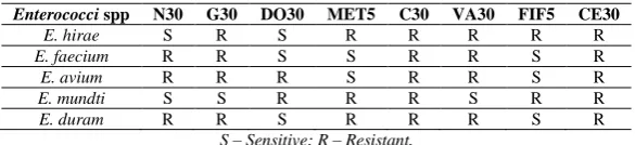
When the ion exchange performed selected fractions were pooled and further subjected in HPLC the sample was made to run for 10 min and the input volume was 10 µl and recorded to 250 nm -320 nm, during the process methanol was used as the solvent system .The enterocin were detected by false array detector at 280 nm, and the peak (267231-height, RT-2.097) was obtained, and it was compared with standard reference enterocin peaks and found significantly (Table7 and Fig 3).
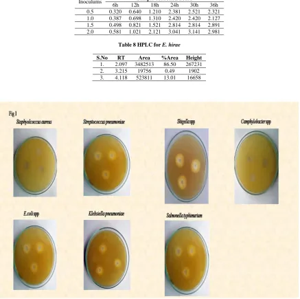
Molecular weight determination in Tricine SDS
591
Table 7 Enterococcus hirae Inoculum Concentration
Inoculums Sample collection hours
[image:7.595.88.526.98.533.2]6h 12h 18h 24h 30h 36h 0.5 0.320 0.640 1.210 2.381 2.521 2.321 1.0 0.387 0.698 1.310 2.420 2.420 2.127 1.5 0.498 0.821 1.521 2.814 2.814 2.891 2.0 0.581 1.021 2.121 3.041 3.141 2.981
Table 8 HPLC for E. hirae
S.No RT Area %Area Height
[image:7.595.115.444.568.690.2]1. 2.097 3482513 86.50 267231 2. 3.215 19756 0.49 1902 3. 4.118 523811 13.01 16658
Fig 1 Antagonistic activity of enterocin SN11 against pathogenic microorganisms
592
Fig. 2b.High pressure liquid chromatographic analysis of enterocin SN11
Fig. 3.Tricine SDS-page analysis of purified enterocin SN11
Probiotic bacteria Enterococcus spp strains were found to be antimicrobial agent with great potential for use in food
industry, attributed by production of enterocin. In the present study, the enterocin produces were enriched in MRS
medium. The isolated organism was identified as E. hirae, E. faecium, E. avium, E. mundtii and E. duram by
biochemical test. Further species level of E.hirae was confined by 16S rRNA sequencing. The isolate strain was
confirmed to be enterocin the activity of the strain through the agar well diffusion methods. E.hirae produced high
enterocin, thereby for the mass production of enterocin for food industry and therapeutic application E.hirae can be
recommended. In the present study, the stability of enterocin was applied under extreme pH, temperature, time duration and different inoculums concentration, the enterocin showed higher production at pH-7.0. The enterocin production was found highest at 37⁰C, and at the higher concentration of inoculum it was found early stage enterocin production higher. Further purification of enterocin was done by cationic ion exchange chromatography and the collected fractions were tested for antimicrobial property. The enterocin showed significant inhibition
against food borne pathogenic and intestinal microorganisms such as Staphyolcoccus aureus, Streptococcus
pneumoniae MTCC-655, E.coli spp MTCC-1583, Klebsiella pneumoniae MTCC-39, Shigella spp MTCC-2957,
Camphylobacter spp and Salmonella typhimurium MTCC 98. Results obtained purification after indicated that enterocin SN11 was highly hydrophobic in nature. The activity increased several purification processes parallel with an increase of specific activity. The spectrum of partially purified enterocin SN11 was the same to that observed with agar well diffusion, but with strong activity against pathogenic microorganisms. The result showed the presence of enterocin component identified by high pressure liquid chromatography with according to the reference
Esther Izquierdo et al., 2008 [31].
The data from the present study suggests to the enterocin SN11 to be a class II a bacteriocin as the molecular weight is >5KDa and is stable at different pH and temperature. This is the first characterization of a class IIa enterocin
produced by animal feed strains of E.hirae. These results are more encouraging as a drug for pharmaceutical, food
593
Acknowledgement
The author sincerely thanks Sophisticated Analytical Instrumentation Facility of Indian Institution of Technology, Madras for providing the instrumentation facilities to this work.
REFERENCES
[1] Cotter PD Cotter, Hill.C, RP Ross, Nature Review Microbiology, 2005, 3:777–788.
[2] Guinane.CM, PD Cotter, C Hill, RP Ross, Journal Applied Microbiology, 2005, l98:1316–1325.
[3]Hancock. REW, HG Sah, Nature Biotechnology, 2006, 24: 1551–1557.
[4] Wu.J.S, Hu , L Cao, Antimicrobial Agents Chemotherapy, 2007, 51:3131–3135.
[5]Cintas.LM, P Casaus, C Herranz, IF Nes , PE Hernandez, Food Science Technology International, 2001, 7:281–
305.
[6]Franz.CMA, MJ van Belkum , WH Holzapfel , H Abriouel , A Galvez, FEMS Microbiology Review, 2007,
31:293–310.
[7] Nes. IF, DB Diep, H Holo, Journal of Bacteriology, 2007, 189:1189–1198.
[8] Martın.M, J Gutierrez, R Criado, C Herranz, LM Cintas, PE Hernandez, Journal of Food Protection, 2006,
69:520–531.
[9] Sanchez.J, A Basanta, B Gomez-Sala, C Herranz, LM Cintas , PE Hernandez, International Journal of Food
Microbiology, 2007, 117:295–305.
[10] Alvarez-Cisneros YM, TR SainzEspunes, C Wacher, FJ Fernandez1, E Ponce-Alquicira, Science Microbial
Pathology Communication Current Research and Technology Advance, 2011.
[11] Nes IF, DB Diep, LS Havarstein, MB Brurberg, V Eijsink , H Holo . Antonie Leeuwenhoek1996, 70:113–128.
[12] Aymerich.TH, LS Holo, M Havarstein, M Hugas, Garriga , IF Nes, Applied Environmental Microbiology,
1996, 62:1676–1682.
[13] Cintas. LM, P Casaus, LV Havarstein, PE Hernandez, IF Nes, Applied Environmental Microbiology, 1997,
63:4321–4330.
[14] Pugsley AP, Microbiology Review, 1993, 57:50–108.
[15] Tomita. H, S Fujimoto,K Tanimoto , Y Ike, Journal of Bacteriology, 1996, 178: 3585–3593.
[16] Worobo. RW, T Henkel, M Sailer, KL Roy, JC Vederas, ME Stiles, Microbiology, 1994, 140:517–526.
[17] Van Belkum. MJ, RW Worobo , ME Stiles, Molecular Microbiology, 1997, 23: 1293–1301.
[18] Gierasch. LM . Biochemistry, 1989, 28:923–930.
[19] Izard. JW , DA Kendall, Molecular Microbiology, 1994, 13:765–773.
[20] Von Heijne. G , Journal of Molecular Biology, 1986, 192:287–290.
[21] Farkas-Himsley, H, R Hill, B Rosen, S Arab, CA Lingwood, Proc Natl Acad Sci, 1995, 92:6996–7000.
[22] Sand. SL , TM Haug, J Nissen-Meyer, O Sand, Journal of Membrane Biology, 2007, 216:61–71.
[23] Koczulla. AR, R Bals, Drugs, 2003, 63:389– 406.
[24] Brotz. H , HG Sahl, Journal Antimicrobial Chemotherapy2000, 46:1–6.
[25] Holt. JG, NR Krig, JT Staley, ST Williams.Bergey’Z, Manual od determinative bacteriology, 9th Edn. Prestons
Street, Baltimore, Maryland 1994, 21202 USA 528-540.
[26] Sathiyavimal S, S Vasantharaj, International Journal of Pharmaceutical Sciences Review and Research, 2013,
16: 75-78.
[ 27] Jagannathan.S Mani KR, Vijayakumar R, Journal of vaccine and vaccination, 2015, 6(1),1-6.
[28] Frantz. CMAP, U Schillinger, WH Holzapfel, International Journal of Food Microbiology, 1996, 29: 255-270.
[29] Glasel. JA, BioTechniques ,1995 18:1.
[30] Schagger. H , G Von Jagow, Annals Biochemistry, 1987 166, 368-379.
[31]Esther Izquierdo, Audrey Bednarczyk, Christine Schaeffer, YiminCai, Eric Marchioni, Alain Van Dorsselaer,
Sad Ennahar. Antigen Ageing and Chemistry , 2008,1917-1923.
[32] Kang. JH, MS Lee MS, Journal of Applied Microbiology, 2005, 98:1169-1176
[33] Shivanandappa KC, Jagannathan S, Pandiyarajan S, Tamilvanan R, Umadevi T, Jeeva Kalaiselvan, Sekar B.