P R O C E E D I N G S
Open Access
Sequence and structural features of binding site
residues in protein-protein complexes:
comparison with protein-nucleic acid complexes
M Michael Gromiha
1,2*, N Saranya
3, S Selvaraj
3, B Jayaram
4, Kazuhiko Fukui
2From
International Workshop on Computational Proteomics
Hong Kong, China. 18-21 December 2010
Abstract
Background:Protein-protein interactions are important for several cellular processes. Understanding the
mechanism of protein-protein recognition and predicting the binding sites in protein-protein complexes are long standing goals in molecular and computational biology.
Methods:We have developed an energy based approach for identifying the binding site residues in protein– protein complexes. The binding site residues have been analyzed with sequence and structure based parameters such as binding propensity, neighboring residues in the vicinity of binding sites, conservation score and
conformational switching.
Results:We observed that the binding propensities of amino acid residues are specific for protein-protein complexes. Further, typical dipeptides and tripeptides showed high preference for binding, which is unique to protein-protein complexes. Most of the binding site residues are highly conserved among homologous sequences. Our analysis showed that 7% of residues changed their conformations upon protein-protein complex formation and it is 9.2% and 6.6% in the binding and non-binding sites, respectively. Specifically, the residues Glu, Lys, Leu and Ser changed their conformation from coil to helix/strand and from helix to coil/strand. Leu, Ser, Thr and Val prefer to change their conformation from strand to coil/helix.
Conclusions:The results obtained in this study will be helpful for understanding and predicting the binding sites in protein-protein complexes.
Background
Protein-protein interactions are important for most of the cellular processes in life. Hence, understanding the mechanism of protein-protein recognition at molecular level is of practical interest and has direct applications to functional genomics. Unravelling the mechanisms of protein-protein recognition is a fundamental problem, which would aid in function prediction and drug design. The availability of structures of numerous protein-pro-tein complexes in Proprotein-pro-tein Data Bank (PDB) enables researchers to analyze the binding sites in terms of
amino acid composition, preference of residues, second-ary structures, solvent accessibility, electrostatic patches, hydrophobic contacts, hydrogen bonding networks and so on [1-3]. The mapping of protein-protein interactions on protein sequences suggests that hotspots can be pre-dicted from amino acid sequences [4]. The concepts of protein-protein interactions in terms of experimental techniques, databases, organization, cooperativity and prediction of protein-protein, protein-ligand and domain interactions have been reviewed in detail earlier [5-7].
Several methods have been proposed for identifying the binding sites in protein-protein complexes based on distance between two residues [8-11]. In our earlier work, we have developed an energy based approach for defining the binding sites in protein-protein complexes
* Correspondence: [email protected]
1
Department of Biotechnology, Indian Institute of Technology Madras, Chennai 600 036, Tamilnadu, India
Full list of author information is available at the end of the article
[12]. In this work, we have analyzed the binding site residues based on sequence and structures of protein-protein complexes. The results showed that the binding site residues have specific preferences at their vicinities and these residues are unique in protein-protein com-plexes. These binding site residues are more conserved than non-binding residues. In addition, several binding and non-binding residues prefer to change their confor-mation from helix to coil, strand to coil and coil to helix/strand. Specifically the residues Glu, Lys and Ser play important roles to conformational switching.
Methods Dataset
We have developed non-redundant datasets of 153 pro-tein-protein hetero dimer complexes from Protein Data Bank that have the sequence identity of less than 25% and solved with better than 3Å resolution [12]. In addi-tion, we have used a benchmark dataset of 124 protein-protein complexes to validate our results [13]. For parison, we have utilized a set of 81 protein-RNA com-plexes [14] and 212 protein-DNA comcom-plexes [15].
Identification of binding site residues
We have calculated the interaction energy between all pairs of atoms in protein-protein complexes using AMBER force field [16]. The interaction energies of all the atoms in a residue have been summed up to assign the interaction energy of a residue. The amino acid resi-dues with interaction energy less than -1 kcal/mol are treated as binding site residues [17].
Binding propensity
The binding propensity (Pbind) for the 20 amino acid
residues in protein-protein complexes is defined as the ratio between the frequency of occurrence of amino acid residues in the binding sites (fb) and in the protein
as a whole (ft). It is calculated using the equation:
Pbind(i) = fb(i)/ft(i) (1)
where, i represents each of the 20 amino acid residues.
Influence of neighboring residues
We have analyzed the influence of neighboring residues of binding sites on various aspects: (i) *B, where * is any residue and B is a binding site residue, (ii) B* and (iii) *B*, which is a tripeptide with the binding site residue at the middle.
Conservation score
We have used the program AL2CO for computing the conservation score for all the residues in receptors and ligands in protein-protein complexes [17]. The target sequence has been compared with non-redundant sequences in SWISS-PROT [18] and multiple sequence
alignment has been performed with ClustalW program [19]. The aligned sequences have been utilized to com-pute the conservation score for all the amino acid residues.
Conformational switching upon complex formation
We have computed the secondary structures of all the residues in free proteins and complexes in a set of 124 protein-protein complexes [13] using DSSP [20]. The secondary structures have been assigned as helix, strand and coil. We have analyzed the conformational changes of residues based on their locations in secondary struc-tures, preferred amino acid residues and binding site residues.
Results and Discussion
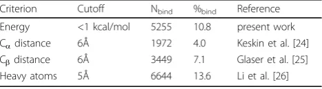
Occurrence of amino acid residues at various ranges of interaction energies
We have identified the binding sites in protein-protein complexes based on interaction energy as explained in Methods section. We observed that 13.9% of the resi-dues have the interaction energy of < -1 kcal/mol and are identified as binding sites in a set of 306 proteins. We have compared the results with those obtained with distance based criteria for defining binding site residues and the data are presented in Table 1. We noticed that only 28% residues are common to each other and the percentage of binding site residues is a balance between those identified with different cutoff distances, indicating the importance of considering the energy between dif-ferent atoms to define the binding residues. In addition, 5.7% of the residues have strong repulsive energies and all these residues have been identified as binding resi-dues in distance based criteria, which are not probable binding residues in protein-protein complexes.
Conservation score for binding site residues in protein-protein complexes
We have computed the conservation score for all the residues and noticed that the binding residues are highly conserved in protein-protein complexes. This observa-tion is consistent with earlier studies reported in the lit-erature [21]. We have estimated the performance of conservation score for identifying the binding sites. We found that the conservation score alone could predict
Table 1 Number and percentage of binding site residues using different methods
Criterion Cutoff Nbind %bind Reference
the binding sites at an average accuracy of 58% with a trade-off between sensitivity and specificity of 59% and 57%, respectively.
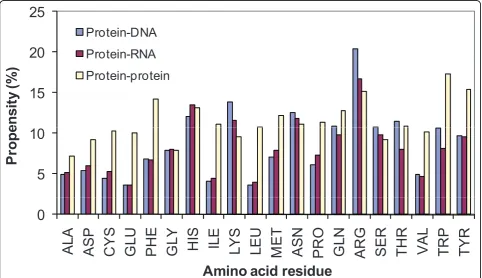
Binding propensity of residues in protein complexes
We have computed the binding propensity in protein-protein complexes and the results are presented in Fig-ure 1. For comparison we have also included the data obtained for protein-RNA and protein-DNA complexes.
We observed that the aromatic as well as positively charged residues highly contribute to interact between the partners in protein-protein complexes, indicating the importance of cation-π, aromatic and electrostatic interactions. The comparison between protein-protein and protein-RNA complexes showed that the residues, Asp, Cys, Glu, Phe, Ile, Leu, Met, Val, Trp and Tyr have remarkably high contribution in protein-protein com-plexes. These residues prefer to form electrostatic, hydrophobic and aromatic interactions in protein-pro-tein complexes. On the other hand, the residues Arg, His, Lys, Asn, Tyr, Gln and Ser highly contribute for the interaction between protein and RNA. Interestingly, these residues belong to positively charged, polar and aromatic groups, which form electrostatic, hydrogen bonds and aromatic interactions with RNA. In protein-DNA complexes, positive charged residues are more dominant than protein-protein and protein-RNA
complexes. Further, polar residues prefer to interact with DNA in the form of hydrogen bonds.
Preference of tripeptides in the vicinity of binding sites
We have set up the following criteria to identify the probable tripeptides for binding: (i) there should be at least three samples and the probability of being in bind-ing sites should be more than 50%. These conditions yielded a set of 208 unique tripeptides among 8000 pos-sibilities. The usage of these tripeptides could predict the binding sites with an accuracy of 72.3% and the cov-erage of 6% of binding sites. The results for selected tri-peptides are presented in Table 2.
We noticed that the central residue of all the tripep-tides CWA, HHE, MNF and WFE are identified as bind-ing site residues. The tripeptides LFP and MRR showed a preference of more than 75%. Although the preference is 66.7% for ITG it has the occurrence of 12 hits and eight of them are binding site residues. The preference is significantly higher than the random choice of 1.3% (10202 binding sites and the total of 8000 tripeptides).
In Table 2, we have also included the preferred tripep-tides at the binding sites of RNA and protein-DNA complexes. The information on tripeptides could identify the binding sites with an accuracy of 78.7% and 71.9% in protein-RNA and protein-DNA complexes. We have compared the preferences of tripeptides at the
20
25
Protein-DNA
Protein-RNA
15
20
ens
it
y (
%
)
Protein RNA
Protein-protein
5
10
Pr
o
p
e
0
AL
A
ASP
CY
S
GL
U
PH
E
GL
Y
HI
S
IL
E
LY
S
LE
U
ME
T
ASN
PR
O
GL
N
AR
G
SER
TH
R
VAL
TR
P
TY
R
A i
id
id
Amino acid residue
interface of protein, RNA and protein-DNA complexes. Interestingly, the preferred tripeptides are unique to protein-protein complexes and none of the tripeptides are common with any of the other com-plexes. This result reveals the existence of different mode of recognition for the protein complexes with other biological molecules.
Importance of sequence specificity revealed from dipeptide preferences
We have analyzed the preference of residues paired with binding site residues on both N and C directions. The preferred residue pairs with *B binding motifs are DW, CW, MW, CM, CY, MR, MY, PF, PH, QW, SW, TH and WN. On the other hand the preferred residue pairs with B* motifs are CW, HH, IW, MW, QM, RR, TF,
WG, WH, WM, WN, YA and YG. Further analysis on the preference of residues on N and C directions of binding sites revealed that the paired amino acids are different on both sides. Specifically, the residues at the N- direction of binding site Trp residue are Ala, and Asp whereas at the C-side are Met, His, Phe, Asp and Val. This result indicates the importance of sequence specificity for binding in protein-protein complexes.
We have compared the specific preferences of dipep-tides in protein-protein, protein-RNA and protein-DNA complexes and the topmost five residue pairs are listed in Table 3. In this table, we included the data obtained with the motifs *B and B*. We observed that the resi-dues mainly paired with Trp at the binding sites in pro-tein-protein complexes. On the other hand, the residues preferred to have pairs with Arg and His in protein-RNA complexes. Interestingly, eight out of ten pairs pre-fer the residue Arg at the binding sites in protein-DNA complexes. This shows the different features of residue pairs at the binding sites for the proteins complexed with proteins, RNA and DNA. Further, we noticed that the residue pair Cys-Trp is common to all the three complexes, which may be a unique pair for binding.
Conformational switching upon complex formation
We have analyzed the residues that change their confor-mation upon binding. We noticed that approximately 7% of residues are involved in conformational changes. The analysis on different secondary structures showed that the changes between regular structures are not favorable, for example, helix to strand and vice versa. Most of the changes are associated with irregular shape or coil. We have also analyzed the preference of amino acid residues to change their conformations upon
Table 2 Preferred tripeptides at the binding sites of protein-protein, protein-RNA and protein-DNA complexes
Tripeptide Nb Nt %bind
Protein-protein
CYS TRP ALA 3 3 100.00
HIS HIS GLU 3 3 100.00
MET ASN PHE 3 3 100.00
TRP PHE GLU 3 3 100.00
ILE TYR GLY 8 12 66.67
LEU PHE PRO 5 6 83.33
MET ARG ARG 4 5 80.00
Protein-RNA
GLY TYR GLY 3 3 100.00
PRO GLY ARG 3 3 100.00
ASP LYS TYR 6 8 75.00
GLY SER THR 3 4 75.00
ILE TYR LYS 8 12 66.67
LYS SER ARG 3 4 75.00
PRO HIS HIS 3 4 75.00
SER ARG LYS 5 7 71.43
VAL GLY SER 6 8 75.00
TYR LYS HIS 5 6 83.33
Protein-DNA
HIS ARG SER 3 3 100.00
SER GLN THR 4 4 100.00
SER TYR GLN 3 3 100.00
GLY MET SER 3 4 75.00
GLY ASN ALA 6 9 66.67
LYS ARG THR 9 14 64.29
GLN SER TYR 3 4 75.00
ARG GLY ASN 5 7 71.43
SER GLN ARG 5 6 83.33
SER THR ILE 5 7 71.43
VAL HIS ASP 3 4 75.00
VAL LYS CYS 5 6 83.33
Nb: number of occurrence at binding sites; Nt: total number of occurrence
Table 3 Topmost five preferred residue pairs at the binding sites of protein-protein, protein-RNA and protein-DNA complexes
Protein-protein Protein-RNA Protein-DNA
*B
ASP TRP CYS TRP SER ARG
CYS TRP HIS ARG GLY ARG
ILE TRP ASN ARG LYS ARG
MET TRP ILE TYR ARG ARG
MET TYR TRP ARG CYS TRP
B*
CYS TRP HIS TRP ARG SER
MET TRP HIS HIS ARG GLY
TRP PHE HIS ARG ASN TRP
TRP HIS LYS HIS ARG LYS
TRP MET MET TRP ARG ASN
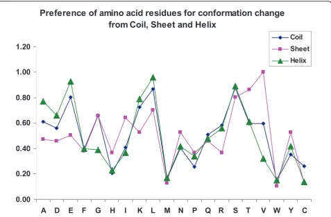
binding in three different secondary structures. The results are shown in Figure 2.
We noticed that the residues Lys, Glu, Leu and Ser have high preference to change their conformation from helix to coil/strand as well as from coil to helix/strand. Further, Leu, Ser, Thr and Val prefer to change their conformation from strand to coil/helix. The analysis of binding site residues showed that the percentage of resi-dues that change their conformation from coil to strand, coil to helix, strand to coil and helix to coil are, 39%, 17%, 17% and 26%, respectively. This result indicates that the proteins tend to form regular secondary struc-tures upon binding, which agrees with the analysis that the binding site residues are located mainly in helix/ strand regions compared with coil [22]. Further, the conformation changes may be necessary for the recogni-tion of protein-protein complexes [23].
We have also analyzed the preference of conforma-tional changes for the neighboring residues of binding sites, N-2, N-1, C+1 and C+2 positions. Interestingly, the preference of next residues (C+1 and N-1 positions) are higher than other positions and the change at the second position on both directions (N-2 and C+2) are similar.
Conclusions
We have developed an energy based approach for iden-tifying the binding sites in protein-protein complexes. The binding sites identified have been further analyzed based on different sequence and structure based para-meters, conservation score, conformational switching and preference of neighboring residues. We observed that the binding residues are significantly highly con-served than the non-binding residues. We have also explored the preferences of residues at the vicinity of binding sites, which showed the importance of sequence specificity. Further, we have analyzed the importance of conformational changes upon complex formation. We noticed that the residues Ser, Leu, Lys, Glu, Thr and Val prefer to change their conformation upon binding. The information obtained in the present study will be useful for understanding and predicting the binding sites of protein-protein complexes.
Acknowledgments
This research was supported by Strategic International Cooperative Program, Japan Science and Technology Agency (JST) and Department of Science and Technology, Government of India.
This article has been published as part ofProteome ScienceVolume 9 Supplement 1, 2011: Proceedings of the International Workshop on
Preference of amino acid residues for conformation change
from Coil, Sheet and Helix
1.20
Coil Sheet
0.80
1.00
Helix
0 40
0.60
0 00
0.20
0.40
0.00
A D
E F
G
H
I
K L
M N
P Q R S T
V W Y C
Computational Proteomics. The full contents of the supplement are available online at http://www.proteomesci.com/supplements/9/S1.
Author details
1
Department of Biotechnology, Indian Institute of Technology Madras, Chennai 600 036, Tamilnadu, India.2Computational Biology Research Center
(CBRC), National Institute of Advanced Industrial Science and Technology (AIST), 2-4-7 Aomi, Koto-ku, Tokyo 135-0064, Japan.3Department of
Bioinformatics, Bharathidasan University, Tiruchirapalli 620024, Tamilnadu, India.4Department of Chemistry and Supercomputing Facility for
Bioinformatics and Computational Biology, Indian Institute of Technology Delhi, New Delhi 110016, India.
Authors’contributions
MMG and KF designed the project. MMG carried out the computations on sequence and structural features. SS and NS are involved in conformational switching. BJ contributed in discussions.
Competing interests
The authors declare that they have no competing interests.
Published: 14 October 2011
References
1. Chakrabarti P, Janin J:Dissecting protein-protein recognition sites.Proteins
2002,47:334-343.
2. Sheinerman FB, Honig B:On the role of electrostatic interactions in the design of protein-protein interfaces.J Mol Biol2002,318:161-177. 3. Kortemme T, Baker D:A simple physical model for binding energy hot
spots in protein-protein complexes.Proc Natl Acad Sci USA2002,
99:14116-14121.
4. Ofran Y, Rost B:Protein-protein interaction hotspots carved into sequences.PLoS Comput Biol2007,3:e119.
5. Shoemaker BA, Panchenko AR:Deciphering protein-protein interactions. Part II. Computational methods to predict protein and domain interaction partners.PLoS Comput Biol2007,3:e43.
6. Keskin O, Gursoy A, Ma B, Nussinov R:Principles of protein-protein interactions: what are the preferred ways for proteins to interact?Chem Rev2008,108:1225-1244.
7. Shaikh SA, Jain T, Sandhu G, Latha N, Jayaram B:From drug target to leads- sketching, A physicochemical pathway for lead molecule design in silico.Current Pharmaceutical Design2007,13:3454-3470.
8. SikićM, TomićS, Vlahovicek K:Prediction of protein-protein interaction sites in sequences and 3D structures by random forests.PLoS Comput Biol2009,5:e1000278.
9. Fariselli P, Pazos F, Valencia A, Casadio R:Prediction of protein–protein interaction sites in heterocomplexes with neural networks.Eur J Biochem
2002,269:1356-1361.
10. Koike A, Takagi T:Prediction of protein-protein interaction sites using support vector machines.Protein Eng Des Sel2004,17:165-173. 11. Ofran Y, Rost B:ISIS: interaction sites identified from sequence.
Bioinformatics2007,23:e13-6.
12. Gromiha MM, Yokota K, Fukui K:Energy based approach for understanding the recognition mechanism in protein-protein complexes.Mol. Biosystems2009,5:1779-1786.
13. Hwang H, Pierce B, Mintseris J, Janin J, Weng Z:Protein-protein docking benchmark version 3.0.Proteins2008,73:705-709.
14. Gromiha MM, Yokota K, Fukui K:Understanding the recognition mechanism of protein-RNA complexes using energy based approach.
Curr Protein Pept Sci2010,11:629-638.
15. Gromiha MM, Fukui K:Scoring Function Based Approach for Locating Binding Sites and Understanding Recognition Mechanism of Protein-DNA Complexes.J Chem Inf Model2011.
16. Cornell WD, Cieplak P, Bayly CI, Gould IR, Merz KM Jr, Ferguson DM, Spellmeyer DC, Fox T, Caldwell JW, Kollman PA:A second generation force field for the simulation of proteins, nucleic acids, and organic molecules.
J Am Chem Soc1995,117:5179-5197.
17. Pei J, Grishin NV:AL2CO: calculation of positional conservation in a protein sequence alignment.Bioinformatics2001,17:700-712.
18. The UniProt Consortium:The universal protein resource (UniProt).Nucleic Acids Res2008,36:D190-195.
19. Larkin MA, Blackshields G, Brown NP, Chenna R, McGettigan PA, McWilliam H, Valentin F, Wallace IM, Wilm A, Lopez R, Thompson JD, Gibson TJ, Higgins DG:ClustalW and ClustalX version 2.Bioinformatics
2007,23:2947-2948.
20. Kabsch W, Sander C:Dictionary of protein secondary structure: pattern recognition of hydrogen-bonded and geometrical features.Biopolymers
1983,22:2577-2637.
21. Konc J, Janezic D:Protein-protein binding-sites prediction by protein surface structure conservation.J Chem Inf Model2007,47:940-4. 22. Gromiha MM, Yokota K, Fukui K:Sequence and structural analysis of
binding site residues in protein-protein complexes.Int J Biol Macromol
2010,46:187-192.
23. Lensink MF, Méndez R:Recognition-induced conformational changes in protein-protein docking.Curr Pharm Biotechnol2008,9:77-86. 24. Keskin O, Tsai CJ, Wolfson H, Nussinov R:A new, structurally
nonredundant, diverse data set of protein-protein interfaces and its implications.Protein Sci2004,13:1043-55.
25. Glaser F, Steinberg DM, Vakser IA, Ben-Tal N:Residue frequencies and pairing preferences at protein-protein interfaces.Proteins2001,43:89-102. 26. Li W, Keeble AH, Giffard C, James R, Moore GR, Kleanthous C:Highly
discriminating protein-protein interaction specificities in the context of a conserved binding energy hotspot.J Mol Biol2004,337:743-59.
doi:10.1186/1477-5956-9-S1-S13
Cite this article as:Gromihaet al.:Sequence and structural features of binding site residues in protein-protein complexes: comparison with protein-nucleic acid complexes.Proteome Science20119(Suppl 1):S13.
Submit your next manuscript to BioMed Central and take full advantage of:
• Convenient online submission
• Thorough peer review
• No space constraints or color figure charges
• Immediate publication on acceptance
• Inclusion in PubMed, CAS, Scopus and Google Scholar
• Research which is freely available for redistribution