Wróbel Grzegorz. Metastatic renal cell carcinoma – a case report. Journal of Education, Health and Sport. 2017;7(9):416-420. elSSN 2391-8306. DOI http://dx.doi.org/10.5281/zenodo.1001661
http://ojs.ukw.edu.pl/index.php/johs/article/view/4926
The journal has had 7 points in Ministry of Science and Higher Education parametric evaluation. Part B item 1223 (26.01.2017).
1223 Journal of Education, Health and Sport eISSN 2391-8306 7
© The Author (s) 2017;
This article is published with open access at Licensee Open Journal Systems of Kazimierz Wielki University in Bydgoszcz, Poland Open Access. This article is distributed under the terms of the Creative Commons Attribution
Noncommercial License which permits any noncommercial use, distribution, and reproduction in any medium, provided the original author(s) and source are credited. This is an open access article licensed under the terms of
the Creative Commons Attribution Non Commercial License (http://creativecommons.org/licenses/by-nc/4.0/) which permits unrestricted, non commercial use, distribution and reproduction in any medium, provided the work is properly cited.
This is an open access article licensed under the terms of the Creative Commons Attribution Non Commercial License (http://creativecommons.org/licenses/by-nc/4.0/) which permits unrestricted, non commercial use, distribution and reproduction in any medium, provided the work is properly cited.
The authors declare that there is no conflict of interests regarding the publication of this paper.
Received: 05.09.2017. Revised 10.09.2017. Accepted: 10.09.2017.
Metastatic renal cell carcinoma – a case report
Grzegorz Wróbel
ORCID iD http://orcid.org/0000-0003-3788-1692
Affiliation Department of Human Anatomy, Faculty of Medicine and Health Sciences, Jan Kochanowski University, Al. IX Wieków Kielc 19 A 25-317 Kielce, Poland;
Phone: 413496965. E-mail: grzegorz.wrobel@ujk.edu.pl Country Poland
Abstract
Renal cell carcinoma (RCC) is not a single uniform entity but a group of related
neoplasms in which the histologic findings, cytogenetic abnormalities, biologic behavior and
disease in a way that is diagnostically very powerful. The case concerns the result imaging
18
F-fluorodeoxyglucose positron emission tomography (PET)-CT to patient 47 years old
(women) is diagnosed with numerous changes in both lungs, the liver and the skeletal system
in the abdominal lymph nodes. Primary change in left kidney is indicated. Metastasis is a
process consisting of cells spreading from the primary site of the cancer to distant parts of the body. Functional imaging, particularly with PET–CT, might improve the accuracy of
diagnosis and provide essential information that could allow clinicians to make more
appropriate therapeutic decisions than they previously could without this technique.
Keywords: positron emission tomography, renal cell carcinoma, anatomic imaging
1. Introduction
The 2004 World Health Organization Classification of adult renal tumors stratifies renal
cell carcinoma (RCC) into several distinct subtypes of which clear cell, papillary and
chromophobe tumors account for 70%, 10%-15%, and 5%, respectively [1]. Preliminary
suspicion of renal tumor is usually put forward on the basis of abdominal ultrasonography (USG), which, when performed by an experienced physician, may indicate the size and
severity of the tumor. The next diagnostic step to confirm the diagnosis of cancer is computed
tomography (CT) with intravenous contrast. When there is suspicion of metastases to the
brain, a tomographic or magnetic resonance imaging of the head is performed. The specific
diagnosis of abnormalities in the skeletal system consists in bone scintigraphy, where after the
administration of an isotopic marker, an image of the entire skeleton is recorded. Suspension
of bone metastases usually requires confirmation in x-ray, tomography or magnetic resonance.
Functional imaging, particularly with PET–CT, might improve the accuracy of diagnosis and
provide essential information that could allow clinicians to make more appropriate therapeutic
decisions than they previously could without this technique [2-5].
2. Case presentation
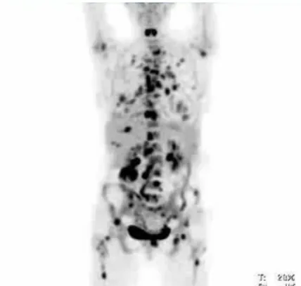
This study was done in the Department of Nuclear Medicine with Positron Emission
Tomography (PET) Unit (Holy Cross Cancer Centre in Kielce). The case concerns the result
imaging 18F-fluorodeoxyglucose positron emission tomography (whole-body 18F-FDG
PET/CT) to patient 47 years old (women) is diagnosed with numerous changes in both lungs,
the liver and the skeletal system in the abdominal lymph nodes. Primary change in left kidney
Figure 1. PET image (from a whole‑body 18F‑FDG‑PET–CT scan) of a 47‑year‑old woman.
3. Discussion
Renal cell carcinoma (RCC) is the most common kidney malignancy and the development of macroscopic metastasis of RCC is the major cause of tumor-associated deaths [6, 7].
Metastasis is a process consisting of cells spreading from the primary site of the cancer to
distant parts of the body. There is some evidence that primary renal cell carcinoma (RCC) and
metastases of RCC exhibit molecular differences that may effect on the biological
characteristics of the tumor [8]. By contrast, the use of PET and PET–CT to stage metastatic
RCC has shown promising results, with sensitivities ranging from 64% to 100%; in the
majority of cases, the sensitivity is close to 100% for metastatic disease [9-11]. Renal cell
5.9% annually until RCC is now the 10th most common in men and 14th most common in
women [12-13]. Renal cancer represents 2%-3% of adult malignancies[12]. The median age
at diagnosis is 65 years with most patients being in the 6th to 8th decade of life[14]. Males are
2 to 3 times as affected as females[1,12,13]. Over the last 65 years, the incidence of RCC has
increased at a rate of 2% per year [12]. Nakajima et al [15] found that RCC (During the whole body phase) that were of higher stage, higher grade, and associated with vascular or
lymphatic invasion showed higher maximum standardized uptake than less aggressive RCC.
Alongi et al. [16] suggested that PET-CT was able to predict disease progression and survival
in patients with recurrent RCC after surgery and so influence clinical decision making.
4. Conclussion
The functional imaging modality of 18F‑FDG‑PET–CT has support for initial staging and
re‑staging in the context of relapse or metastasis in RCC.
5. References
1. Eble J. N., Sauter G., Epstein J. I., Sesterhenn I. A. (2004). World Health Organization Classification of Tumours. Pathology and Genetics of Tumours of the Urinary System
and Male Genital Organs. Lyon: IARC Press.
2. Federle M. P., Jeffrey R. B., Woodward P. J. et al. (2009) Diagnostic Imaging:
Abdomen, Published by Amirsys. Lippincott Williams & Wilkins.
3. McPhee S. J., Papadakis M. A. (2008) Current Medical Diagnosis and Treatment.
McGraw-Hill Professional.
4. Tanagho E. A., McAninch J. W. (2004) Smith's general urology. McGraw-Hill
Medical.
5. Ng C. S., Wood C. G., Silverman P. M. et al. (2008) Renal cell carcinoma: diagnosis,
staging, and surveillance. AJR Am J Roentgenol. 191 (4), 1220-32.
doi:10.2214/AJR.07.3568
6. Semeniuk-Wojtaś A., Stec R., Szczylik C. (2016) Are primary renal cell carcinoma and metastases of renal cell carcinoma the same cancer? Urol Oncol, 34(5), 215-20.
doi: 10.1016/j.urolonc.2015.12.013
7. Bielecka Z. F., Czarnecka A. M., Szczylik C. (2014) Genomic analysis as thefirst
step toward personalized treatment in renal cell carcinoma. Front Oncol 4, 194.
8. Low G., Huang G., Fu W., Moloo Z., Girgis S. (2016) Review of renal cell carcinoma
and its common subtypes in radiology. World J Radiol. 8(5), 484-500
9. Townsend, D. W., Beyer, T. (2002) A combined PET/CT scanner: the path to true
10.Seto, E., Segall G. M., Terris, M. K. (2000) Positron emission tomography detection of
osseous metastases of renal cell carcinoma not identified on bone scan. Urology 55,
286.
11. Aide, N. et al. (2003) Efficiency of [(18)F]FDG PET in characterising renal cancer
and detecting distant metastases: a comparison with CT. Eur. J. Nucl. Med. Mol. Imaging 30, 1236–1245.
12.Motzer R. J., Agarwal N., Beard C., Bhayani S., Bolger G. B., Carducci M. A., Chang
S. S., Choueiri T. K., Hancock S. L., Hudes G. R., et al. (2011). Kidney cancer. J Natl
Compr Canc Netw. 9, 960–977.
13.Siegel R. L., Miller K. D., Jemal A. Cancer statistics, 2016. CA Cancer J Clin. 2016
66(1), 7-30. doi: 10.3322/caac.21332
14.Zhang J ., Lefkowitz R. A., Bach A. (2007) Imaging of kidney cancer. Radiol
Clin North Am 2007; 45: 119-147 DOI: 10.1016/j.rcl.2006.10.011
15.Nakajima R, Abe K, Kondo T, Tanabe K, Sakai S. Clinical role of early
dynamic FDG-PET/CT for the evaluation of renal cell carcinoma. Eur Radiol
2015; Epub ahead of print [PMID: 26403580 DOI: 10.1007/s00330-015-4026-3]
16.Alongi P., Picchio M., Zattoni F., Spallino M., Gianolli L., Saladini G., Evangelista L. Recurrent renal cell carcinoma: clinical and prognostic value of
