DOI: 10.1002/ejic.200700419
Aqueous Reduction of [Cp*
2W
2O
5]: Characterization of the Triangular
Clusters [Cp*
3W
3O
4(OH)
2]
2+and [Cp*
3W
3O
6]
+– Comparison with
Molybdenum
Chiara Dinoi,
[a]Pelin Sözen,
[b]Gülnur Taban,
[b]Deniz Demir,
[b]Funda Demirhan,*
[b]Petr Prikhodchenko,
[c]Jenny Gun,
[c]Ovadia Lev,
[c]Jean-Claude Daran,
[a]and
Rinaldo Poli*
[a]Keywords:Tungsten / O ligands / Metal clusters / Density functional calculations
Zinc reduction of Cp*2W2O5in an acidified water–methanol
medium affords the green 3-electron trinuclear cluster [Cp*3W3O4(OH)2]2+, isolated and crystallographically
char-acterized as the triflate salt,1. Upon exposure to air, this com-plex is oxidized to a related 2-electron cluster, [Cp*3W3O6]+,
which was crystallized and characterized in three different salts, 2–4. The 3-electron cluster exhibits a near-tetragonal frozen-glass EPR spectrum, and it shows evidence of coup-ling of the unpaired electron to only one of the three W
Introduction
Aqueous organometallic chemistry is attracting growing interest because of its “green” impact[1–3] and because of
its potential in catalytic[4,5] and biomedical applications.[6]
Although this area is widely explored for low-valent sys-tems, usually upon appending hydrophilic functionalities to the ligand periphery to increase solubility in an aqueous environment,[7–9]studies of high-oxidation organometallics
are still rather scarce.[10–12]For redox-active metals, these
studies are particularly interesting, as they may open the way to electrocatalytic applications.[12]
In a recent contribution,[13]we reported that the zinc
re-duction of Cp*2Mo2O5in a strongly acidic (CF3COOH or
CF3SO3H) mixed H2O/MeOH medium affords the
trinu-clear mixed-valence (Mo313+) cluster [Cp*3Mo3O2(OH)4]2+.
The ion adopts a nearly equilateral triangular shape; each edge is bridged by two ligands. The distortion of the
tri-[a] Laboratoire de Chimie de Coordination, UPR CNRS 8241 liée par convention à l’Université Paul Sabatier et à l’Institut National Polytechnique de Toulouse,
205 Route de Narbonne, 31077 Toulouse Cedex, France Fax: +33-561553003
E-mail: [email protected]
[b] Celal Bayar University, Faculty of Sciences & Liberal Arts, De-partment of Chemistry,
45030 Muradiye-Manisa, Turkey
[c] Laboratory of Environmental Chemistry, The Casali Institute of Applied Chemistry, The Hebrew University of Jerusalem, Jerusalem 91904, Israel
Supporting information for this article is available on the WWW under http://www.eurjic.org or from the author.
atoms. The two-electron cluster is diamagnetic. An electro-spray ionization MSnstudy revealed a stepwise expulsion of
neutral [Cp*WO2] units as the main fragmentation process.
DFT calculations unveiled the intimate details of the elec-tronic structures of these complexes and fully rationalized the structural and spectroscopic properties.
(© Wiley-VCH Verlag GmbH & Co. KGaA, 69451 Weinheim, Germany, 2007)
angle is caused by one edge that is bridged by two OH groups, whereas the other two edges are bridged by one O atom and one OH group. As a consequence, the former edge is slightly longer than the latter ones. Although the number of bridging OH versus O groups was not revealed directly by the X-ray structural analysis, it was suggested indirectly by the same technique through the existence of short hydrogen-bonding contacts (or lack thereof) between the Mo3O6core and the two CF3CO2and CF3SO3anions,
and this was confirmed by comparison with a DFT geome-try-optimized model, by EPR spectrometry, and by mag-netic susceptibility measurements.
In this contribution, we examine the parallel behavior of the pentamethylcyclopentadienyltungsten system and re-port the isolation and characterization of two new redox-related tritungsten oxido clusters, [Cp*3W3O6]+ and
[Cp*3W3O4(OH)2]2+.
Results and Discussion
The [Cp*3W3O4(OH)2]2+Cluster
The reduction of [Cp*2W2O5] was carried out under the
same conditions previously used for the reduction of [Cp*2Mo2O5], namely by metallic zinc in an acidic H2O/
Mo analogue, a series of color changes was observed over a period of three days, yielding a final green solution through an intermediate orange-red color. Crystallization of the final green product was successful in the case of the triflic acid reaction, revealing the nature of the compound as [Cp*3W3O4(OH)2](CF3SO3)2·2H2O (1). Thus, the
reac-tion stoichiometry is as shown in Equareac-tion (1). The oxi-dation state of tungsten in the final product is +5, yielding a 3-electron W315+cluster. Compound1is quite soluble in
CH2Cl2 and its solutions are air sensitive, and they turn
rapidly to orange when exposed to the atmosphere. The na-ture of the orange product is shown in the next section.
(1)
[image:2.595.227.535.98.693.2]Figure 1. An ORTEP view of the dication in compound [Cp*3W3O4(OH)2](CF3SO3)2·2H2O (1).
Figure 2. An ORTEP view of compound1that highlights the H-bonding interactions between dication, anions, and interstitial water molecules. The Cp* ligands bonded to the W3 cluster are omitted for clarity [symmetry codes: (i) 3/2 –x,y– 1/2, 1 –z; (ii) 1/2 –x,y– 1/2, –z].
Table 1. Selected bond lengths and angles for the trinuclear clusters in all compounds.[a]
Bond lengths [Å]
1 2[b] 3 4
W1–W2 2.7416(3) 2.6565(4) 2.6685(5) 2.6637(8) W1–W3 2.6545(3) 2.6505(5) 2.6657(5) 2.6614(9) W2–W3 2.7485(3) 2.6849(5) 2.6606(8) W1–O11 1.882(4) 1.958(5) 1.980(6) 1.917(8) W1–O12 1.905(4) 1.975(4) 1.967(6) 1.931(8) W1–O21 2.076(3) 1.967(4) 1.949(6) 1.958(8) W1–O22 2.009(3) 1.952(4) 1.955(5) 1.973(8) W1–CG1 2.034(2) 2.0507(3) 2.054(4) 2.0555(6) W2–O11 1.979(4) 1.948(5) 1.937(6) 1.935(8) W2–O13 2.038(4) 1.968(5) 1.944(8) W2–O21 2.045(3) 1.960(4) 1.954(5) 1.966(8) W2–O23 2.001(4) 1.977(5) 1.967(8) W2–CG2 2.003(2) 2.0400(4) 2.068(7) 2.0549(6) W3–O12 2.003(4) 1.960(6) 1.955(8) W3–O13 2.080(4) 1.998(5) 1.936(8) W3–O23 1.872(3) 1.962(6) 1.945(8) W3–O22 1.899(3) 1.963(6) 1.952(8) W3–CG3 2.050(2) 2.053(8) 2.0510(6)
Bond angles [°]
W2–W1–W3 61.216(8) 60.075(7) 60.443(14) 59.95(2) W1–W3–W2 60.955(7) 59.828(13) 60.07(2) W1–W2–W3 57.829(7) 59.843(7) 59.728(13) 59.98(2) CG1–W1–O11 111.29(11) 113.53(13) 113.2(3) 112.5(2) CG1–W1–O12 112.01(11) 113.00(13) 112.7(3) 110.3(2) CG1–W1–O21 114.19(10) 112.67(13) 113.6(3) 115.7(2) CG1–W1–O22 113.22(10) 112.96(13) 113.2(3) 114.0(2) O11–W1–O12 93.18(15) 84.6(2) 85.1(2) 87.2(3) O11–W1–O22 134.61(16) 134.02(18) 133.6(2) 133.4(3) O12–W1–O22 78.29(14) 76.75(19) 76.8(2) 74.9(3) O11–W1–O21 73.59(14) 77.6(2) 75.7(2) 76.5(3) O12–W1–O21 133.66(15) 133.80(19) 133.6(2) 134.0(3) O22–W1–O21 80.67(14) 85.72(19) 86.6(2) 85.8(3) CG2–W2–O11 114.72(10) 113.40(14) 112.0(3) 114.0(2) CG2–W2–O23 113.47(10) 114.0(3) 113.2(2) CG2–W2–O13 114.14(10) 112.61(13) 111.5(2) 112.9(2) CG2–W2–O21 115.30(10) 114.1(2) 113.9(2) O11–W2–O23 131.80(14) 85.23(19) 133.9(2) 132.8(3) O11–W2–O13 87.65(15) 86.2(2) 87.0(3) O23–W2–O13 72.14(14) 77.16(19) 76.0(2) 75.4(3) O11–W2–O21 72.32(15) 76.6(2) 75.9(3) O23–W2–O21 88.19(14) 86.2(2) 84.9(3) O13–W2–O21 130.57(14) 133.2(3) 134.4(2) 133.2(3) CG3–W3–O23 111.55(11) 114.9(3) 114.5(2) CG3–W3–O22 112.91(10) 112.07(18) 113.7(2) CG3–W3–O12 112.40(10) 111.4(3) 111.6(2) CG3–W3–O13 113.53(10) 114.06(18) 111.5(2) O23–W3–O22 93.60(15) 86.4(2) 86.6(3) O23–W3–O12 134.86(15) 134.8(3) 133.7(2) 133.9(3) O22–W3–O12 78.58(15) 75.6(2) 75.0(3) O23–W3–O13 73.76(15) 76.8(2) 75.9(3) O22–W3–O13 133.32(15) 133.7(2) 134.8(3) O12–W3–O13 79.89(15) 85.6(2) 87.7(3) W1–O11–W2 90.44(15) 85.70(18) 86.3(2) 87.5(3) W1–O12–W3 85.55(14) 85.12(17) 85.9(2) 86.5(3) W2–O13–W3 83.74(13) 84.91(17) 85.8(2) 86.6(3) W2–O21–W1 83.39(13) 85.5(2) 85.5(3) W3–O22–W1 85.53(14) 85.9(2) 85.6(3) W3–O23–W2 90.36(15) 85.2(2) 85.5(3)
[image:2.595.54.288.239.456.2] [image:2.595.91.248.502.685.2]The structure of the dication is shown in Figure 1, whereas Figure 2 illustrates the arrangement of the anions and the interstitial H2O molecules relative to the trimetallic
cluster. Relevant bonding parameters are collected in Table 1. The H atoms on the bridging OH groups could not be directly located from the diffraction data, but their presence is clearly demonstrated by the H-bonding net-work. Hydrogen bond contacts for compound 1are avail-able in the Supporting Information. As shown in Figure 2, the bridging atoms O13 and O21 establish short contacts with atoms O100 and O200 of the interstitial water mole-cules. The latter, in turn, establish short contacts with two different atoms of triflate anions: O100 with O6 but also with O4 of a second asymmetric unit; O200 with O3 but also with O1 of a third asymmetric unit. Close inspection of the difference Fourier map obtained after refinement of all heavy atoms revealed in fact the position of the H atoms on the interstitial H2O molecules. This observation excludes
the alternative formulation as a neutral [Cp*3W3O6]
com-plex with two interstitial CF3SO3H molecules, because in
the latter case only one of the two H2O protons would point
towards an oxygen atom of the trifluoromethylsulfonate group, whereas the other one would point towards the bridging oxido group of the W3 cluster. Although these
considerations establish the presence of H atoms on O13 and O21, they provide no information on the absence or presence of H atoms on the other bridging O atoms, O11, O12, O22, and O23. This point will be addressed in further detail later by inspection of the spectroscopic properties and by comparison of the experimental (X-ray diffraction) and computed (DFT) bonding parameters.
[image:3.595.147.458.553.717.2]A most informative structural feature is the variation of the W–W distances: the two bonds bridged by one O atom and one OH group, W1–W2 [2.7416(3) Å] and W2–W3 [2.7485(3) Å], are almost 0.1 Å longer than the bond bridged by two O groups, W1–W3 [2.6545(3) Å]. In ad-dition, the W–OH distances [in the range 2.038–2.080 Å; average 2.06(2) Å] are statistically longer than the W–O dis-tances [in the range 1.872–2.009 Å; average 1.94(6) Å]. Furthermore, the W–(µ–OH)–W angles [83.39(13) and
Figure 3. A view of the relative orientation of the cation and anion in the structure of [Cp*3W3O6](CF3SO3) (2). Only one of the two symmetry-related orientations of the trifluoromethylsulfonate anion and of the Cp*(2) ligand are shown for the sake of clarity.
83.73(13)°] are smaller than the W–(µ-O)–W angles in the W(µ-O)2W moiety [average 85.54(14)°]. The W–(µ-O)–W
angles in the W(µ-O)(µ-OH)W moiety are even larger [average 90.40(15)°]. This is certainly a secondary effect re-lated to the longer W–W distance.
The [Cp*3W3O6]+Cluster
The Triflate Structure – Compound 2
As stated above, exposure of solutions of compound1to air rapidly yields orange-red solutions. Crystallization from this oxidized solution yielded red crystals of compound2, which was shown by X-ray analysis to correspond to [Cp*3W3O6](CF3SO3). This formulation is consistent with
the spectroscopic properties and with a comparison be-tween the experimental (X-ray diffraction) and computed (DFT) structural parameters, as shown in a later section. The cation is a mixed-valence W3V,V,VIcluster, namely a
2-electron cluster. Thus, the transformation generating this cluster can be described as in Equation (2).
(2)
Like compound1, compound2is soluble in CH2Cl2, but
it is also fairly soluble in MeOH/H2O solvent mixtures.
These solutions are relatively stable in air, but not indefi-nitely so, to ultimately yield colorless solutions, which ap-parently occurs faster at high pH values.1H NMR
spectro-scopic monitoring in MeOD/D2O reveals the formation
of a single resonance (δ = 2.05 ppm) attributed to [Cp*WO3]–[14]at neutral or basic pH, whereas several other
passes through atom W2 and through the midpoint of the W1–W1i bond. Therefore, there is a 50:50 distribution of
two symmetry-related orientations of the Cp* ligand bonded to atom W2. In addition, the triflate anion is also located on a special position (anotherC2axis, orthogonally
cutting through the C–S bond), yielding again a 50:50 dis-tribution of two symmetry-related orientations. In spite of this problem, the structural model refined to quite accept-able final residuals, yielding relatively precise bond lengths and angles.
The crucial question concerns the absence of H atoms on the bridging O atoms. No residual electron density that could be assigned to such atoms was visible from the final difference Fourier map. However, even more convincing is the absence of close contacts between the two ions. The anion is located on the same plane as the W3triangle,
sepa-rated from it by one of the Cp* ligands, see Figure 3. Two of the Cp* methyl H atoms are in close contact with the anion: H11B···O2 2.448, H11B···F11 2.506, H12C···O11 2.428, H12C···F12 2.540 Å. It may be reasonably argued that, if any OH group was present on the triangular W3
cluster, this would establish H-bonding contacts with the triflate anion, as observed for the reduced analogue 1(see above). Rather, the space above and below the W3triangle,
in close contact with the bridging O atoms, is occupied by CH3 groups of the [Cp*3W3O6]+ ions located in other
asymmetric units.
Two Trifluoroacetate Structures – Compounds 3 and 4
Before discussing the structural parameters within the [Cp*3W3O6]+ ion in more detail, we introduce two
ad-ditional compounds related to2. These were obtained from the same synthetic procedure shown above but by using CF3COOH in place of triflic acid. Although the green
product could not be obtained in single crystalline form, red crystals of the oxidation product were obtained after exposure to air. From two batches, crystallized under slightly different conditions, two different crystals contain-ing the same [Cp*3W3O6]+ion were obtained: [Cp*3W3O6
]-[Zn3(µ3-O)(µ-CF3COO)4(CF3COO)(CF3COOH)(H2O)3]·
CF3COOH (3) and [Cp*3W3O6]2[Zn(H2O)6](CF3COO)2
-(CF3COOHOOCCF3)2·4H2O (4). The bonding parameters
for the tritungsten cluster of each compound are collected in Table 1, together with those of compounds1and2.
Compound 3 contains a complex, apparently unprece-dented trinuclear zinc anion, as well as an interstitial CF3COOH molecule as shown in Figure 4. The closest
rela-tives appear to be Zn3(µ3-O)(µ-CF3COO)xclusters with x
= 5[15]or 6,[16]and with similar frameworks and geometries.
The anion in compound 3 contains one four-coordinate (Zn2) and two six-coordinate (Zn1, Zn3) zinc ions joined together by a triply bridging ligand O4. One carboxylate ligand bridges atoms Zn1 and Zn2, a second one bridges atoms Zn2 and Zn3, two additional ones bridge atoms Zn2 and Zn3, and a fifth one is terminally bonded to the four-coordinate Zn2 atom. The latter one is disordered among two different positions, one of which features a H-bond to the aqua ligand identified by atom O2 [O71a···O2 =
2.70(2) Å]. The other position features two weaker H-bond-ing interactions, the first one with the same aqua ligand [O71···O2 = 3.11(3) Å] and the second one (not shown in Figure 4) with atom O92 of a coordinated acid ligand [O71···O92 = 3.02(2) Å]. The latter ligand is terminally bonded to atom Zn3 through the carbonyl oxygen atom O91, whereas the OH functionality of this ligand (O92) is engaged as a proton donor in H-bonding with the triply bridging oxygen atom O4. The most relevant H-bonding contacts in compound3are supplied in the Supporting In-formation. The coordination sphere of six-coordinate Zn1 is completed by two water molecules (O1 and O2), whereas that of six-coordinate Zn3 is completed by a third water molecule (O3). The interstitial acid is a proton donor to the OH group of the coordinated acid (O92) and an acceptor from one of the aqua ligands (O2). The anion also estab-lishes H-bonding interactions, as a proton donor, with the W3cation: three stronger ones through the aqua ligands O1
and O3 (with atoms O21, O23, and O13i), plus two weaker
ones through the aqua ligands O3 (with atom O12i) and O2
(with atom O22), see Figure 5. H-bonding interactions are established on both opposite sides of the triangular W3face,
which results in zig–zag 1D H-bonded chains (see Fig-ure S1, Supporting Information). The most significant bonding parameters of the [Zn3(µ3-O)(µ-CF3COO)4
-(CF3COO)(CF3COOH)(H2O)3]– anion are collected in
Table 2.
Figure 4. A view of the [Zn3(µ3-O)(µ-CF3COO)4(CF3 COO)-(CF3COOH)(H2O)3]–anion and the interstitial CF3COOH mole-cule in compound3. Only one component (C71a, C72a, and O71a) of the disordered CF3COO group is shown for the sake of clarity.
Compound 4 features a [Zn(H2O)6]2+ ion sitting on an
inversion center, plus an isolated CF3COO– ion, a
homo-conjugate CF3COO···H···OOCCF3– ion, and two water
molecules in general positions, in addition to the [Cp*W3O6]+ ion, also located in a general position. The
three bridging O atoms on one face of the W3triangle (O21,
O22, and O23) are acceptors for H-bonds with the aqua ligands of the [Zn(H2O)6]2+cation, see Figure 6. The
oppo-site face of the W3triangle (atoms O11, O12, and O13) does
[image:4.595.306.546.396.568.2]Figure 5. A view of the arrangement between the [Zn3(µ3-O)(µ -CF3COO)4(CF3COO)(CF3COOH)(H2O)3]– anion, the interstitial CF3COOH molecule, and the [Cp*3W3O6]+cation in compound3. The cation Cp* ligands and the aqua H atoms have been removed and only the coordination spheres of the Zn atoms are shown for clarity [symmetry code: (i) 1/2 +x, 3/2 –y,z– 1/2].
Table 2. Relevant bond lengths and angles for the [Zn3(µ3-O)(µ -CF3COO)4(CF3COO)(CF3COOH)(H2O)3]–anion of compound3.
Bond lengths [Å]
Zn1–O1 2.047(6) Zn2–O81 1.966(7)
Zn1–O41 2.050(6) Zn2–O62 1.970(6)
Zn1–O51 2.065(7) Zn3–O3 2.068(6)
Zn1–O4 2.076(6) Zn3–O52 2.070(6)
Zn1–O12 2.098(7) Zn3–O4 2.092(6)
Zn1–O61 2.229(6) Zn3–O82 2.108(6)
Zn2–O72 1.953(7) Zn3–O91 2.122(6)
Zn2–O4 1.954(5) Zn3–O43 2.132(6)
Bond angles [°]
O4–Zn1–O1 172.8(2) O4–Zn3–O31 170.9(2) O4–Zn1–O2 91.6(3) O4–Zn3–O43 94.4(2) O4–Zn1–O41 99.5(2) O4–Zn3–O52 90.6(2) O4–Zn1–O51 93.4(2) O4–Zn3–O82 100.6(2) O4–Zn1–O61 93.7(2) O4–Zn3–O91 92.0(2) O1–Zn1–O2 85.7(3) O3–Zn3–O43 76.6(2) O1–Zn1–O41 87.3(3) O3–Zn3–O52 89.0(3) O1–Zn1–O51 88.0(3) O3–Zn3–O82 88.4(2) O1–Zn1–O61 79.4(3) O3–Zn3–O91 88.8(3) O2–Zn1–O41 92.8(3) O43–Zn3–O52 94.5(3) O2–Zn1–O51 168.1(3) O43–Zn3–O82 164.8(3) O2–Zn1–O61 84.7(3) O43–Zn3–O91 87.2(3) O41–Zn1–O51 97.0(3) O52–Zn3–O82 87.7(3) O41–Zn1–O61 166.7(3) O52–Zn3–O91 176.8(3) O51–Zn1–O61 84.2(3) O82–Zn3–O91 89.9(3) O4–Zn2–O62 112.7(2) Zn1–O4–Zn2 111.2(3) O4–Zn2–O72 115.0(3) Zn1–O4–Zn3 112.1(2) O4–Zn2–O81 112.3(3) Zn2–O4–Zn3 111.0(3) O62–Zn2–O72 107.4(3)
O62–Zn2–O81 103.6(3) O72–Zn2–O81 105.0(3)
[Zn(H2O)6]2+cation are H-bond donors towards the
inter-stitial water molecules (O111ito O2W and O113ito O1W)
and to the free CF3COO–ion (O112 to O51). The other O
atom of the latter (O52) is a H-bond acceptor from O1W. The homoconjugate anion, CF3COOHOOCCF3–, also
es-tablishes H-bonds as acceptor with both interstitial water molecules. The relevant H-bonding parameters are provided
[image:5.595.46.287.331.649.2]in the Supporting Information. The H-bonding network yields an arrangement of the structure in 2D layers (see Fig-ure S2, Supporting Information).
[image:5.595.307.542.515.715.2]Figure 6. A view of the arrangement between the [Cp*3W3O6]+, [Zn(H2O)6]2+, CF3COO–, and CF3COO···H···OOCCF3–ions, and the two interstitial H2O molecules, in the structure of compound 4. The cation Cp* ligands and all H atoms have been removed for clarity [symmetry code: (i) –x, –y, –z].
Description of the [Cp*3W3O6]+Structure
The [Cp*3W3O6]+ cluster has identical bonding
param-eters, within experimental error, in the structures of com-pounds 2, 3, and 4, see Table 1. A representative view is shown in Figure 7. These parameters are significantly dif-ferent from those of the dication of compound 1. In par-ticular, the three W–W distances are essentially equivalent, and they are placed in the narrow range 2.650–2.685 Å, with averages of 2.655(3) Å for 2, 2.673(10) Å for 3, and 2.662(2) Å for4or 2.663(10) Å for the entire set. This value is very close to the distance of the W–W bond bridged by
two O ligands in compound1. The higher symmetry of the Cp*3W3O6 scaffold in the monocation, approaching ideal
D3h symmetry, is also revealed by the small spread of the
W–O distances [range 1.918–1.998 Å; averages 1.960(10) Å for 2, 1.964(16) Å for 3, 1.948(17) Å for 4; global average 1.957(16) Å] and W–O–W angles [range 84.9–87.5°; averages 85.2(4)° for2, 85.8(4)° for3, 86.2(8)° for4; global average 85.7(7)°]. These values are also close to those of the W(µ-O)2W moiety in compound1.
Spectroscopic Identification
According to the formulation of the two clusters, derived from the X-ray analyses shown above, as [Cp*3W3O4
-(OH)2]2+for the green derivative and [Cp*3W3O6]+ for the
red-orange derivatives, the first cluster has three metal elec-trons and should therefore be paramagnetic, whereas the second one has only two, which should be located in a sym-metric metal–metal bonding combination,[13] and should
therefore be diamagnetic. These expectations are in agree-ment with the experiagree-ment.
Compound1does not exhibit any strong1H NMR
spec-troscopic signal. In contrast, it has an EPR resonance, al-though this is clearly visible only at low temperatures (⬍200 K). The frozen glass spectrum in CH2Cl2solution is
shown in Figure 8. The overall shape is close to that ex-pected for tetragonal symmetry, though a slight splitting of the perpendicular component is clearly visible. This is in nice agreement with the asymmetry of the bridge system, which introduces a slight perturbation to the threefold sym-metry of the W3 triangle as also indicated by the W–W
distances. The perpendicular component does not have suf-ficient resolution to reveal the 183W satellites. The parallel
[image:6.595.307.542.239.382.2]component, conversely, shows them quite clearly (see en-largement in Figure 8b). This feature was satisfactorily sim-ulated as the sum of three Lorentzians, plus a quadratic function to account for the baseline drift. The satellites correspond to ca. 15 % of the total intensity, in agreement with coupling of the unpaired electron with only one of the three cluster W atoms (theoretical values of 14.4, 24.7, and 31.6 % are expected for coupling to 1, 2, or 3 equiv. W atoms, respectively). This observation will be fully rational-ized on the basis of the computational results (vide infra).
Figure 8. EPR spectrum of a frozen CH2Cl2solution of compound 1at 120 K: (a) experimental spectrum; (b) enlargement of the 3800– 4050 G range; (c) simulation of (b), see text.
Contrary to compound 1, compounds2–4do not show any EPR signal. They exhibit instead an NMR resonance at δ= 2.28 ppm in CDCl3, consistent with the equivalence
of the three Cp* ligands. Because this single1H NMR
spec-troscopic resonance is not conclusively proving the chemical constitution of the isolated bulk material, further charac-terization was sought by mass spectrometry (MS).
An electrospray ionization (ESI) MS study of compound
4in CH2Cl2gives a very clean isotopic cluster, which agrees
with the theoretical isotopic distribution of [Cp*3W3O6]+,
see Figure 9. The same pattern is also observed in MeOH and in MeOH/H2O mixtures. Note the absence of
[image:6.595.304.546.436.692.2]fragmen-tation peaks, which reflects the robustness of the molecular
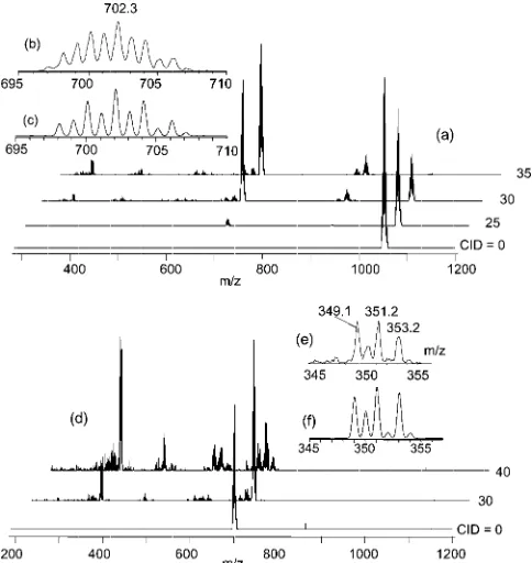
[image:6.595.60.279.575.707.2]Figure 9. MS (ESI) of a CH2Cl2solution of compound4. An ex-pansion of the molecular ion isotopic cluster is shown in part (b) and the theoretical simulation in part (c).
scaffold. To learn about the fragmentation pattern, the colli-sion-induced decay (CID) of this ion was investigated by MSn. As shown in Figure 10(a), the ion displays a major
decay pathway by expulsion of a [Cp*WO2] fragment with
transformation into [Cp*2W2O4]+(isotope cluster centered
at m/z = 702). However, the isotope pattern [Figure 10(b) with simulation in Figure 10(c)] reveals the presence of a second species one mass unit lighter. Therefore, elimination of a [Cp*WO2H] unit, with the presumed involvement of a
CH3 group and generation of a tetramethylfulvene ligand,
constitutes a competitive fragmentation pathway. Further trapping of these species and new CID in an MS3
experi-ment, see Figure 10(d), is less selective but the major decay pathway is once again expulsion of a [Cp*WO2] fragment
from [Cp*2W2O4]+, yielding [Cp*WO2]+ (isotope cluster
centered at m/z= 351). The latter ion is not produced in a major pathway by direct expulsion of [Cp*2W2O4] from the
trinuclear species.
DFT Calculations
Model compounds of the triangular W3 clusters,
ob-tained by replacing the Cp* ligands with the simpler Cp ring, i.e. [Cp3W3O4(OH)2]2+ (5) for compound 1 and
[Cp3W3O6]+ (6) for compounds 2–4, were subjected to
geometry optimization to find further supporting evidence for their stoichiometry (notably the number of OH vs. O groups) and to investigate their electronic structure. The calculations were run with no symmetry restrictions. Both calculations converged smoothly, giving final geometries in remarkably good agreement with the corresponding experi-mental ones for the Cp* derivatives (see Table 3). The only slight discrepancy consists of slightly longer bond lengths from the calculations relative to the experiment (W–O dis-tances by 0.02–0.05 Å; W–W disdis-tances by 0.06–0.10 Å), as is typically observed for DFT calculations.
The geometrical trends for the less symmetric dihy-droxido cluster 5 (see Scheme 1) are perfectly reproduced. Particularly notable is the difference between the W–W and W–W⬘ distances. The dioxido-bridged (W–W) bond is shorter than the two oxidohydroxido-bridged (W–W⬘) bonds by about 0.1 Å, like in the experimental structure. The lengths of different types of W–O bonds also have the same trend in the calculated and experimental structures (b⬎d⬎a⬎c, for the labeling, refer to Scheme 1), as do the different types of W–OH bonds (e⬎f). Finally, the two W– CG distances are longer than the W⬘–CG⬘distance.
Excellent agreement is also found between the optimized geometry for the [Cp3W3O6]+ cluster and the
experimen-tally observed structure for the Cp* analogue. In particular, the geometry is now close to the ideal D3hsymmetry, with
[image:7.595.303.545.98.501.2]three very similar W–W distances. Note that the average optimized W–W distance in6is essentially identical to the shorter W–W distance for the dioxido-bridged bond in 5. The same phenomenon is observed in the experimental structures. The three W–CG distances are also essentially equivalent.
Table 3. Comparison of the DFT optimized geometries (distances [Å], angles [°]) for the model [Cp3W3O4(OH)2]2+(5) and [Cp3W3O6]+ (6) complexes with the X-ray structures of the Cp* analogues.[a]
Parameter[b,c] [(C5R5)3W3O4(OH)2]2+ R = H (DFT) (5) R = Me (X-ray)[d]
W–W 2.723 2.6545(3)
W–W⬘ 2.839, 2.853 2.745(6)
W–(µ-O) (a) 1.920, 1.914 1.902(4) W–(µ-O) (b) 2.049, 2.054 2.005(4) W–(µ-O) (c) 1.894, 1.896 1.877(5) W⬘–(µ-O) (d) 2.012, 2.018 1.990(11) W–(µ-OH) (e) 2.148, 2.152 2.078(4) W⬘–(µ-OH) (f) 2.091, 2.091 2.042(7)
W–CG 2.096, 2.099 2.043(14)
W⬘–CG⬘ 2.060 2.003(2)
W⬘–W–W⬘ 57.17 57.829(7)
W–W⬘–W⬘ 61.66, 61.17 61.08(13) W–(µ-O)–W (a–b) 86.60, 86.60 85.54(14) W–(µ-O)–W⬘(c–d) 93.16, 93.60 90.40(15) W–(µ-OH)–W⬘(e–f) 84.09, 84.49 83.6(3) CG-W–(µ–O) (a) 112.28, 113.29 112.5(4) CG–W–(µ-O) (b) 113.86, 115.23 112.8(4) CG–W–(µ-O) (c) 110.54, 111.46 111.4(3) CG–W⬘–(µ-O) (d) 113.85, 115.97 114.1(12) CG–W–(µ-OH) (e) 115.33, 116.00 113.9(7) CG⬘–W⬘–(µ-OH) (f) 116.11, 116.37 114.7(12)
Parameter [(C5R5)3W3O6]+
R = H (DFT) (6) R = Me (X-ray)[e]
W–W 2.725, 2.732, 2.717 2.663(10) W-(µ-O) 1.946, 2.014, 1.932, 1.957(16)
2.038, 1.919, 2.047, 1.919, 2.054, 1.918, 2.047, 1.937, 2.037
W–CG 2.108, 2.109, 2.110 2.053(7) W–W–W 59.71, 60.01, 60.28 60.0(2) W-(µ-O)–W 86.99, 85.95, 86.63, 85.7(7)
87.03, 87.05, 86.24
CG–W-(µ-O) 114.55, 113.05, 113(1) 112.90, 114.23,
112.56, 114.42, 114.56, 112.92, 113.39, 114.48, 112.47, 113.93
[a] The experimental parameters (X-ray structures) that are chemi-cally and geometrichemi-cally equivalent are averaged; for the individual parameters, see Table 1. [b] Label W refers to the two geometrically equivalent W atoms; W⬘ refers to the unique atom sitting on the idealC2symmetry axis; CG and CG⬘are the centers of gravity of the rings bonded to atoms W and W⬘, respectively. [c] For the labels assigned to geometrically different W–O and W–OH bonds (a–f), refer to Scheme 1. [d] From the structure of compound 1. [e] Averaged values from the structures of compounds2–4.
Scheme 1. Labeling of geometrically different W–O bonds in the structure of [Cp3W3O4(OH)2]2+. The idealC
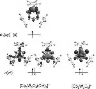
The electronic structure analysis is particularly interest-ing. The qualitative interaction diagram for the metal-based orbitals in this type of molecular scaffold was previously discussed[17,18] and is reproduced in Scheme 2 for
conve-nience. The relevant metal-based molecular orbitals calcu-lated for the two systems are shown in Figure 11, whereas more extensive data are available in the Supporting Infor-mation. For system 6 – a 2-electron W3V,V,VIcluster – the
two metal electrons are located in the a(z2) orbital, as
ex-pected. This orbital interaction insures the metal–metal at-tractive force. The additional electron in system 5 – a 3-electron W3V,V,Vcluster – is located in one of the two
orbit-als derived from thee(xy) set. Because the ideal symmetry is reduced from D3htoC2by the addition of the two
pro-tons, the e(xy) set splits (e1 ofa type +e2 of b type) and
the unpaired electron is located in the symmetrica compo-nent. Note that, whereas the antisymmetric (b) component must be nil on the unique tungsten atom (W⬘) (see Support-ing Information), there is no symmetry restriction for the composition of the a-type orbital. However, the composi-tion of this orbital is such that the contribucomposi-tion from the two symmetry-equivalent W atoms is negligible, whereas atom W⬘contributes greatly. Thus, the spin density is very much localized on the W⬘atom (0.911), whereas it is
[image:8.595.87.253.551.705.2]negli-Scheme 2. Qualitative metal–metal orbital interactions for a Cp3M3L6molecule of idealD3hsymmetry.
Figure 11. Filled metal-based molecular orbitals for systems5(left) and6(right). For the former, both spin orbitals resulting from the spin unrestricted calculation are shown.
gible on the two W atoms (0.028 and 0.036), in perfect agreement with the measured relative intensity of the satel-lites in the EPR spectrum (vide supra). In this respect, it should be noted that the related molybdenum complex [Cp*3Mo3O2(OH)4]2+ – a 5-electron Mo3IV,IV,V cluster –
was shown to have the same ordering for the metal-based molecular orbitals.[13] Therefore, the unpaired electron for
that complex is located in the antisymmetric borbital and couples to only two Mo nuclei, again consistent with the experimental evidence from EPR spectroscopy.
Comparison with Molybdenum and Redox Considerations
The reduction process reported here parallels that re-ported recently for the reduction of Cp*2Mo2O5.[13]
How-ever, the Mo system leads to a deeper reduction with gener-ation of a 5-electron cluster, the trinuclear Mo3IV,IV,V
[Cp*3Mo3O2(OH)4]2+complex. This product has the same
molecular architecture as the two trinuclear W clusters de-scribed here, differing only by the number of protons and electrons. This difference agrees with the general trend of greater stability for higher oxidation states upon descending the same group of transition metals. It may be predicted that, by treatment with suitable oxidizing agents (or at a suitable electrochemical potential in an electrochemical ex-periment) the 5-electron Mo cluster could produce a 3-elec-tron or a 2-elec3-elec-tron cluster with the same structure found in the present study for the W system. Conversely, suitable reductive treatment of the W system could afford a 5-elec-tron system such as the previously reported Mo cluster. Other electronic configurations such as 4-electron MIV,V,V
could also exist. Given that proton gains/losses are associ-ated with these transformations (at least between the two types of W clusters reported in the present contribution), it may also be expected that the redox potential is pH depend-ent in a protic environmdepend-ent. A detailed analysis of the elec-trochemical behavior of these clusters is beyond the scope of the present investigation and may be carried out at a later time.
Previous studies of the Cp*2Mo2O5system by a coupled
electrochemical–electrospray mass spectrometry methodol-ogy, by using a flow-through electrochemical cell,[19,20]
re-vealed a very rich redox behavior, including the formation of trinuclear clusters that are related to the isolated cluster. We have now attempted to carry out a similar investigation for Cp*2W2O5, but no sufficient quantities of soluble
prod-ucts were revealed by the mass spectrometric analysis. At the potential at which the Mo system started to yield re-ductive processes, the W system showed no activity, whereas the rapid formation of a film on the surface of the electrode, which blocked any further electrochemical transformation, was observed at lower potentials. Therefore, it seems that the Cp*2W2O5is more difficult to reduce than the Mo
Conclusions
Like its Mo analogue,[13]the compound [Cp*
2W2O5] can
be reduced in an aqueous medium to a triangular metal– metal bonded cluster. However, only 2- and 3-electron clus-ters are obtained for W, whereas the Mo system undergoes a deeper reduction to yield a 5-electron cluster. Like for the Mo cluster, the oxido/hydroxido asymmetry in the bridge system splits the degeneracy of the “e”-type frontier orbit-als, as indicated by the EPR spectrum of the 3-electron clus-ter and confirmed by the DFT calculations. The variability of the cluster electrons for this system will be further ex-plored for applications in organometallic chemistry and ca-talysis.
Experimental Section
General Procedures:All preparations and manipulations were car-ried out with Schlenk techniques under an oxygen-free argon atmo-sphere. All glassware was oven-dried at 120 °C. Solvents were dried by standard procedures and distilled under nitrogen prior to use. 1H NMR spectra were recorded with a Bruker AM 250. Chemical
shifts are expressed in ppm downfield from Me4Si. Coupling con-stants are given in Hertz. Mass spectra were recorded with a Finni-gan (San Francisco, USA) LCQ quadrupole ion-trap mass spec-trometer equipped with an electrospray-ionization (ESI) interface. The ESI was operated in the positive ion mode with a spray voltage of 4.5 kV. The capillary voltage was 20 V and the source tempera-ture was 100 °C. Mass spectra were obtained by scanning the mass analyzer fromm/z100 to 2000 with 5 total microscans. Maximum inject time was 400 ms. The analyzer was operated at a background pressure of 2⫻10–5Torr. In all experiments, helium was introduced at an estimated pressure of 1 mTorr to improve the ion-trapping efficiency and as collision gas for the collision-induced dissociation events. The compounds were isolated in the ion trap with an isola-tion width of 12m/zunits and activated by using increased collision energy to obtain collision-energy-dissociation profiles. EPR mea-surements were carried out at the X-band microwave frequency with a Bruker ESP300 spectrometer. The spectrometer frequency was calibrated with diphenylpicrylhydrazyl (DPPH, g = 2.0037). The starting compound, [Cp*2W2O5], was prepared as described in the literature.[14]
Synthesis and Crystallization of [Cp*3W3O4(OH)2](CF3SO3)2· 2H2O (1):Metallic zinc (20 mesh, 0.415 g, 6.34 mmol) was added to a solution of [Cp*2W2O5] (50 mg, 0.070 mmol) in MeOH/H2O (1:1, 6 mL). The mixture was acidified with CF3SO3H (10 drops) and stirred under an atmosphere of argon at r.t. for 3 d, during which time it changed from a yellow solution to a green suspension. The mixture was filtered, and the solid was extracted with THF (0.5 mL). The addition of Et2O (1.5 mL) to the filtrate yielded the product as a green precipitate (0.047 g, 72 %). A single crystal for X-ray analysis was obtained by diffusion of an Et2O layer into a THF solution at r.t. EPR (X-band, CH2Cl2, 120 K): g⬜= 1.89; g储
= 1.72 (aW储= 114 G).
Synthesis and Crystallization of [Cp*3W3O6](CF3SO3) (2):A sam-ple of compound1(61 mg, 0.044 mmol) was dissolved in CH2Cl2 and stirred for 1 h at r.t. in open air. The color of the green solution turned orange. The solvent was evaporated to dryness to afford an orange power, which was recrystallized from CH2Cl2/pentane to give red crystals (25 mg, 47 % yield).1H NMR (250 MHz, CDCl
3):
δ= 2.28 ppm.
Reduction of [Cp*2W2O5] by Zinc in the Presence of CF3COOH – Formation of Compounds 3 and 4:Metallic zinc (20 mesh, 0.415 g, 6.34 mmol) was added to a solution of [Cp*2W2O5] (50 mg, 0.070 mmol) in MeOH/H2O (1:1, 6 mL). The mixture was acidified with CF3COOH (10 drops) and stirred under an atmosphere of argon at r.t. for 5 d during which time it changed from a yellow solution to a green suspension. The mixture was filtered, and the solid was dried under vacuum (31 mg). Attempts to produce single crystals from this material failed as only powdery precipitates were obtained. Red single crystals of compound 3were eventually ob-tained from CH2Cl2/hexane after exposure of the solution to air. 1H NMR (250 MHz, CD2Cl2):δ= 2.26 ppm.
In a parallel identical procedure, from one of the numerous attempts to crystallize the green solid, the yellow mother solution of the green powdery precipitate was recovered, dried, and the yel-low-orange residue was set again for crystallization from THF/hex-ane to yield single crystals of compound 4.1H NMR (250 MHz, CDCl3):δ= 2.28 ppm. MS (ESI, CH2Cl2):m/z= 1053 (envelope) [M]+(see Results and Discussion section).
X-Ray Diffraction Studies:A single crystal of each compound was mounted under inert perfluoropolyether at the tip of a glass fiber and cooled in the cryostream of an Oxford-Diffraction XCALI-BUR CCD diffractometer. Data were collected by using the mono-chromatic MoKα radiation (λ = 0.71073). The structures were
solved by direct methods (SIR97)[21]and refined by least-squares procedures onF2by using SHELXL-97.[22]All H atoms attached to carbon were introduced at idealized positions and treated with the riding model. All H atoms attached to oxygen were either lo-cated on difference Fourier syntheses or their coordinates were cal-culated on the basis of hydrogen bonding geometry and energy considerations.[23]They were then treated as riding on their parent O atoms. In compound Cp*3W3O6(CF3SO3), the cation, Cp*3W3O6, is arranged around a twofold axis resulting in one of the Cp* to be disordered over two positions. The CF3SO3anion is also disordered and arranged around another twofold axis. For both the disordered parts, the atomic positions and anisotropic thermal parameters were restrained to reasonable values using the tools available within SHELXL-97.[22]The drawing of the mole-cules was realized with the help of ORTEP3.[24]Crystal data and refinement parameters are shown in Table 4. CCDC-643997 to -644000 contain the supplementary crystallographic data for this paper. These data can be obtained free of charge from the Cam-bridge Crystallographic Data Centre via www.ccdc.cam.ac.uk/ data_request/cif.
Computational Details:The DFT calculations were carried out on model systems where the Cp* ligands were replaced on the simpler Cp rings. The starting geometries were based on the crystallograph-ically determined structures and no symmetry restrictions were im-posed. The geometries were fully optimized and the resulting min-ima of the potential energy surface (PES) were verified by the posi-tive value of all second derivaposi-tives of the energy. The calculations were performed by using the B3LYP three-parameter hybrid den-sity functional method of Becke[25]and the standard LANL2DZ basis set, which included the Hay and Wadt effective core potentials (ECP) for the tungsten atoms,[26]as implemented in the Gaussian03 suite of programs.[27]The calculation on the open-shell system5 was carried out by using the spin-unrestricted formulation. The value of⬍S2⬎resulting from the calculation is 0.7575, indicating negligible spin contamination.
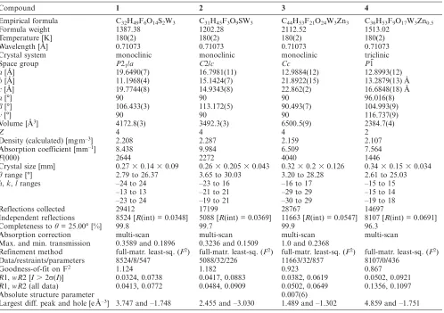
Table 4. Selected crystallographic and refinement parameters for all compounds.
Compound 1 2 3 4
Empirical formula C32H49F6O14S2W3 C31H45F3O9SW3 C44H53F21O24W3Zn3 C36H53F9O17W3Zn0.5
Formula weight 1387.38 1202.28 2112.52 1513.02
Temperature [K] 180(2) 180(2) 180(2) 180(2)
Wavelength [Å] 0.71073 0.71073 0.71073 0.71073
Crystal system monoclinic monoclinic monoclinic triclinic
Space group P21/a C2/c Cc P1¯
a[Å] 19.6490(7) 16.7981(11) 12.9884(12) 12.8993(12)
b[Å] 11.1968(4) 15.1424(7) 21.8922(15) 13.2879(13) Å
c[Å] 19.7744(8) 14.9343(8) 22.862(2) 16.6848(18) Å
α[°] 90 90 90 96.016(8)
β[°] 106.433(3) 113.172(5) 90.493(7) 104.993(9)
γ[°] 90 90 90 116.737(9)
Volume [Å3] 4172.8(3) 3492.3(3) 6500.5(9) 2384.7(4)
Z 4 4 4 2
Density (calculated) [mg m–3] 2.208 2.287 2.159 2.107
Absorption coefficient [mm–1] 8.438 9.984 6.509 7.564
F(000) 2644 2272 4040 1446
Crystal size [mm] 0.27⫻0.14⫻0.09 0.26⫻0.205⫻0.043 0.32⫻0.2⫻0.126 0.34⫻0.15⫻0.034
θrange [°] 2.79 to 26.37 3.65 to 30.03 3.20 to 28.28 2.61 to 25.03
h,k,lranges –24 to 24 –23 to 16 –16 to 17 –15 to 15
–13 to 13 –21 to 21 –29 to 29 –15 to 14
–23 to 24 –19 to 21 –30 to 29 –19 to 18
Reflections collected 29412 17199 28767 14697
Independent reflections 8524 [R(int) = 0.0348] 5088 [R(int) = 0.0369] 11663 [R(int) = 0.0547] 8107 [R(int) = 0.0691]
Completeness toθ= 25.00° [%] 99.8 99.7 99.9 96.3
Absorption correction multi-scan multi-scan multi-scan multi-scan
Max. and min. transmission 0.3589 and 0.1896 0.3236 and 0.1509 1.0 and 0.2368
Refinement method full-matr. least-sq. (F2) full-matr. least-sq. (F2) full-matr. least-sq. (F2) full-matr. least-sq. (F2) Data/restraints/parameters 8524/8/547 5088/32/226 11663/32/857 8107/0/436
Goodness-of-fit on F2 1.124 1.182 0.923 0.867
R1,wR2 [I⬎2σ(I)] 0.0324, 0.0738 0.0417, 0.0883 0.0382, 0.0619 0.0502, 0.0921 R1,wR2 (all data) 0.0413, 0.0772 0.0484, 0.0909 0.0502, 0.0649 0.1356, 0.1097
Absolute structure parameter 0.007(6)
Largest diff. peak and hole [e Å–3] 3.747 and –1.748 2.455 and –3.030 1.489 and –1.302 4.859 and –1.751
and representative molecular orbitals for systems5and6(5 pages), H-bonding parameters of1,3, and4.
Acknowledgments
We are grateful to the European Commission for funding of this work through the AQUACHEM Research Training Network (Pro-ject no. MRTN-CT-2003-503864). We also thank CINES and CICT (project CALMIP) for a grant of free computer time. Supple-mental travel support was provided by a Bosphorus bilateral Program of Integrated Actions, co-sponsored by the French Minis-try of Foreign Affairs in France and by TUBITAK in Turkey [TBAG-U/142(105T256)].
[1] T. Chan, L. Li, Y. Yang, W. Lu, ACS Symp. Ser.2002,819, 166–177.
[2] F. Joó,Acc. Chem. Res.2002,35, 738–745. [3] I. T. Horvath,Acc. Chem. Res.2002,35, 685. [4] D. Sinou,Top. Curr. Chem.1999,206, 41–59. [5] D. Sinou,Adv. Synth. Catal.2002,344, 221–237.
[6] G. E. Jaouen, J. Organomet. Chem. 1999, 589 (special issue: bioorganometallic chemistry).
[7] P. Kalck, F. Monteil,Adv. Organomet. Chem.1992, 34, 219– 284.
[8] B. E. Hanson,Coord. Chem. Rev.1999,186, 795–807. [9] C. Muller, D. Vos, P. Jutzi, J. Organomet. Chem. 2000, 600,
127–143.
[10] A. Bino, F. A. Cotton, Z. Dori,J. Am. Chem. Soc.1981,103, 243–244.
[11] A. Bino, M. Ardon, E. Shirman,Science2005,308, 234–235. [12] R. Poli,Chem. Eur. J.2004,10, 332–341.
[13] F. Demirhan, B. Çagatay, D. Demir, M. Baya, J.-C. Daran, R. Poli,Eur. J. Inorg. Chem.2006, 757–764.
[14] C. Dinoi, G. Taban, P. Sözen, F. Demirhan, J.-C. Daran, R. Poli,J. Organomet. Chem.2007,692, 3743–3749.
[15] A. L. Grzesiak, F. J. Uribe, N. W. Ockwig, O. M. Yaghi, A. J. Matzger,Angew. Chem. Int. Ed.2006,45, 2553–2556. [16] J. S. Seo, D. Whang, H. Lee, S. I. Jun, J. Oh, Y. J. Jeon, K. Kim,
Nature2000,404, 982–986.
[17] P. Kubácek, R. Hoffmann, Z. Havlas,Organometallics1982,1, 180–188.
[18] T. A. Albright, J. K. Burdett, M. H. Whangbo,Orbital Interac-tions in Chemistry, John Wiley & Sons, New York,1985. [19] J. Gun, A. Modestov, O. Lev, D. Saurenz, M. A. Vorotyntsev,
R. Poli,Eur. J. Inorg. Chem.2003, 482–492.
[20] J. Gun, A. Modestov, O. Lev, R. Poli,Eur. J. Inorg. Chem.2003, 2264–2272.
[21] A. Altomare, M. Burla, M. Camalli, G. Cascarano, C. Giacov-azzo, A. Guagliardi, A. Moliterni, G. Polidori, R. Spagna,J. Appl. Crystallogr.1999,32, 115–119.
[22] G. M. Sheldrick, SHELXL97: Program for Crystal Structure Refinement, University of Göttingen, Göttingen, Germany, 1997.
[27] M. J. Frisch, G. W. Trucks, H. B. Schlegel, G. E. Scuseria, M. A. Robb, J. R. Cheeseman, J. Montgomery, J. A., T. Vreven, K. N. Kudin, J. C. Burant, J. M. Millam, S. S. Iyengar, J. Tom-asi, V. Barone, B. Mennucci, M. Cossi, G. Scalmani, N. Rega, G. A. Petersson, H. Nakatsuji, M. Hada, M. Ehara, K. Toyota, R. Fukuda, J. Hasegawa, M. Ishida, T. Nakajima, Y. Honda, O. Kitao, H. Nakai, M. Klene, X. Li, J. E. Knox, H. P. Hratch-ian, J. B. Cross, C. Adamo, J. Jaramillo, R. Gomperts, R. E. Stratmann, O. Yazyev, A. J. Austin, R. Cammi, C. Pomelli, J. W. Ochterski, P. Y. Ayala, K. Morokuma, G. A. Voth, P. Sal-vador, J. J. Dannenberg, V. G. Zakrzewski, S. Dapprich, A. D.
Daniels, M. C. Strain, O. Farkas, D. K. Malick, A. D. Rabuck, K. Raghavachari, J. B. Foresman, J. V. Ortiz, Q. Cui, A. G. Ba-boul, S. Clifford, J. Cioslowski, B. B. Stefanov, G. Liu, A. Lia-shenko, P. Piskorz, I. Komaromi, R. L. Martin, D. J. Fox, T. Keith, M. A. Al-Laham, C. Y. Peng, A. Nanayakkara, M. Challacombe, P. M. W. Gill, B. Johnson, W. Chen, M. W. Wong, C. Gonzalez, J. A. Pople, Gaussian 03, Revision B.04, Gaussian, Inc., Pittsburgh PA,2003.
![Table 1. Selected bond lengths and angles for the trinuclear clustersin all compounds.[a]](https://thumb-us.123doks.com/thumbv2/123dok_us/8138813.244223/2.595.227.535.98.693/table-selected-bond-lengths-angles-trinuclear-clustersin-compounds.webp)
 (2)](https://thumb-us.123doks.com/thumbv2/123dok_us/8138813.244223/3.595.147.458.553.717/figure-view-relative-orientation-cation-anion-structure-cp.webp)
![Figure 4.Acule in compoundof the disordered CFviewofthe[Zn3(µ3-O)(µ-CF3COO)4(CF3COO)-(CF3COOH)(H2O)3]– anion and the interstitial CF3COOH mole- 3](https://thumb-us.123doks.com/thumbv2/123dok_us/8138813.244223/4.595.306.546.396.568/figure-acule-compoundof-disordered-cfviewofthe-cooh-interstitial-cooh.webp)
![Figure 5. A view of the arrangement between the [Zn3CFCF(µ3-O)(µ-3COO)4(CF3COO)(CF3COOH)(H2O)3]– anion, the interstitial3COOH molecule, and the [Cp*3W3O6]+ cation in compound 3.The cation Cp* ligands and the aqua H atoms have been removedand only the coordination spheres of the Zn atoms are shown forclarity [symmetry code: (i) 1/2 + x, 3/2 – y, z – 1/2].](https://thumb-us.123doks.com/thumbv2/123dok_us/8138813.244223/5.595.46.287.331.649/arrangement-interstitial-molecule-compound-removedand-coordination-forclarity-symmetry.webp)
![Table 3. Comparison of the DFT optimized geometries (distances[Å], angles [°]) for the model [Cp3W3O4(OH)2]2+ (5) and [Cp3W3O6]+(6) complexes with the X-ray structures of the Cp* analogues.[a]](https://thumb-us.123doks.com/thumbv2/123dok_us/8138813.244223/7.595.303.545.98.501/table-comparison-optimized-geometries-distances-complexes-structures-analogues.webp)