C A S E R E P O R T
Open Access
Three different situations and approaches in the
management for anomalous origin of the right
coronary artery from the left coronary sinus: case
report
Woon Heo
1, Ho-Ki Min
1*, Do Kyun Kang
1, Hee Jae Jun
1, Youn-Ho Hwang
1and Hyung Chae Lee
2Abstract
Anomalous origin of the right coronary artery from the left coronary sinus is rare but potentially dangerous if any ischemic signs are present. Multiple therapeutic options were advocated so far. We experienced three different situations and surgical approaches to these anomalies, and reviewed retrospectively. For the first case, we made a neo-ostium on the right sinus of Valsalva and anastomosed with the right coronary artery after arteriotomy. For the second and third cases, we applied coronary artery bypasses emergently: patient 2 the gastroepiploic artery during off-pump coronary artery bypass and patient 3 the left internal thoracic artery during surgery for acute aortic dissection. For the better outcomes, it is important to understand anatomic and hemodynamic characteristics of each patient and select the surgical options considering each characteristic.
Keywords:Coronary artery anomaly,Anomaly
Background
Anomalous origin of the right coronary artery from the left sinus of Valsalva (ARCA) is a rare congenital ano-maly but a common cause of sudden death in the young [1]. It is commonly identified incidentally by angiog-raphy, during cardiac operation, or at autopsy [2]. The fact that these patients are usually asymptomatic but could initially present with sudden death can make their management challenging. We report 3 successful surgi-cal outcomes in patients with ARCA in different situa-tions and approaches.
Case presentation
Three Koreans with ARCA who had different characte-ristics were performed operations in our institute, and the medical records were reviewed retrospectively. The characteristics and operative procedures were descri-bed in Table 1. Two cases (patient 1 and patient 2) were
diagnosed preoperatively and one case (patient 3) was identified incidentally during surgery.
Patient 1>
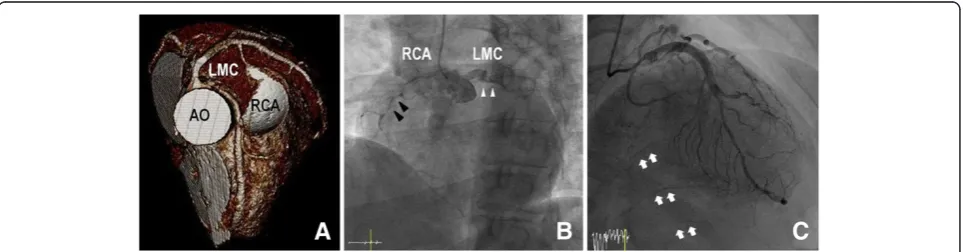
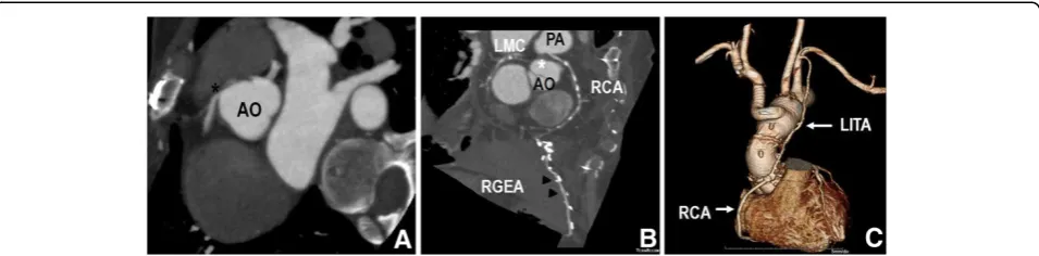
A 39-year-old woman visited out-patient department with exertional chest discomfort for 2 months. Pre-operative work-ups including cardiac enzymes, electro-cardiogram, nuclear perfusion scan, and echocardiogram were carried out and the results were normal without any ischemic signs. Computed tomography (CT) revealed ARCA (Figure 1A). We thought that panic symptoms on physical exertion were caused due to ischemia and de-cided for a surgical correction. Under cardiopulmonary bypass and cardioplegic arrest, a neo-ostium was created on the right coronary sinus at the nearest site from the right coronary artery (RCA). After that, a right coronary arteriotomy was made on its opposite site. Anastomosis between them was performed (Figure 2A). The postopera-tive course was uneventful and she remains asymptomatic for 22 months.
* Correspondence:minhoki@naver.com
1Department of Thoracic and Cardiovascular Surgery, Haeundae Paik
Hospital, Inje University College of Medicine, 875 (Jwadong) Haeundae-ro, Haeundaegu, Busan 612-030, Korea
Full list of author information is available at the end of the article
Patient 2>
A 61-year-old woman was emergently admitted for chest pain and diagnosed with acute myocardial infarction. Cardiac catheterization revealed that patient had triple vessel disease concomitant with ARCA (Figure 1B), and chest CT confirmed ARCA. An emergent off-pump cor-onary artery bypass was performed. The internal thor-acic arteries (ITAs) and the right gastroepiploic artery (RGEA) were prepared. Then, the resected right ITA was anastomosed toin situ the left ITA in a “Y” config-uration. The distal anastomoses were constructed in the following sequence: the distal left ITA to the left anterior descending artery (LAD) and the right ITA to the two obtuse marginal branches sequentially. Subsequently, the distal RCA was revascularized with the in situ RGEA (Figure 2B). The postoperative course was uneventful, and she remains asymptomatic for 14 months.
Patient 3>
A 44-year-old man was emergently admitted for intract-able chest pain. He had suffered from intermittent chest pain for several months. Echocardiogram revealed hypo-kinesia in the territory of the RCA. Subsequent cardiac catheterization revealed that there were no significant le-sions on the left coronary systems, but the RCA ostium was invisible. Also it showed collateral circulation to the
distal RCA via septal branches of LAD (Figure 1C). Add-itional aortography showed a contrast filling to false lumen via an intimal tear. Transesophageal echocardiog-raphy and chest CT confirmed acute type A aortic dis-section, and he underwent emergent surgery. During the surgery, ARCA with slit-like ostium was incidentally de-tected. The ascending aorta and part of the aortic arch were replaced, and the RCA was bypassed by the left ITA with proximal ligation (Figure 2C). The postopera-tive course was uneventful, and he remains asymptom-atic for 6 months.
Although difficult to estimate the exact prevalence, pub-lished data suggest that the incidence of anomalous origin of the coronary artery from opposite sinus (AOCA) may be around 0.1 ~ 0.3% up to 1.07%. ARCA is estimated to be six to ten times more common than anomalous origin of the left coronary artery (ALCA) [1,3].
Patients are usually asymptomatic. However, it could cause exertional syncope, angina, palpitations, and even sudden cardiac death (SCD) [4]. Thus, AOCA has con-cluded that it is a potentially dangerous anomaly and should be considered to apply aggressive treatment if pa-tients have any ischemic signs [3,5]. It has been described that SCD in ALCA happens more common than ARCA [3]. However, we believe that physicians can encounter major adverse cardiac events happening within ARCA
Figure 1Pre-operative images. (A)Three dimensional volume rendering of contrast-enhanced CT scan shows the anomalous RCA shared the same sinus with the left coronary system, with its attenuated proximal portion passing between the aorta and pulmonary trunk in patient 1. The left coronary system is patent.(B)Coronary angiogram shows anomalous origin of the right coronary artery (black arrowheads) from left sinus of Valsalva, next to the left (white arrowheads) coronary arteries in patient 2. Also it is noted that severe stenotic lesions are present on the RCA.
patients more frequently than ALCA patients since there are more absolute number of cases of ARCA than ALCA.
The diagnosing ARCA is often made incidentally, be-cause many tests including physical exam, ECG and exer-cise stress testing are generally unremarkable for diagnosis of ARCA [4]. In a review of the literature by Basso and colleagues, they concluded that many tests including 12-lead ECG, maximal effort stress ECG, and myocardial per-fusion scintigraphy would be unlikely to provide clinical evidence of myocardial ischemia and would be unreliable for clinical recognition of these anomalies. They also men-tioned that premonitory symptoms including syncope or chest pain occurred not uncommonly in about one third shortly before sudden death [4]. In patient 1, we thought that panic symptoms on physical exertion were caused due to ischemia and decided for a surgical correction des-pite of negative functional tests. Fortunately, recently multi-detector CT scanners provide excellent spatial re-solution allowing visualization of the coronary anatomy, all coronary artery anomalies become easy to diagnose [6]. We believe that much more AOCA cases could be detected faster and easier than ever after using a multi-detector CT scanner as a diagnosing tool. Also, mag-netic resonance angiography can be used to identify anomalous vessels [3]. However, when emergency surgery is required, it is difficult to perform multi-detector CT scanner or magnetic resonance angiography in all pa-tients because of unstable hemodynamics and the time re-quired for the scan [7]. In our cases, multi-detector CT scanner or magnetic resonance angiography could not be performed because of an emergency situation and un-stable hemodynamics in patient 2 and because ARCA was incidentally diagnosed during surgery in patient 3.
Ultimately, it is important to understand the patho-physiology of AOCA treatment to manage this condition
effectively. The proposed mechanisms include slit-like ostium, acute angulation, impinged or spastic intramural proximal portion, and flattening of the interarterial seg-ment from compression between the great vessels [2,8,9].
Multiple therapeutic options have been suggested. Ac-cording to medical journals, some authors described some efficacy of medical therapy with nitrates, calcium or β -blockade, or anti-arrythmic drugs [2,10]. However, for AOCA, original problem of this condition cannot be possibly corrected by medication. Therefore, we are sug-gesting that this issue deserves consideration. Percuta-neous coronary angioplasty has also been advocated by some physicians and reported feasible short-term results [11,12]. However, in our opinions, these reports have failed to establish a widely accepted consensus due to its rareness and no long-term results. Furthermore, this can-not effectively treat ostial issues that might be present, and leaves a long stent length that could be prone to late occlusion even with drug-eluting stent [13]. If it would show superior or equal advantages to surgical correction from long-term results, it might be a feasible strategy.
Multiple surgical options were advocated including coronary artery bypass (CABG), coronary reimplantation, pulmonary artery translocation, neo-ostium formation, and unroofing of the intramural segment [14]. Surgical strategy of choice for ARCA is quite controversial. Coron-ary reimplantation is one of the most physiologically bene-ficial procedures, but is technically difficult, and stenosis may occur at the anastomotic site. CABG is technically feasible, but the arterial conduit has a competitive flow problem if no stenotic lesions are present on the natural coronary artery. Also, a vein conduit may be problematic if the patient is young because of long-term patency. Some authors reported that favorable results were achieved with unroofing procedures [15]. However, it may cause valve
may be expected to have a long-term patency compared to the vein graft because of high grade stenosis. To the best of our knowledge, this report is the first to describe in situ RGEA to RCA bypass in a case of ARCA. How-ever, a longer follow-up period should be required to evaluate a long-term patency.
In case of patient 3, ARCA was incidentally diagnosed during surgery. If not recognized right after preoperative evaluations, it could result in serious complications peri-operatively. Firstly, ARCA could be injured iatrogenically due to its abnormal course and position. So when inci-dentally detected in the operative field, careful dissection and suture are essential to avoid iatrogenic injury. Sec-ondly, it could be a matter how to achieve appropriate myocardial protection. When a patient has a slit-like or flap-like opening, cardioplegia could not be delivered ef-fectively by direct cannulation of the coronary ostium, which could result in inadequate myocardial protec-tion. In this situation, retrograde cardioplegia delivery would be an alternative strategy. In our case, we could not deliver direct antegrade cardioplegia through the RCA ostium because of its slit-like opening and suspi-ciously proximal stenosis of the RCA on preoperative angiogram. We carefully dissected the aortic root and the proximal RCA. After the RCA was ligated and di-vided at the level of its origin, a coronary probe was passed into the RCA to confirm patency. Then ante-grade cardioplegia was infused into the RCA using a small cannula.
Conclusions
ARCA is a potentially dangerous anomaly and should treat aggressively if any ischemic signs are present and surgery is a treatment of choice for ARCA. For the bet-ter outcomes, it is important to understand anatomic and hemodynamic characteristics of each patient and ap-propriately select the surgical options considering each characteristic.
Consent
Written informed consent was obtained from the patients for publication of this case report and all accompanying images. A copy of the written consent is available for re-view by the Editor-in-Chief of this journal.
Authors’contributions
WH: participated in the management and wrote the manuscript. HM: performed surgery on the patient and revised the manuscript. DK: revised the manuscript. HJ: revised the manuscript. YH: revised the manuscript. HS: revised the manuscript. SK: revised the manuscript. HL: revised the manuscript. IR: revised the manuscript. All authors read and approved the final manuscript.
Authors’information
HM - Is a surgeon of the Thoracic and Cardiovascular Surgery department at the Haeundae Paik Hospital where the patient underwent operation. He is also an assistant professor at the Inje University College of Medicine in Busan, Korea.
Acknowledgement
We greatly appreciate the assistance of the staff of the Department of Thoracic and Cardiovascular Surgery, Haeundae Paik Hospital, Inje University College of Medicine.
Author details
1Department of Thoracic and Cardiovascular Surgery, Haeundae Paik
Hospital, Inje University College of Medicine, 875 (Jwadong) Haeundae-ro, Haeundaegu, Busan 612-030, Korea.2Department of Thoracic and
Cardiovascular Surgery, National Masan Tuberculosis Hospital, Busan, Korea.
Received: 27 May 2013 Accepted: 25 November 2013 Published: 23 January 2014
References
1. Angelini P:Coronary artery anomalies–current clinical issues: definitions, classification, incidence, clinical relevance, and treatment guidelines. Tex Heart Inst J2002,29(4):271–278.
2. Yuan SM, Tager S, Raanani E:Anomalous origin of the right coronary artery from the left coronary sinus.Chang Gung Med J2009,32(4):455–458. 3. Penalver JM, Mosca RS, Weitz D, Phoon CK:Anomalous aortic origin of
coronary arteries from the opposite sinus: a critical appraisal of risk.BMC Cardiovasc Disord2012. www.biomedcentral.com/1471-2261/12/83. 4. Basso C, Maron BJ, Corrado D, Thiene G:Clinical profile of congenital
coronary artery anomalies with origin from the wrong aortic sinus leading to sudden death in young competitive athletes.J Am Coll Cardiol 2000,35:1493–1501.
5. Taylor AJ, Rogan KM, Virmani R:Sudden cardiac death associated with isolated congenital coronary artery anomalies.J Am Coll Cardiol1992, 20(3):640–647.
6. Ten Kate GJ, Weustink AC, Feyter PJ:Coronary artery anomalies detected by msct-coronary angiography in the adult.Neth Heart J 2008,16:369–375.
7. Morimoto H, Mukai S, Obata S, Hiraoka T:Incidental single coronary artery in an octogenarian with acute type A aortic dissection.Interact Cardiovasc Thorac Surg2012,15(2):307–330.
8. Yanagawa B, Alghamdi AA, Chen RB, Amankwaa A, Verma S:Coronary artery bypass graft for anomalous right coronary artery.J Card Surg2011, 26(1):44–46.
9. Davies JE, Burkhart HM, Dearani JA, Suri RM, Phillips SD, Warnes CA, Sundt TM 3rd, Schaff HV:Surgical management of anomalous aortic origin of a coronary artery.Ann Thorac Surg2009,88(3):844–847.
11. Rudan D, Todorovic N, Starcevic B, Raguz M, Bergovec M:Percutaneous coronary intervention of an anomalous right coronary artery originating from the left coronary artery.Wien Klin Wochenschr2010, 122(15–16):508–510.
12. Hong LF, Luo SH, Li JJ:Percutaneous coronary intervention with anomalous origin of right coronary artery: case reports and literature review.J Geriatr Cardiol2013,10(2):205–209.
13. Fedoruk LM, Kern JA, Peeler BB, Kron IL:Anomalous origin of the right coronary artery: right internal thoracic artery to right coronary artery bypass is not the answer.J Thorac Cardiovasc Surg2007,133:456–460. 14. Ho JS, Strickman NE:Anomalous origin of the right coronary artery from
the left coronary sinus: case report and literature review.Tex Heart Inst J 2002,29:37–39.
15. Romp RL, Herlong JR, Landolfo CK, Sanders SP, Miller CE, Ungerleider RM, Jaggers J:Outcome of unroofing procedure for repair of anomalous aortic origin of left or right coronary artery.Ann Thorac Surg2003, 76(2):589–596.
doi:10.1186/1749-8090-9-21
Cite this article as:Heoet al.:Three different situations and approaches in the management for anomalous origin of the right coronary artery from the left coronary sinus: case report.Journal of Cardiothoracic Surgery
20149:21.
Submit your next manuscript to BioMed Central and take full advantage of:
• Convenient online submission
• Thorough peer review
• No space constraints or color figure charges
• Immediate publication on acceptance
• Inclusion in PubMed, CAS, Scopus and Google Scholar
• Research which is freely available for redistribution