Is there any change in the pattern of bacterial translocation
with increased time of the obstruction?
Sajjad Noorshafiee
1, Ghodratollah Maddah
2, Kiarash Ghazvini
3,
Saeed Niazmand
4, Monavvar Afzal Aghaee
5, Samaneh Sajadi
6,
Mohsen Abdollahi
7, Abbas Abdollahi
81
General Surgeon, Endoscopic and Minimally Invasive Surgery Research Center, Mashhad University of Medical Sciences, Mashhad, Iran;
2
Associate Professor of Surgery, Endoscopic and Minimally Invasive Surgery Research Center, Mashhad University of Medical Sciences, Mashhad, Iran;
3
Assistant Professor, Department of Microbiology and Virology, Faculty of Medicine, Mashhad University of Medical Sciences, Mashhad, Iran;
4
Assistant Professor, Cardiovascular Research Center, Department of Physiology, Faculty of Medicine, Mashhad University of Medical Sciences, Mashhad, Iran;
5
Assistant Professor of Community Medicine, Surgical Oncology Research Center, Mashhad University of Medical Sciences, Mashhad, Iran;
6
Internist, Surgical Oncology Research Center, Mashhad University of Medical Sciences, Mashhad, Iran;
7
General Practitioner, Surgical Oncology Research Center, Mashhad University of Medical Sciences, Mashhad, Iran;
8
Associate Professor of Surgery, Surgical Oncology Research Center, Mashhad University of Medical Sciences, Mashhad, Iran.
Received: November 9, 2014 Revised: December 1, 2014 Accepted:January 2, 2015
Abstract
Introduction: Bacterial translocation is defined as the passage of bacteria from intestinal tract to the extraintestinal organs such as the peritoneum and blood circulation. The aim of this study is to examine bacterial translocation (regarding type of bacteria and effect of time of obstruction on bacterial translocation) from intestinal lumen to the peritoneum and viscera in acute, simple mechanical, small bowel obstruction in rats.
Methods: In this cohort study, thirty female Wistar rats were divided into three groups with two subgroups, each sub-group containing 5 rats. The 1st sub-group consisted of two sham-operated and non-operated control subsub-groups. The 2nd group was the IO-24 group, and the 3rd group was the IO-48 group in which the interval between producing intestinal obstruction and the second laparotomy was 24 h and 48 h respectively. Each subgroup was divided into two subgroups of partial and complete obstruction. The data was analyzed using Fisher’s exact test and K2 test in SPSS.
Results: The most common types of bacteria were E. coli in aerobic culturing and bacteroid in anaerobic culturing. However, as the time of obstruction increased, the pattern of bacterial translocation changed to anaerobic bacteria.
Conclusions:Our study showed that with increased time of obstruction, pattern of bacterial translocation changed from aerobic to anaerobic. Enterococci were the most common type of bacteria in an aerobic group.
Key Words: Intestinal obstruction; Bacterial translocation; Rats
@2014 Journal of Surgery and
Trauma; Birjand University of Medical Sciences Journal Office, Ghaffari Ave., Birjand, I.R. Iran Tel: +985632443041 (5533) Fax: +985632440488 Po Bax 97175-379 Email: [email protected]
Correspondence to:
Abbas Abdollahi MD, Surgical Oncology Research Center, Faculty of Medicine, Mashhad University of Medical Sciences, Mashhad, Iran.;
Telephone Number: +98-511-8022677 Email Address:[email protected]
Introduction
Recent extensive research has recognized a function of gastrointestinal tract other than simple digestion, absorption and excretion of food, namely intestinal barrier function, which describes the ability of the gut epithelium to separate potentially harmful luminal contents such as bacteria and endotoxins from the closely regulated internal milieu of the human body [1].
Intestinal barrier dysfunction can lead to the invasion of intestinal anti-genes and toxins to blood circulation. It then causes the release of systemic mediators to the circulation, which in turn, activate or stimulate cellular immune system and finally result in systemic inflammation and multiple organ failure [2]. Bacterial translocation is most often found with intestinal manipulation, obstruction, open fractures and burns; it may be also observed in cases with sepsis, multiple trauma, and ileus [3].
The aim of this study is to evaluate bacterial translocation (in terms of type of bacteria and effect of time of obstruction in bacterial translocation) from intestinal lumen to the peritoneum and viscera in acute simple mechanical small bowel obstruction in rats.
Methods
The experiment was conducted on 30 female Wistar rats weighing 200-250 g. The sample size was estimated by the Fisher's exact test for the level of significance set for five percent and the power of at least 80 percent. The NCSS & PASS software was used to estimate the sample size. Therefore, we had 30 rats. The animals were kept
in a 25±20℃ temperature in Mashhad University's
animal lab with a 12 hr light/dark cycle. They were fed with standard diet and tap drinking water ad libitum for 48 hours and checked every 24 hour. The rats were randomly assigned into six groups (three main groups with two subgroups in each of them) (n=5 in each subgroup).
Group 1 or the control group consisted of two subgroups; in the 1st subgroup, the rats were only controlled, and nothing else was done; in the 2nd subgroup or sham, laparotomy only (LO) was performed, and the abdomen was closed without performing any further surgical procedure.
Group 2 was the IO-24 group in which the interval between producing intestinal obstruction
complete intestinal obstruction, while in the 2nd
subgroup (i.e., PIO-24), partial intestinal
obstruction was exercised. Then, the abdomen was closed and again laparotomy was performed within the next 24 hours.
And group 3 was the IO-48 group in which the second laparotomy was performed 48 h after producing intestinal obstruction. This group contained the 1st subgroup of complete intestinal obstruction (CIO-48) and the 2nd subgroup of partial intestinal obstruction (PIO-48).
Sampling of blood and peritoneal fluid was performed to determinate the type of bacteria in all groups after 24 and 48 hours respectively.
Surgery technique
At first, the rats underwent general anesthesia with Ketamine and Xylazine. A mixture of 0.2 mg Ketamine 10% (=0.2 cc) and 1 mg Xylazine 10% (=0.1 cc) was intraperitoneally injected using an insulin syringe. In 2-3 min, the rats underwent generally anesthetized. Heart pulse rate and respiratory rate were monitored during surgery.
The abdomen was gently shaved and then the rats were transferred to the surgery table. With the rats in supine position and under sterile condition, the abdomen was opened through a midline incision. At first, the small intestine was evaluated for any anatomic abnormality which may exclude the rats from the study. Terminal ileum was detected through cecum, and a defect was made in the mesenteryand then a complete or
partial obstruction was performed using
laparoscopic clip (Horizon Company) which was applied at approximately 2 cm to the ileocecal valve.
In complete obstruction, the clip was fastened as tight as to create complete obstruction with no pressure on intestinal vessels, which may result in ischemia. For partial intestinal obstruction, the small bowel at 2 cm proximal to the distal ileum was incompletely closed using a laparoscopic clip. This type of closure allows some liquid contents and gas to pass through the point of obstruction, whereas complete obstruction impedes the passage of all bowel contents. Then, intestines were returned to the abdomen and the wound or incision was sutured with nylon 3-0 at two layers. The sterile condition was maintained throughout the study.
No rats died during the surgery. The rats were
Environmental light was adjusted according to the condition (12-h light, 12-h darkness). Then, the rats were again transferred to the laboratory for the second stage of surgery.
Mortality was recorded and the rats were generally anesthetized under the same conditions as the previous surgery. Under sterile conditions with the rats in supine position, the abdomen was opened through the previous incision. Upon entering the abdominal cavity, the intestines were evaluated in terms of the presence of dilatation, which was suggestive of the success of the first surgery.
The intestines were also evaluated in the view of perforation, ischemia signs, and necrosis. In case of any of these signs, the rats were excluded from the study. No rat was excluded from the study since none of the signs were observed. Normal saline was poured into the abdominal cavity using a tiny sampler, and then the samples were taken by the sampler. This sampling was also performed in dead rats. The samples were transferred to blood agar plates for aerobic and
anaerobic culturing. Then, a clamshell
thoracotomy was performed and blood was directly drawn from the heart and cultured in tryptic soy broth (TSB). The samples were incubated in aerobic and anaerobic conditions for 48 and 96 hours respectively. The cultures were evaluated after incubation. Type of bacteria was detected and the number of colonies was counted on plates. Type of bacteria grown on TSB was also detected.
The data were analyzed through descriptive statistics, including frequency, mean and standard deviation, etc. In addition, the Fisher’s exact test was used to compare the groups and subgroups based on different variables.
Results
All the samples of blood cultures were positive except for those of the control group. Because of the liquidness of culture media, it was only possible to determine the type of bacteria but not to enumerate it. The results of blood cultures are shown in Table 1.
Evaluation of the aerobic and anaerobic samples of peritoneum culture showed that all the samples were positive, regardless of their type. The
most common aerobic bacteria was E-coli (in IO-24 and IO-48, both partial and complete), while the most common type of anaerobic bacteria was bacterioid. The results are shown in Table 2. It indicates a significant difference between the rate of aerobic and anaerobic bacteria in the peritoneal culture of each subgroup (p<0.01).
Table 1: Results of blood culture
Table 2: Results of peritoneal and blood culture and the mean bacterial growth rate
Group Peritoneum P-value Blood
Anaerobic Aerobic
Control Negative Negative -- Negative
Sham 2 3 -- Negative
PI0-24 203±31 426±22 0.0002 Positive CI0-24 459±53 700±36 0.0011 Positive PI0-48 83±19 228±34 0.0011 Positive CI0-48 46±13 167±26 0.0007 Positive
The type and count of bacteria such as the aerobic and anaerobic were evaluated in all the groups. The Fisher's exact test showed a significant difference in the ratio of aerobic bacteria in 4 subgroups (p<0.0001). The results shown in Table 3 indicate a significant difference in all types of bacteria.
Finally, the overall mortality was reported in each group. The highest rate of mortality was found in CIO-48 group (Table 4). Comparison of the survival rate in IO-24 group using Fisher's exact test showed no significant difference between type of intestinal obstruction and mortality status (dead and survived rats) (p=0.4444). Also, no significant difference was found between type of intestinal obstruction and mortality status in IO-48 (p=0.206).
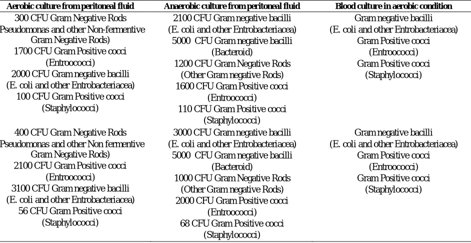
The culture results of the samples taken from the dead rats were reviewed which are shown in Table 5.
Group Result Type of bacteria Control Negative Negative
LO Negative Negative
PI0-24 Positive E. coli, bacterioid, CI0-24 Positive E. coli, bacterioid PI0-48 Positive E. coli, bacterioid CI0-48 Positive Enterococci, E. coli
Table 3: The mean and standard deviation of bacteria (blood and peritoneum)
Type of bacteria
CI0-48 PI0-48 CI0-24 PI0-24
P-value* A n a e r o b ic F r e q . (P e r c e n t) A e r o b ic F r e q . (P e r c e n t) A n a e r o b ic F r e q . (P e r c e n t) A e r o b ic F r e q . (P e r c e n t) A n a e r o b ic F r e q . (P e r c e n t) A e r o b ic F r e q . (P e r c e n t) A n a e r o b ic F r e q . (P e r c e n t) A e r o b ic F r e q . (P e r c e n t)
E. coli 43 (35.8%) 23 (3.4%) 304 (31%) 279 (83.3%) 1026 (36.6%) 1180 (85.7%) 787 (24.9%) 856 (84.2%) 0.003
Entrococci 39 (32.5%) 550 (80.5%) 74 (7.6%) 43 (12.8%) 142 (5.1%) 141 (10.2%) 104 (3.3%) 129 (12.7%) <0.0001
Bacterioid 50 (7.3%) 0 541 (55.2%) 0 1636 (58.3%) 0 1235 (39.1%) 0 <0.0001
Pseudomonas 38 (31.7%) 60 (8.8%) 0 0 0 56 (4.1%) 0 27 (2.7%) <0.0001
Miscellaneous 0 0 61(6.2%) 13(3.9%) 0 0 1035 (32.7%) 5 (0.5%) <0.0001
Total 140 (100%) 67 (100%) 980 (100%) 335 (100%) 2804 (100%) 1377 (100%) 3161 (100%) 1617(100%) <0.0001 * The p-value is for comparing the ratio of aerobic and anaerobic bacteria between the four groups using the Fisher’s exact test. The test was performed for any and all types of bacteria
Table 4: The mortality rate in different groups of rats
Table 5: Culture results of the samples taken from the dead rats
Aerobic culture from peritoneal fluid Anaerobic culture from peritoneal fluid Blood culture in aerobic condition 300 CFU Gram Negative Rods
(Pseudomonas and other Non-fermentive Gram Negative Rods)
1700 CFU Gram Positive cocci (Entroococci)
2000 CFU Gram negative bacilli (E. coli and other Entrobacteriacea)
100 CFU Gram Positive cocci (Staphylococci)
2100 CFU Gram negative bacilli (E. coli and other Entrobacteriacea)
5000 CFU Gram negative bacilli (Bacteroid)
1200 CFU Gram Negative Rods (Other Gram negative Rods) 1600 CFU Gram Positive cocci
(Entroococci) 110 CFU Gram Positive cocci
(Staphylococci)
Gram negative bacilli (E. coli and other Entrobacteriacea)
Gram Positive cocci (Entroococci) Gram Positive cocci
(Staphylococci)
400 CFU Gram Negative Rods (Pseudomonas and other Non fermentive
Gram Negative Rods) 2100 CFU Gram Positive cocci
(Entroococci)
3100 CFU Gram negative bacilli (E. coli and other Entrobacteriacea)
56 CFU Gram Positive cocci (Staphylococci)
3000 CFU Gram negative bacilli (E. coli and other Entrobacteriacea)
5000 CFU Gram negative bacilli (Bacteroid)
1000 CFU Gram Negative Rods (Other Gram negative Rods) 2000 CFU Gram Positive cocci
(Entroococci) 68 CFU Gram Positive cocci
(Staphylococci)
Gram negative bacilli (E. coli and other Entrobacteriacea)
Gram Positive cocci (Entroococci) Gram Positive cocci
(Staphylococci)
Time Survival Total P-value
Died Not died
Control Type Control 0 5 5 -
LO 0 5 5
IO-24 Type
PIO 0 5 5
0.4444
CIO 2 3 5
Total 2 13 15
IO-48 Type
PIO 1 4 5
0.206
CIO 4 1 5
Total 5 5 10
Discussion
Bacterial translocation is the passage of toxic products and endotoxin from gastrointestinal tract to extra-intestinal sites such as the mesenteric lymph nodes, liver, spleen, kidney, and blood circulation [4].
Three mechanisms have been suggested for BT including [1] bacterial overgrowth and ecological disturbances of gastrointestinal system, [2] increased permeability in intestinal mucosal barrier, and [3] immune defense dysfunction [5, 6].
Both aerobic and anaerobic bacteria are usually found following intestinal obstruction. Our study indicated that with increased time of the obstruction, pattern of bacterial translocation changed from aerobic to anaerobic according to culture results in 24 and 48 hours after performing the obstruction. It was also found that E. coli was the most common type of bacteria in the IO-24 group, whereas enterococcus was the most common type of bacteria in the IO-48 group. The phenomenon of microbial synergy in these infections is well characterized.
It has been postulated that facultative organisms function in part to lower the oxidation-reduction potential in the microenvironment and that this change allows the propagation of obligate anaerobes [7]. Although further studies are needed to explain the reason for the change in the pattern of bacterial translocation, we believe that obstruction makes some changes in bacterial flora of the intestine in addition to tissue damage and partial ischemic and necrosis which facilitate growth of anaerobic bacteria. The predominance of some bacteria among clinical isolates suggests that they possess one or more factors that enhance their ability to cause disease. Typically, virulence factors associated with anaerobes confer the ability to evade host defenses, adhere to cell surfaces, produce toxins and/or enzymes, or display surface structures that contribute to pathogenic potential.
Other studies have reported E. coli as the most common type of bacteria in aerobic group and enterococcus in the anaerobic group [8, 9].
On the other hand, it seems that in our study, increased number of bacteria in IO-24 compared to IO-48 group may be influenced by the condition that the immune system requires several hours to adjust bacterial translocation.
BT has been shown to occur in various patient populations ranging from patients with colorectal
cancer, pancreatitis, intestinal obstruction,
cholestasis, to those receiving parental nutrition
and cases of malnutrition, with little evidence for the latter. It is also reported in elective surgical patients [6].
The intestinal epithelium changes related to obstruction are similar to those of the intestinal ischemia which include mitochondrial destruction, decreased capacity of the cell to produce energy and to preserve the equilibrium and structure of the mucosal epithelium; so, BT is also observed in ischemic patients [10, 11].
Our study showed similar BT in peritoneum and blood as well as after 24 and 48 hours of the obstruction. There was no difference between groups of partial and complete obstruction.
The role of immunological system in protecting the intestinal barrier function to avoid BT is known. Peyer's patches in the small intestine, along with lymphocytes, macrophages, and local IgA, develop an immune defense system [12].
In our study, BT was detected in all cases, and E. coli was the most common bacterium observed in both blood and peritoneal fluid. E. coli has also been frequently seen in other studies. Fernando et al. reported BT in 86% of the cases, while Berg et al. reported BT in 100% of the cases [13, 14].
In patients with the intestinal obstruction, the
effect of manipulation and milking for
decompression of the intestine has been evaluated. It is hypothesized that manipulation may cause paralytic ileus after surgery and may compromise the intestinal motility. Since, after resolving the obstruction and after 24 hours, the intestinal motilitybecomes normal, so in these patients, BT is not decreased with milking [15].
Systemic changes in patients with intestinal obstruction, that is associated with BT, may consequently result in the systemic inflammatory response syndrome. Therefore, these patients experience an elevated level of acute phase inflammatory markers such as the C-reactive protein (CRP) in the blood. Thus, CRP can be considered a predictor of vascular compromise during intestinal obstruction [16].
The relationship between bacterial
translocation and survival rate has been confirmed by the increased rate of mortality following BT. Our study showed similar mortality rate following obstruction in all groups. No difference was observed between mortality rate of cases with partial or complete obstruction and that of groups of IO-24 or IO-48.
Different methods are used to prevent BT and its systemic complications. Given the role of growth hormone (GH) in stimulating the mucus secretions, it is used as a drug in the prevention of BT. The protective effect of GH is related to the decreased
rate of BT. Also, vitamin C and
somatostatinanalogus are used in patients with BT, which lead to decreased rate of BT from 100% in the control group to 43% in the experimental group [17, 18].
Besides, myosin light chain kinase is used to avoid BT and protect intestinal mucosal barrier. In these cases, mucus histology including villous structure is maintained and the rate of mucosal damage such as villous blunting and epithelial sloughing is decreased. In addition, mucosal TNF (tumor necrosis factor) level is decreased and the rate of bacterial over growth and translocation is decreased [19].
Besides, saccharomyces cerevisiae strain UFMG 905 significantly protects the intestinal mucosal barrier. In case of using this material, measuring the level of interlukine-10 and IGA determines that the rate of immunological function is increased and thus BT is decreased. In addition, measuring the level of blood uptake of 99m TC-DTPA determines the rate of permeability [20].
Conclusions
In conclusion, bacterial translocation is an active process by which bacteria pass through the normally impermanent intestinal mucosal barrier and into lymph nodes or the systemic circulation. In this study, BT occurred both systemically (to the blood) and locally (to the peritoneum). No significant difference was noted between groups of partial and complete obstruction; however, pattern of bacterial translocation changed from aerobic to anaerobic with increased time of the obstruction.
Enterococcus was the most common type of bacteria in anaerobic group. It was found that in the IO-24 group, E. coli was the most common type of bacteria, whereas in the IO-48 group, the most common type of bacteria was enterococcus.
Acknowledgements
The results described in this paper formed part of a thesis submitted by the first author for a postgraduate degree in general surgery. We sincerely acknowledge Ms. M. Hassanpour for editing the manuscript.
References
1.MacFie J, Reddy BS, Gatt M, Jain PK, Sowdi R, Mitchell CJ. Bacterial translocation studied in 927 patients over 13 years. Br J Surg. 2006;93(1):87-93.
2.Generoso SV, Viana ML, Santos RG, Arantes RM, Martins FS, Nicoli JR, et al. Protection against increased intestinal permeability and bacterial translocation induced by intestinal obstruction in mice treated with viable and heat-killed Saccharomyces boulardii. Eur J Nutr. 2011;50(4):261-9.
3.Hanna N, Bialowas C, Fernandez C. Septicemia Secondary to Ileus in Trauma Patients: A Human Model for Bacterial Translocation. South Med J. 2010;103(5):461-3.
4.Berg RD. Bacterial translocation from the gastrointestinal tract. Adv Exp Med Biol. 1999;473:11-30.
5.Zapata-Sirvent RL, Larocca A, Piñate S, Antequera R, González RC, del Médico P, et al. Factors involved in bacterial translocation in an experimental model of intestinal obstruction. G E N. 1989;43(3):185-93. [Spanish]
6.Macfie J. current status of bacterial translocation as a cause of surgical sepsis. Br Med Bull.2004;71:1-11.
7.Cohen-Poradosu R, Kasper DL. Anaerobic Infections: General Concepts. In: Mandell GL, Bennett JE, Dolin R, editors. Mandell, Douglas, and Bennett's Principles and Practice of Infectious Diseases. 7th ed. Philadelphia: Elsevier Inc, Churchill Livingstone; 2010.pp:3083-5.
8.Berber I, Aydin C, Cevahir N, Yenisey C, Gumrukcu G, Kocbil G, et al. Tempol reduces bacterial translocation after ischemia/reperfusion injury in a rat model of superior mesenteric artery occlusion. Surg Today. 2009;39(5):407-13.
9.Gun F, Salman T, Gurler N, Olgac V. Effect of probiotic supplementation on bacterial translocation in thermal injury. Surg Today. 2005;35(9):760-4.
10.Antequera R, Bretaña A, Cirac A, Brito A, Romera MA, Zapata R. Disruption of the intestinal barrier and bacterial translocation in an experimental model of intestinal obstruction. Acta Cient Venez. 2000;51(1):18-26.
11.Samel S, Keese M, Kleczka M, Lanig S, Gretz N, Hafner M, et al. Microscopy of bacterial translocation during small bowel obstruction and ischemia in vivo–a new animal model. BMC Surg. 2002;2:6.
12.Van Leeuwen PA, Boermeester MA, Houdijk AP, Ferwerda CC, Cuesta MA, Meyer S, et al. Clinical significance of translocation. Gut. 1994;35(1 suppl):S28-34.
13.Zanoni FL, Benabou S, Greco KV, Moreno ACR, Cruz JWMC, Filgueira FP, et al. Mesenteric microcirculatory dysfunctions and translocation of indigenous bacteria in a rat model of strangulated small bowel obstruction. Clinics (Sao Paulo). 2009;64(9):911-9.
14.Akçay MN, Capan MY, Gündogdu C, Polat M, Oren D. Bacterial translocation in experimental intestinal obstruction. J Int Med Res. 1996;24(1):17-2.
15.Törer N, Nursal TZ, Tufan H, Can F, Bal N, Tarim A, et al. Effect of manual bowel decompression (milking) in the obstructed small bowel. Am J Surg. 2008;195(6):807–13.
16.El-Awady SI, El-Nagar M, El-Dakar M, Ragab M, Elnady G. Bacterial translocation in an experimental intestinal obstruction model. C-reactive protein reliability? Acta Cir Bras. 2009;24(2):98-106.
17.Balik AA, YildIrgan MI, Kilic A, Gündogdu C, Erdogan F. Effects of Growth Hormone on Bacterial
Translocation Due to Intestinal Obstruction. Turk J Med Sci.2000; 30:209-12.
18.Akyildiz M, Ersin S, Oymaci E, Dayangaç M, Kapkac M, Alkanat M. Effects of somatostatin analogues and vitamin C on bacterial translocation in an experimental intestinal obstruction model of rats. J Invest Surg. 2000;13(3):169-73.
19.Wu CC, Lu YZ, Wu LL. Role of myosin light chain kinase in intestinal epithelial barrier defects in a rat model of bowel obstruction. BMC Gastroenterol. 2010;10:39.
20.Generoso SV, Viana M, Santos R, Martins FS, Machado JA, Arantes RM, et al. Saccharomyces cerevisiae strain UFMG 905 protects against bacterial translocation preserves gut barrier integrity and stimulates the immune system in a murine intestinal obstruction model. Arch Microbiol. 2010;192(6):477-84.