RESEARCH
Development of a microarray-based
method for allergen-specific IgE and IgG4
detection
Guzel Feyzkhanova, Sergei Voloshin, Olga Smoldovskaya, Alla Arefieva, Marina Filippova, Viktor Barsky,
Ludmila Pavlushkina, Veronika Butvilovskaya, Alexei Tikhonov, Yuri Reznikov and Alla Rubina
*Abstract
Background: sIgE and sIgG4 detection is necessary for more accurate and effective type I hypersensitivity diagnos-tics and the estimation of disease development. Typically, the analyses of these antibodies are performed separately with the help of various specialized systems. The aim of this study was to develop a microarray-based method for the simultaneous quantitative detection of sIgE and sIgG4 to the most common allergens in a single sample.
Methods: A quantitative method for the simultaneous detection of sIgE and sIgG4 was developed based on the technology of hydrogel microchips previously designed at Engelhardt Institute of Molecular Biology, Russian Acad-emy of Sciences (EIMB RAS). The microarray contained gel pads with immobilized allergens and gel pads that allow for the obtaining of sIgE and sIgG4 internal calibration curves for each allergen during the assay. The possibility of the simultaneous detection of sIgE and sIgG4 was developed using the corresponding Cy5 and Cy3 fluorescent dyes. Results: The multiplex immunoassay method using hydrogel microarrays developed in this study allowed the quantitative detection of sIgE and sIgG4 to 31 allergens from different groups in a single assay. A comparison of the microarray with the existing plate-based analogues (i.e., ALLERG-O-LIQ and sIgG4 ELISA) was performed by analysing 152 blood serum samples and by evaluating Pearson correlation coefficients, ROC analysis, and Passing-Bablok linear regression results.
Conclusion: The implementation of this method in allergy diagnostics will provide the possibility of simultaneously performing primary patient screening and obtaining additional information concerning the severity of the allergies and the choice of an appropriate therapy.
Keywords: Immunoassay, Allergy diagnostics, Microarrays, sIgG4, Biochips
© The Author(s) 2017. This article is distributed under the terms of the Creative Commons Attribution 4.0 International License (http://creativecommons.org/licenses/by/4.0/), which permits unrestricted use, distribution, and reproduction in any medium, provided you give appropriate credit to the original author(s) and the source, provide a link to the Creative Commons license, and indicate if changes were made. The Creative Commons Public Domain Dedication waiver (http://creativecommons.org/ publicdomain/zero/1.0/) applies to the data made available in this article, unless otherwise stated.
Background
The incidence of allergic diseases steadily increases year after year. One or more allergic signs are detected in 30–40% of the world population, and the number of patients suffering from immunoglobulin-related food intolerance (so-called food allergies) has reached 240– 550 million people [1].
According to the classification of Coombs and Gell [2], IgE is assumed to be a key marker of type I
hypersensitivity for in vitro diagnostics. Fc region of IgE coupled with allergen has the ability to bind to FcεRI receptor on the basophils and mast cells membranes, that elicits their subsequent activation. This process explains the rapid effects of allergen-sIgE complexes in the forma-tions of allergic rhinitis, asthma, urticaria and anaphylac-tic reactions [3].
However, the presence of sIgE in the blood is not an absolute marker for the presence of clinical manifesta-tions of immunological failure [4]. In certain cases, the manifestation of allergy symptoms can be suppressed by the presence of immunoglobulin G, which acts as a “blocking antibody”. Typically, such blocking properties
Open Access
*Correspondence: [email protected]
are distinctive hallmarks of the IgG4 antibody subclass [5]. According to the number of studies sIgG4 level as well as the ratio sIgE/sIgG4 is associated with likelihood of allergic symptoms reporting [6, 7] and can improve the prediction of tolerance to some allergens [8]. There-fore, sIgG4 detection together with sIgE measurement gives more inclusive information for in vitro analysis interpretation.
The sIgG4 level is also used to monitor allergen-specific immunotherapy (SIT) because successful therapy is char-acterized by an increase in sIgG4 and a decrease in sIgE [5, 9]. Thus, patient management strategy, particularly the selection of appropriate drugs for SIT, depends not only on the early recognition of the allergens that cause hypersensitivity but also on the monitoring of sIgE and sIgG4 levels [10]. Currently, determination of sIgE in clinical lab is generally performed by numerous single-plex and multisingle-plex tests [11]. Amongst the most useful tools for multiplex sIgE determination are microarrays [12], which allow for the identification of a plurality of analytes during a single analysis of a blood serum sample.
Test systems for the determination of sIgG4 levels in the serum are less widespread. Given that the IgG4 serum concentration on average is greater than the IgE con-centration, these tests often require an additional sam-ple preparation stage, i.e., the pre-dilution of the serum prior to analysis. Consequently, the handling of two sepa-rate tests is required for the parallel detection of sIgE and sIgG4 in the same serum sample. This requirement complicates the diagnoses of patients with suspected allergies. To solve this problem, an microarray approach based on developing antibodies that are specific to cer-tain classes of immunoglobulins and labelled with vari-ous dyes was proposed [13]. The main advantage of this approach is the ability to multiply the number of defined parameters within a single analysis. In comparison with conventional ELISA microarray format allows to detect sIgE and sIgG4 to the number of allergens, including those sensibilization to which was not suspected and was not exhibited because of the blocking antibodies. Fur-thermore such testing with microarrays requires appreci-ably less amount of biomaterial.
The study conducted by Rubina et al. in cooperation with Fooke-Achterrath [14] demonstrated the possibility of simultaneous sIgE and sIgG4 detection using hydrogel biochips. As a result of this approach, in this paper we have developed a method for the simultaneous quantita-tive analysis of allergen-specific immunoglobulins E and G4 to 31 allergens belonging to different groups. The cur-rent study presents the analytical characteristics of the developed method and comparison to the established reference methods after assay of serum samples from patients and healthy donors.
Methods Samples
In our research, we included surplus blood serum sam-ples that remained after routine diagnostic procedures. The samples were obtained from healthy donors (control group) and patients who were referred for sIgE determi-nation by allergologists/pulmonologists for the diagno-sis and monitoring of the following disease states: atopic dermatitis, asthma, urticaria, and rhinitis. The object of our research was an age-diverse group (5–65 years old) from the Moscow population.
In total, 152 serum samples were analysed in this study. Among these, 82 samples were from adult patients with suspected allergic diseases, and 15 samples were from healthy donors; these samples were provided by the Fed-eral State Budgetary Institution Polyclinic No. 1 of the Business Administration for the President of the Russian Federation. Additionally, 45 sera samples from children with suspected allergic diseases and 10 sera samples from healthy children who did not have allergies were pro-vided by the Filatov Moscow City Pediatric Clinic No. 13. The conditions of blood sampling, the delivery, and storage of the samples were identical. The blood for serum isolation was collected via a puncture of the median cubital vein, the serum was separated from the blood corpuscles by centrifugation (3000 rpm, 10 min) within the first 2 h after blood sampling. All of the sam-ples were exposed to a single refrigeration at −45 °C. Samples were delivered in insulated containers with ice packs.
Design and manufacture of the microarrays
Table 1 provides a list of the allergens (GREER, USA) that were immobilized in the microarray gel pads. The micro-array contained elements with allergens belonging to the main groups, i.e., pollen, household, epidermal, food, fungi, and insect venoms.
Preparation of the dye‑conjugated antibodies
Two microliters of Cy5 or Cy3 N-hydroxysuccinimide ester solution (GE Healthcare, UK) (2 mg/ml in dimeth-ylformamide) were added to 75 µl of anti-IgE (Bethyl, USA) or anti-IgG4 (Fitzgerald, USA) antibody solution (1 mg/ml in 0.01 M bicarbonate buffer, pH 9.5). The reac-tion was performed at 22 °C for 1 h in the dark with stir-ring (550 rpm). Purification of the antibody conjugate from the unreacted dye (Cy5 or Cy3) was performed by gel filtration on a Micro Bio-Spin column (Bio-Rad, USA) filled with Sephadex G-25 Coarse (Sigma, USA) and equilibrated with PBS (0.01 M phosphate buffer, pH 7.2, 0.15 M NaCl) buffer. The dye/protein molar ratio of the final conjugates was determined spectrophotometrically to be 3.
Analysis of sIgE and sIgG4 on the microarrays
Each microarray was incubated with sixty-five micro-liters of blood serum at 37 °C for 20 h. After wash-ing in PBST (PBS, 0.1% Tween-20 (Sigma, USA)) for 20 min, 50 µl of developing solution containing anti-IgE-Cy5 and/or anti-IgG4-Cy3 (working concentrations of 2.5 and 1.5 µg/ml, respectively) was applied to the
microarray, and the microarray was incubated at 37 °C for 1 h in the dark. After a final washing in PBST for 30 min, the registration of the fluorescence signals was performed.
Measurement of the fluorescence intensities of the microarray gel pads
The registration of fluorescence signals was performed using a two-wavelength microarray analyser (EIMB RAS) based on the concept of digital wide-field fluorescence microscopy. While working with the fluorescent dyes, excitation was provided by laser diodes that emitted light at 532 nm (Cy3) or 655 nm (Cy5). The fluorescence inten-sities of the gel elements were registered by a CCD cam-era using interference filters of 607 ± 35 nm for Cy3 and 716 ± 22 nm for Cy5. The analyser operation, the analyses of the fluorescent images and the calculations of the sIgE and sIgG4 concentrations were performed with ImageAs-say software (EIMB RAS). A standard method of the gel element fluorescence calculation that has been previously described [17] was employed. The final fluorescence of each data point was calculated as the median value of the four fluorescence signals obtained from the repeats.
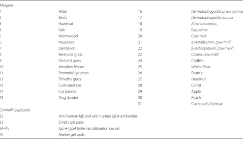
Table 1 List of gel pads in the microarray
a Purified allergen components
Allergens
1 Alder 16 Dermatophagoides pteronyssinus
2 Birch 17 Dermatophagoides farinae
3 Hazelnut 18 Alternaria tenius
4 Oak 19 Egg white
5 Wormwood 20 Cow milk
6 Mugwort 21 α-lactalbumin, cow milka
7 Dandelion 22 β-lactoglobulin, cow milka
8 Bermuda grass 23 Casein, cow milka
9 Orchard grass 24 Codfish
10 Meadow fescue 25 Wheat flour
11 Perennial rye grass 26 Peanut
12 Timothy grass 27 Hazelnut
13 Cultivated rye 28 Carrot
14 Cat dander 29 Apple
15 Dog dander 30 Peach
31 Cockroach, German
Controlling gel pads
32 Anti-human IgE and anti-human IgG4 antibodies
33 Empty gel pads
34–45 IgE + IgG4 (internal calibration curve)
Processing and interpretation of the results
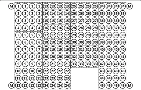
The concentrations of antibodies that were immobi-lized in the microarray gel pads 34–45 (Fig. 1) were chosen and arranged in ascending order so that after
analysing the fluorescent signals from rows 34–45, the entire ranges of the signal intensities for sIgE (up to 100 IU/ml) and sIgG4 (up to 2500 ng/ml) were covered.
Fig. 1 Design of the microarray for the simultaneous detection of sIgE and sIgG4 to 31 allergens. The numbers in the circles correspond to the allergen numbers in Table 1
Each manufactured lot of microarrays was calibrated using characterized standard blood sera-based samples including a zero sample (PBS, 0.1% PVA, and 0.1% PVP). For each allergen, the sIgE and sIgG4 concentrations of the standard samples were ascribed to the relative fluo-rescence intensity of each gel pad that was used for cali-bration curve plotting.
Treatment with a mixture of fluorescently labelled conjugates of anti-IgE-Cy5 and anti-IgG4-Cy3 resulted in the formation of binary complexes with correspond-ing conjugates in the gel pads 34–45. Accordcorrespond-ing to the fluorescent signals from these gel pads and the attrib-uted concentrations of sIgE and sIgG4, internal calibra-tion curves were constructed for each of the allergens. The determinations of the sIgE and sIgG4 concentrations were performed according to the fluorescent signals from the gel pads with the immobilized allergens in relation to the internal calibration curves.
Analysis of IgE and IgG4 using reference methods
The analyses of the serum samples were performed using the Specific IgE REAST (ALLERG-O-LIQ) and Specific IgG4 ELISA (Dr. Fooke Laboratorien GmbH, Germany) test systems according to the procedures described in the manufacturer’s instructions. Data processing was per-formed with the ALLERG-O-Win software (Dr. Fooke Laboratorien, GmbH).
Determination of the analytical characteristics Dilution test
The linearity of the method was evaluated via the analy-sis of blood serum samples that had been diluted 2, 4, 8, and 16 times. The dilutions were performed with the zero sample.
Detection limit
The detection limits for sIgE and sIgG4 were determined via serial dilutions of two samples that contained signifi-cant amounts of sIgE to pollen (grey alder, birch, meadow fescue, timothy grass), cat dander, and cow milk and sIgG4 to cat dander, dog dander, cow milk, wheat, pea-nut, and hazelnut. The detection limit for each allergen was established as the concentration associated with the fluorescence value that was two standard deviations larger than the average value of the tenfold measured flu-orescent signal of the zero sample.
Within‑run precision
The evaluation of the within-run precision of the method was performed via an analysis of blood serum samples containing sIgEs and sIgG4s to various allergens. The
assay was performed using 10 repeats for each sample. The samples were chosen such that the concentrations of sIgE and sIgG4 covered the entire dynamic ranges of the measurements.
Comparison with other methods: correlation and regression analysis
For a number of allergens, the Pearson correlation coeffi-cients r [18] of the concentrations obtained by the micro-array and commercial test systems were determined. Passing–Bablok regression analyses [19], ROC curve anal-ysis, sensitivity and specificity were also performed using the MedCalc program, version 16.4.3. The parameters of the regression line were determined as (Y = A + B × X), where the intercept is A, the slope is B, and the associated 95% confidence intervals were calculated.
Results and discussion
Design of the microarray and analysis procedure
The three-dimensional hydrogel microarray produced at EIMB RAS was used as an analytical instrument for the development of a multiplex simultaneous quantitative assay of sIgEs and sIgG4s for 31 allergens in blood serum samples.
The microarray structure and lists of the immobilized allergen extracts and purified components are provided in Fig. 1 and Table 1. The allergens were chosen with con-sideration of the frequencies of allergen reactions in Cen-tral Russia, which mainly corresponds to the frequencies in Central and Northern Europe [20]. The allergens belonged to different groups that included pollen, indoor allergens and food allergens.
In addition to allergens, the structure of the microarray was enlarged with gel pads with immobilized immuno-globulins E and G4, which were used to plot an internal calibration curve (after development with Cy5- and Cy3-labeled anti-human antibodies). The internal calibration curve was used to control for the development system activity during the assay.
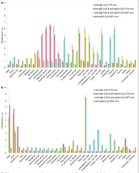
Mutual influence of Cy3‑ and Cy5‑labeled antibodies on the developing system
analysis of the hydrogel microarrays, which indicates the propriety of the selected developing system.
Analytical characteristics of the method
The accuracy of the developed method was evaluated via an estimation of analytical characteristics includ-ing the dilution linearity, detection limit and within-run precision.
Dilution linearity
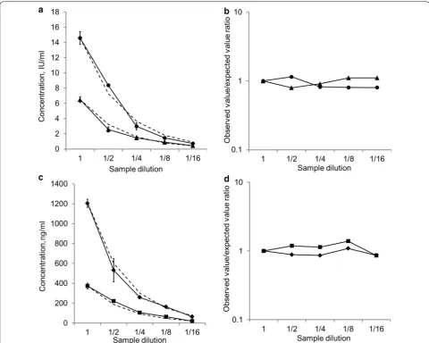
This test was performed via the serial dilution of a sample with a high analyte concentration and follow-up compar-isons of the experimental concentrations with the esti-mated concentrations.
A blood serum sample containing 14.58 IU/ml sIgE to birch pollen, 6.49 IU/ml sIgE to meadow fescue, 1205 ng/ ml sIgG4 to cow milk and 373 ng/ml sIgG4 to peanut was diluted 2, 4, 8, and 16 times with the zero sample. Fig-ure 4a, c illustrate the decreasing concentration-dilution curves for the sIgE and sIgG4 measurements, respec-tively. The dashed line depicts the corresponding curves for the expected concentrations. The experimental/ expected concentration ratios for the different dilutions are provided in Fig. 4b, d for sIgE and sIgG4, respectively.
As illustrated in the figures, the experimentally deter-mined sIgE concentrations differed from the calculated concentration by no more than ±10%. The experimental/ expected concentration ratios for sIgG4 were also in the
range of 0.9–1.1. The presence of the regular pattern dur-ing the serum dilution indicates the absence of a matrix effect, i.e., a lack of interference from different serum components. Consequently, the mean per cent linearities for both sIgE and sIgG4 for the serum were in the range of 90–110%, which meets with the requirements for immunoassay methods.
Detection limit
The detection limits were determined via serial dilution of the serum samples. For example, the reliable detected concentrations for the serum sample with sIgE concen-trations to meadow fescue (15.14 IU/ml), timothy grass (11.11 IU/ml), cat dander (3.36 IU/ml), and cow milk (2.38 IU/ml) were 0.24 IU/ml, 0.17 IU/ml, 0.21 IU/ml, and 0.15 IU/ml, respectively. The same sample contained 708 ng/ml sIgG4 to cat dander and 432 ng/ml sIgG4 to peanut. The reliable sIgG4 concentrations after dilution were 11 and 14 ng/ml, respectively.
For all allergens, the limit of sIgE detection did not exceed 0.25 IU/ml. This value is above the detection limit of the existing methods (0.1 IU/ml for ImmunoCAP and Immulite) but still below the internationally accepted cut-off concentration for allergodiagnostics (0.35 IU/ml). The detection limit for the sIgG4 concentration did not exceed 100 ng/ml.
Within‑run precision
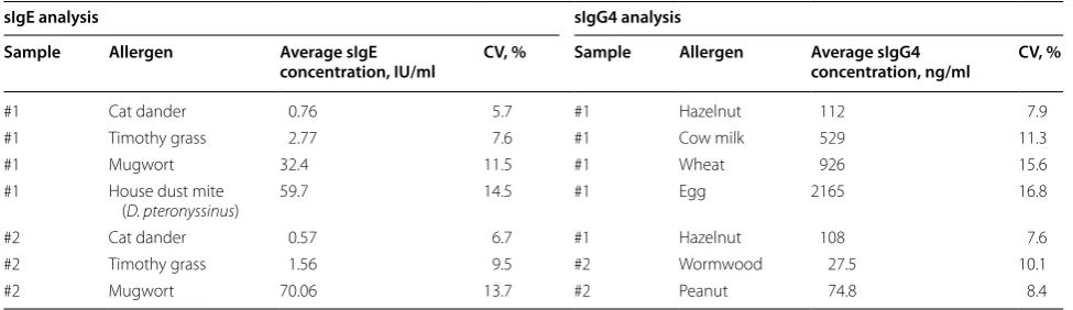
Table 2 provides the average concentrations that were determined in the assay of the 2 serum samples in 10 rep-etitions and the calculated coefficients of variation for the different allergens. As shown in the table, the within-run
precision did not exceed 15% for sIgE or 17% for sIgG4 determinations in the measured concentration range.
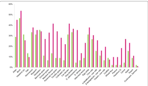
The results of the comparison between the hydrogel microarrays and ELISA
The serum samples (n = 127) were obtained from atopic patients from 2 age groups, i.e., age 0.5–17 years (n = 45) and age 18–74 years (n = 82). Figure 5 illustrates the distribution of increased (≥0.35 IU/ml) sIgE concentra-tions for different allergens for the patients based on the experimental data. For the comparative evaluation of the multiplex simultaneous sIgE and sIgG4 immunoassay on the hydrogel microarrays, analyses of 152 serum samples (127 atopic patients and 25 healthy donors) for different allergens were performed with both the microarrays and the reference methods for the sIgE (ALLERG-O-LIQ) and sIgG4 (sIgG4 ELISA) determinations. For each aller-gen, no fewer than 10 serum samples were analysed using ALLERG-O-LIQ and an sIgG4 ELISA.
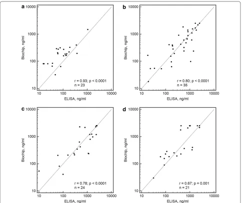
Scatter diagrams of the results obtained from the anal-yses of sIgE concentrations to birch pollen, mugwort pol-len, timothy grass polpol-len, and dog dander on the hydrogel microarrays and ALLERG-O-LIQ are provided in Fig. 6. The scatter diagrams of the results for the sIgG4 con-centrations to dog dander, cow milk, wheat flour, and hazelnut in the samples as measured on the hydrogel microarrays and the sIgG4 ELISA are provided in Fig. 7.
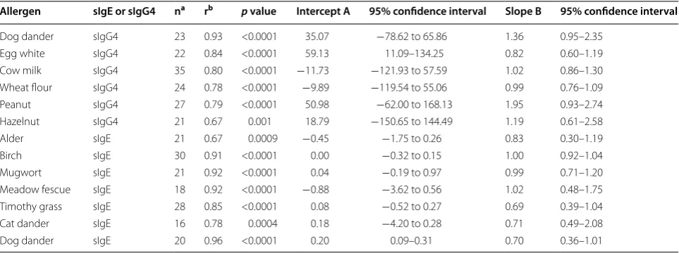
Pearson’s correlation coefficients r and the sample sizes n for the analysed allergens are provided in Table 3. For the remaining allergens, sufficient sample sizes were not obtained for the determination of correlation coefficients with p < 0.01.
Table 2 Results of the within-run precision test
The average sIgE and sIgG4 concentrations were obtained in a simultaneous immunoassay of the microarrays, and the corresponding coefficients of variation for the 2 serum samples analysed in 10 repetitions are provided
sIgE analysis sIgG4 analysis
Sample Allergen Average sIgE
concentration, IU/ml CV, % Sample Allergen Average sIgG4 concentration, ng/ml CV, %
#1 Cat dander 0.76 5.7 #1 Hazelnut 112 7.9
#1 Timothy grass 2.77 7.6 #1 Cow milk 529 11.3
#1 Mugwort 32.4 11.5 #1 Wheat 926 15.6
#1 House dust mite
(D. pteronyssinus) 59.7 14.5 #1 Egg 2165 16.8
#2 Cat dander 0.57 6.7 #1 Hazelnut 108 7.6
#2 Timothy grass 1.56 9.5 #2 Wormwood 27.5 10.1
The correlation coefficients observed in the compari-sons between the microarray and reference methods for the different allergens were in the ranges of 0.68–0.93 for the sIgE analyses and 0.67–0.96 for the sIgG4 analy-ses. The obtained values are similar to the values of the correlation coefficients that have been calculated in com-parisons of immunoassay methods in other works (for example, r = 0.525–0.979 in [21] and r = 0.60–0.98 in [22]). For some allergens the significant dispersion of the results led to the small correlation coefficients. This fact can be explained by the distinct component compounds of the protein allergen extracts produced by numerous manufacturers, by the variety of protein modifications that occur during the immobilization process, as well as the wide range of developing anti-human antibodies that can interact with sIgE populations with different effica-cies [23].
Passing-Bablok regression analyses yielded the inter-cept A and slope B for each allergen. The values are
presented in Table 3. For all except two cases (sIgG4 to egg white and sIgE to dog dander), the 95% confidence intervals for the intercept included 0, and for all cases, the 95% confidence intervals for the slope included 1.
ROC analysis for sIgE detection was performed with the data differentiated through the common sIgE cut-off of 0.35 IU/ml (with disease: ≥0.35 IU/ml sIgE with the reference method; without disease: <0.35 IU/ml sIgE with the reference method). The optimum cut-off for the described microarray-based method as defined by the Youden’s J statistic-associated criterion was 0.52 IU/ ml. For this cut-off the sensitivity was 87.6%, the specific-ity was 90.6%, and the diagnostic accuracy was 87%. The area under the curve (AUC) was 0.931 that corresponded to high accuracy test [24]. In general, the results observed following the application of different immunoassay meth-ods cannot be inter-convertible because of significant differences that inevitably appear during clinical sample assays [25, 26]; however, in our case, it may be said that
for these concretely analysed allergens, there is no over-estimation or underover-estimation of the sIgE and sIgG4 measurements compared to the employed reference methods, i.e., ALLERG-O-LIQ and sIgG4 ELISA.
Conclusion
A microarray for the multiplex quantification of the concentrations of sIgE and sIgG4 to 31 allergens from different groups in serum samples was developed. The
simultaneous detection of sIgE and sIgG4 was made possible via the use of a developing system with two fluorescent dyes. This method allows for the obtain-ing of sIgE and sIgG4 levels in common units without the construction of an external calibration curve. The
analytical characteristics of the method satisfy the requirements that are applicable to immunofluorescent test systems.
The usage of this method in allergy diagnostics pro-vides the possibility of both performing primary patient
screening and obtaining the additional information that is necessary for allergy severity evaluation and therapy selection.
Abbreviations
EIMB RAS: Engelhardt Institute of Molecular Biology, Russian Academy of Sciences; sIgE: specific immunoglobulin E; sIgG4: specific immunoglobulin G4; SIT: allergen-specific immunotherapy; Cy5: cyanine 5; Cy3: cyanine 3; PBS: phosphate-buffered saline; PBST: phosphate-buffered saline with Tween® 20; PVA: polyvinyl alcohol; PVP: polyvinylpyrrolidone.
Authors’ contributions
AR, GF, and OS planned the experiments. YR and LP selected the samples according to the clinical histories. VB adopted the microarray analyser for the two-wavelength operation and provided technical support. SV, OS, and AT performed the analyses of the samples using the commercial test systems. AA, GF, and VD performed the analyses of the samples on the microarray. GF, OS, and MP analysed and interpreted the collected data. AR, SV, AA, GF, and YuR were involved in the manuscript preparation. AZ, VB, MP, and LP criti-cally reviewed the manuscript. All authors have read and approved the final manuscript.
Acknowledgements
This work was supported by the Russian Science Foundation, Grant No. 14-50-00060.
Competing interests
The authors declare that they have no competing interests.
Consent for publication
The authors give their consent for the publication of these research results.
Received: 16 August 2016 Accepted: 13 December 2016
References
1. World Allergy Organization White Book on Allergy: Update 2013. World Allergy Organization, 2013.
2. Coombs RR, Gell PG. Classification of allergic reactions responsible for clinical hypersensitivity and disease. Clin Asp Immunol. 1975;3:761–81. 3. Grimbaldeston MA, Metz M, Yu M, Tsai M, Galli SJ. Effector and potential
immunoregulatory roles of mast cells in IgE-associated acquired immune responses. Curr Opin Immunol. 2006;18:751–60.
4. Bousquet J, Anto JM, Bachert C, Bousquet PJ, Colombo P, Crameri R, Daeron M, Fokkens W, Leynaert B, Lahoz C, Maurer M, Passalacqua G, Valenta R, Van Hage M, Van Ree R, Daeron M, Fokkens W, Leynaert B, Lahoz C, Maurer M, Passalacqua G, Valenta R, Van Hage M, Van Ree R. Factors responsible for differences between asymptomatic subjects and patients presenting an IgE sensitization to allergens. A GA2LEN project. Allergy Eur J Allergy Clin Immunol. 2006;61:671–80.
5. Greenberger PA. Immunotherapy update: mechanisms of action. Allergy Asthma Proc. 2002;23:373–6.
6. Burnett M, Wegienka G, Havstad S, Kim H, Johnson CC, Ownby D, Zoratti E. Relationship of dog- and cat-specific IgE and IgG4 levels to allergic symptoms on pet exposure. J Allergy Clin Immunol Pract. 2013;1:350–3.
7. Santos AF, James LK, Bahnson HT, Shamji MH, Couto-Francisco NC, Islam S, Houghton S, Clark AT, Stephens A, Turcanu V, Durham SR, Gould HJ, Lack G. IgG4 inhibits peanut-induced basophil and mast cell activation in peanut-tolerant children sensitized to peanut major allergens. J Allergy Clin Immunol. 2015;135:1249–56.
8. Vazquez-Ortiz M, Pascal M, Jiménez-Feijoo R, Lozano J, Giner MT, Alsina L, Martín-Mateos MA, Plaza AM. Ovalbumin-specific IgE/IgG4 ratio might improve the prediction of cooked and uncooked egg tolerance development in egg-allergic children. Clin Exp Allergy. 2014;44:579–88.
9. Bullock RJ, Barnett D, Howden MEH. Immunologic and clinical responses to parenteral immunotherapy in peanut anaphylaxis—a study using IgE and IgG4 immunoblot monitoring. Allergol Immunopathol (Madr). 2005;33:250–6.
10. Stylianou E, Ueland T, Borchsenius F, Michelsen AE, Øvstebø R, Mollnes TE, Skjønsberg OH, Aukrust P. Specific allergen immunotherapy: effect on IgE, IgG4 and chemokines in patients with allergic rhinitis. Scand J Clin Lab Invest. 2016;76:118–27.
11. Matricardi PM, Kleine-Tebbe J, Hoffmann HJ, Valenta R, Hilger C, Hofmaier S, Aalberse RC, Agache I, Asero R, Ballmer-Weber B, Barber D, Beyer K, Biedermann T. EAACI molecular allergology user’s guide. Pediatr Allergy Immunol. 2016;27(suppl23):1–250.
12. Passalacqua G, Melioli G, Bonifazi F, Bonini S, Maggi E, Senna G, Triggiani M, Nettis E, Rossi RE, Vacca A, Canonica GW. The additional values of microarray allergen assay in the management of polysensitized patients with respiratory allergy. Allergy. 2013;68:1029–33.
Table 3 Microarray: ALLERG-O-LIQ and microarray—sIgG4 ELISA comparisons by Passing-Bablok regression
a Sample size
b Pearson correlation coefficient
Allergen sIgE or sIgG4 na rb p value Intercept A 95% confidence interval Slope B 95% confidence interval
• We accept pre-submission inquiries
• Our selector tool helps you to find the most relevant journal
• We provide round the clock customer support
• Convenient online submission
• Thorough peer review
• Inclusion in PubMed and all major indexing services
• Maximum visibility for your research
Submit your manuscript at www.biomedcentral.com/submit
Submit your next manuscript to BioMed Central
and we will help you at every step:
13. Renault NK, Gaddipati SR, Wulfert F, Falcone FH, Mirotti L, Tighe PJ, Wright V, Alcocer MJC. Multiple protein extract microarray for profiling human food-specific immunoglobulins A, M, G and E. J Immunol Methods. 2011;364:21–32.
14. Rubina AY, Feizkhanova GU, Filippova MA, Talibov VO, Fooke-Achterrath M, Zasedatelev AS. Multiplex assay of allergen-specific and total immuno-globulins of E and G classes in the biochip format. Dokl Biochem Biophys. 2012;447:289–93.
15. Rubina AY, Dementieva EI, Stomakhin AA, Darii EL, Pan’kov SV, Barsky VE, Ivanov SM, Konovalova EV, Mirzabekov AD. Hydrogel-based protein microchips: manufacturing, properties, and applications. Biotechniques. 2003;34:1008–22.
16. Feyzkhanova GU, Filippova MA, Talibov VO, Dementieva EI, Maslennikov VV, Reznikov YP, Offermann N, Zasedatelev AS, Rubina AY, Fooke-Achter-rath M. Development of hydrogel biochip for in vitro allergy diagnostics. J Immunol Methods. 2014;406:51–7.
17. Rubina AY, Filippova MA, Feizkhanova GU, Shepeliakovskaya AO, Sidina EI, Boziev KhM, Laman AG, Brovko FA, Vertiev YV, Zasedatelev AS, Grishin EV. Simultaneous detection of seven staphylococcal enterotoxins: develop-ment of hydrogel biochips for analytical and practical application. Anal Chem. 2010;82:8881–9.
18. Armitage P, Berry G, Matthews JNS, editors. Statistical methods in medical research. Oxford: Blackwell Science Ltd; 2002.
19. Passing H, Bablok W. A new biometrical procedure for testing the equality of measurements from two different analytical methods. Application of linear regression procedures for method comparison studies in Clinical Chemistry, Part I. Clin Chem Lab Med. 1983;21:709–20.
20. Reznikov YP, Topoleva TS, Goryackina LA. Composition of “Russian aller-gologic panel” for in vitro diagnosis of atopia in children and adolescents. J “Pediatria” Named After GN Speransky. 2001;80:48–50.
21. Lee YW, Sohn JH, Lee J-H, Hong C-S, Park J-W. Allergen-specific IgE meas-urement with the IMMULITE 2000 system: intermethod comparison of detection performance for allergen-specific IgE antibodies from Korean allergic patients. Clin Chim Acta. 2009;401:25–32.
22. Plebani M, Bernardi D, Basso D, Borghesan F, Faggian D. Measurement of specific immunoglobulin E: intermethod comparison and standardiza-tion. Clin Chem. 1998;44:1974–9.
23. Cox L. Overview of serological-specific IgE antibody testing in children. Curr Allergy Asthma Rep. 2011;11:447–53.
24. Akobeng AK. Understanding diagnostic tests 3: receiver operating char-acteristic curves. Acta Paediatr. 2007;96(5):644–7.
25. Graham F, Bégin P, Paradis L, Lacombe-Barrios J, Paradis J, Des Roches A. Comparison of ImmunoCAP and Immulite serum specific IgE assays for the assessment of egg allergy. Allergy Asthma Clin Immunol. 2016;12:29. 26. Lee J-HJ-S, Park KH, Kim H-S, Kim KW, Sohn MH, Kim C-H, Lee J-HJ-S, Hong