P R I M A R Y R E S E A R C H
Open Access
Assessment of potential anti-cancer stem cell
activity of marine algal compounds using an
in vitro
mammosphere assay
Jo-Anne de la Mare
1, Jason N Sterrenberg
1, Mugdha G Sukhthankar
1, Maynard T Chiwakata
2, Denzil R Beukes
2,
Gregory L Blatch
1,3and Adrienne L Edkins
1*Abstract
Background:The cancer stem cell (CSC) theory proposes that tumours arise from and are sustained by a
subpopulation of cells with both cancer and stem cell properties. One of the key hallmarks of CSCs is the ability to grow anchorage-independently under serum-free culture conditions resulting in the formation of tumourspheres. It has further been reported that these cells are resistant to traditional chemotherapeutic agents.
Methods:In this study, the tumoursphere assay was validated in MCF-7 cells and used to screen novel marine algal compounds for potential anti-cancer stem cell (CSC) activityin vitro.
Results:MCF-7 breast cancer cells were observed to generate tumourspheres or mammospheres after 3-5 days growth in anchorage-independent conditions and an apparent enrichment in potential CSCs was observed by an increase in the proportion of CD44high/CD24lowmarker-bearing cells and Oct4 expression compared to those in the bulk population grown in regular adherent conditions. Using this assay, a set of algal metabolites was screened for the ability to inhibit mammosphere development as a measure of potential anti-CSC activity. We report that the polyhalogenated monoterpene stereoisomers RU017 and RU018 isolated from the red algaPlocamium cornutum, both of which displayed no cytotoxicity against either adherent MCF-7 breast cancer or MCF-12A non-transformed breast epithelial cells, were able to prevent MCF-7 mammosphere formationin vitro. On the other hand, neither the brown algal carotenoid fucoxanthin nor the chemotherapeutic paclitaxel, both of which were toxic to adherent MCF-7 and MCF-12A cells, were able to inhibit mammosphere formation. In fact, pre-treatment with paclitaxel appeared to enhance mammosphere formation and development, a finding which is consistent with the reported resistance of CSCs to traditional chemotherapeutic agents.
Conclusion:Due to the proposed clinical significance of CSC in terms of tumour initiation and metastasis, the identification of agents able to inhibit this subpopulation has clinical significance.
Keywords:Mammosphere assay, Cancer stem cells, Halogenated monoterpenes
Background
The cancer stem cell theory challenges the traditional monoclonal models of cancer development and has revolutionized the way we think about the origin and progression of cancers [1,2]. This theory states that tumours consist of a heterogenous population of cells
that is initiated and maintained by a subpopulation of cells with both cancer and stem cell characteristics [2,3]. Most importantly the theory purports that cancer stem cells (CSCs) are able to undergo assymetrical division, meaning that they can both self-renew to produce more cancer stem cells and differentiate to give rise to the various cell types within a tumour [1,4].
The first solid tumour stem cells were identified in breast cancer, where it was demonstrated that a CD44+CD24- marker-bearing subpopulation could re-generate a tumour from as little as 100 cells, whereas * Correspondence:a.edkins@ru.ac.za
1The Biomedical Biotechnology Research Unit (BioBRU), Department of
Biochemistry, Microbiology and Biotechnology, Rhodes University, P. O. Box 94, Grahamstown 6140, South Africa
Full list of author information is available at the end of the article
© 2013 de la Mare et al.; licensee BioMed Central Ltd. This is an Open Access article distributed under the terms of the Creative Commons Attribution License (http://creativecommons.org/licenses/by/2.0), which permits unrestricted use, distribution, and reproduction in any medium, provided the original work is properly cited.
tens of thousands of cells from the bulk population failed to do so [5]. Breast cancer stem cells (BCSCs) have been isolated from both primary patient samples and cell lines such as MCF-7 and SKBR3 [5-8]. These CSCs can be isolated in a number of ways. Originally BCSCs were identified and extracted from the bulk population using fluorescence-activated cell sorting (FACS) based on their specific cell surface markers, in this case CD44+CD24-lin- [5,8]. Recently, breast cancer stem-like cells have also been isolated based on their functional characteristics; in particular their ability to grow anchorage-independently in serum-free conditions [7,9]. In these culture conditions non-stem cancer cells undergo anoikis, a programmed cell death associated with loss of adhe-sion, thus selecting for the CSC-like subpopulation [10,11]. These CSC-like cells form tumourspheres or mammospheres in suspension in vitro, have been shown to be capable of in vivo tumour formation at limiting cell dilutions and express high levels of stem cell markers such as Oct4 [7,9,12].
While attention in the past decades has turned to-wards marine natural products as a source of lead anti-cancer compounds, marine algae have received considerably less attention in terms of their potential for bioactive metabolites than other marine organisms such as sponges, Cnidarians and cyanobacteria [13]. In addition, very few studies of the biological activity of algal metabolites go beyond the standardin vitro cyto-toxicity screening tests [14,15]. Recently, a number of polyhalogenated monoterpene compounds were iso-lated from the red algaePlocamium suhrii, Plocamium cornutum and Plocamium corallorhiza collected from the South African coastline, which were cytotoxic to oesophageal and breast cancer cells in vitro [16,17]. We report that two polyhalogenated monoterpenes isolated from Plocamium cornutum red algae inhibit MCF-7 mammosphere formationin vitro, while having no adverse effects on either the bulk MCF-7 popula-tion or non-transformed MCF-12A breast epithelial cells.
Results
Paclitaxel and fucoxanthin, but not the monoterpenes RU017 and RU018, are toxic towards breast cancer and non-transformed breast epithelial cellsin vitro
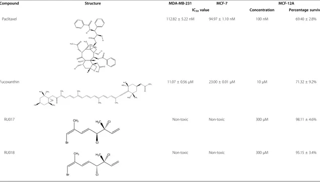
The differential toxicity of the algal polyhalogenated monoterpenes RU017 and RU018, as well as the carot-enoid fucoxanthin (FXN) and the chemotherapeutic pac-litaxel (Ptx), against breast cancer and non-transformed breast epithelial cells was assessed by MTT assay. In this assay, Ptx was found to decrease the percentage survival of immortalized non-transformed breast epithelial cells (MCF-12A) to approximately 70% at a concentration equivalent to the IC50 value of the compound in both
metastatic (MDA-MB-231) and non-metastatic (MCF-7) breast cancer cellsin vitro(Table 1). In the case of FXN, the metastatic MDA-MB-231 cells were more suscep-tible to the carotenoid than non-metastatic MCF-7 cells, while MCF-12A breast epithelial cells displayed a mod-erate susceptibility to the compound at a concentration of 10 μM. For the halogenated monoterpenes RU017 and RU018, neither of the compounds was toxic to MCF-7 or MDA-MB-231 breast cancer or MCF-12A non-transformed breast epithelial cell lines, even at a concentration of 300μM (Table 1).
MCF-7 breast cancer cells form mammospheres in anchorage-independent serum-free culture conditions The mammosphere assay was carried out for the MCF-7 breast cancer cell line in order to enrich for cancer stem cell-like cells, which are able to grow in anchorage-independent serum-free conditions [11]. The develop-ment of non-adherent tumourspheres or mammospheres [11] under these conditions was observed over the course of one week. As depicted in Figure 1, the single-cell suspension seeded on Day 0 had formed small, ir-regular cell “clumps” by Day 1 (Figure 1Ai), which had developed into small suspended colonies representative of mammospheres by Day 3 (Figure 1Aii). By Day 5, the mammospheres had increased in size to approximately 100 μM (0.1 mm) in diameter and displayed a more regular spherical three-dimensional shape (Figure 1Aiii). After seven days growth in anchorage-independent conditions, the mammospheres, while remaining roughly the same size as Day 5, began to exhibit different morphologies (Figure 1Aiv). The most striking feature of the Day 7 cultures was the formation of hollow mammospheres; empty bubble-like structures surrounded by one or more cells or small mammospheres attached to what appeared to be an outer membrane of the hollow spherical bodies (Figure 1Aiv, inset bottom right). The number of mammospheres per well increased steadily from Day 1 to Day 7 (Figure 1B), while the number of single cells and clusters containing one to three cells were observed to decrease with increased incubation time (data not shown).
Table 1 Differential cytotoxicity screening of paclitaxel and novel algal compounds against breast cancer and non-transformed breast epithelial cells
in vitro
Compound Structure MDA-MB-231 MCF-7 MCF-12A
IC50value Concentration Percentage survival
Paclitaxel 112.82 ± 5.22 nM 94.97 ± 1.10 nM 100 nM 69.40 ± 2.8%
Fucoxanthin 11.07 ± 0.56μM 23.00 ± 0.01μM 10μM 71.32 ± 9.2%
RU017 Non-toxic Non-toxic 300μM 98.11 ± 4.6%
RU018 Non-toxic Non-toxic 300μM 95.15 ± 3.4%
de
la
Mare
et
al.
Cancer
Cell
Internationa
l
2013,
13
:39
Page
3
of
14
http://ww
w.cancerci.com
/content/1
Figure 1breast cancer cells form cancer stem cell-enriched mammospheres under serum-free culture conditions. A) Alterations in number, size and shape of mammospheres was observed over the course of seven days. Photographs were taken at Day 1 (i), Day 3 (ii), Day5 (iii) and Day 7 (iv). Images are representative of at least three randomly selected fields. Insets show specific structural features of MCF-7
Mammosphere-derived cells (anchorage-independent) also displayed increased expression of the stem cell marker Oct3/4 compared to those derived from the bulk adherent population, as assessed by Western analysis (Figure 1E).
The polyhalogenated monoterpene stereoisomers RU017 and RU018 inhibit the formation of MCF-7 mammospheres The effect of the marine algal polyhalogenated mono-terpenes RU017 and RU018, as well as the carotenoid FXN, on the formation and development of MCF-7 mammospheres was assessed by addition of these com-pounds to the culture medium either at seeding or after four days growth in anchorage-independent conditions (Figure 2). The compounds were added at a concentration equivalent to the IC50 values in
MDA-MB-231 cells as determined by MTT assay (Table 1). In the case of the non-toxic compounds RU017 and RU018, a concentration of 300 μM was selected. This high concentration of the compounds was not, how-ever, toxic to the MCF-7 breast cancer or MCF-12A breast epithelial cell lines under regular adherent con-ditions (Table 1). However, under non-adherent condi-tions, both of the latter compounds prevented the formation of MCF-7 mammospheres, leaving only sin-gle cells or clusters of two or three cells in the treated samples by Day 3, which were further decreased by Day 6 (Figures 2Ab and 2Ac). In comparison, MCF-7 cells treated with the vehicle control (dimethyl sulphoxide, DMSO) formed distinct mammospheres by Day 3 (Figure 2Aa ii). The carotenoid FXN caused a statistically significant decrease in the sphere forming efficiency (SFE) both at Day 3 and Day 6 (Figure 2B) and resulted in smaller mammospheres in general compared to the control (Figure 2Ad vs. Figure 2Aa). The compound was, however, unable to inhibit mammosphere formation completely (Figures 2Ad and 2B).
For all of the algal compounds tested, the effect of the compounds on sphere forming efficiency when added at Day 4 differed from that obtained when the compounds were added upon seeding of the MCF-7 cells (Figure 2A iii vs. iv). When added to existing MCF-7 mammospheres on Day 4, none of the compounds screened were able to re-move the existing MCF-7 mammospheres or prevent their further development (Figures 2A iv and 2B). The addition of FXN after four days resulted in the formation of much larger, irregularly shaped mammospheres than those ob-served in the DMSO-treated MCF-7 cells (Figure 2Ad iv vs. Figure 2Aa iv). In contrast to the effects when added upon seeding, neither of the stereoisomers RU017 or RU018 were able to eliminate the MCF-7 mammospheres or affect their further development when added at Day 4 (Figures 2Ab iv and 2Ac iv, respectively).
The WST-1 cell proliferation assay was carried out on treated mammospheres (treated both at seeding and on Day 4) after eight days growth in anchorage-independent culture conditions. The percentage survival values for each sample were calculated relative to the DMSO-treated vehicle control (taken as 100%) after 8 days growth in anchorage-independent conditions when equal numbers of cells are seeded, and are indicated in Figure 2C. While FXN produced a (non-significant) re-duction in cellular survival to 70% when added upon seeding, the compound was unable to decrease cell survival when added four days after seeding (107% sur-vival relative to DMSO-treated cells). The halogenated monoterpenes RU017 and RU018 both resulted in statis-tically significant decreases in cellular survival (53% and 44%, respectively) when added at Day 0, while the slight reduction in survival (to 84% and 77%, respectively), when added at Day 4 was not statistically significant.
In order to further characterize the effect of RU017 and RU018 on sphere forming efficiency as observed in Figure 2B, untreated mammospheres were dissociated and seeded into regular anchorage-dependent growth conditions, followed by treatment with either of the algal compounds and assessed for viability using an MTT assay. In this assay, it was found that the algal com-pounds had a small but statistically significant effect on cell viability when compared to the vehicle control, but that neither were able to reduce cell viability to below 78% (Figure 2D). This minor reduction in viability was similar to that observed when cells were treated with the monoterpenes in anchorage-independent mammosphere conditions after four days growth, but differed from that observed when cells were treated upon seeding into anchorage-independent culture conditions (Figure 2C).
The inhibitory effect of the marine algal compounds RU017 and RU018 on MCF-7 mammosphere formation in vitrois dose-dependent
The effects of the algal compounds RU017, RU018 and FXN on the formation and development of MCF-7 mammospheres were more thoroughly investigated by determining whether the observed alterations to the mammospheres were dose-dependent. In addition, the effect of various concentrations of the chemotherapeutic agent, Ptx, on sphere forming efficiency was assessed. For both of the monoterpene stereoisomers, RU017 and RU018, the inhibition of MCF-7 mammosphere forma-tion appeared to be dose-dependent (Figures 3A i and ii, respectively; Figure 3B). In each case, treatment with 50
μM, but not 25μM, of the compounds had a significant effect on the number (Figure 3B) and size of the MCF-7 mammospheres formed after six days, although the mammospheres treated with 25μM were observed to be more irregular in shape when compared to the
DMSO-de la Mareet al. Cancer Cell International2013,13:39 Page 5 of 14
treated control (Figures 3Ab and 3Ac, i and ii respect-ively). The latter concentrations of RU017 and RU018 did not, however, reduce cellular viability of the treated mammospheres compared to the DMSO control as de-termined by WST-1 assay (Figure 3C). For both haloge-nated monoterpenes, treatment with 100 μM appeared to inhibit mammosphere formation, resulting only in small cell clumps (Figure 3Ad, i and ii, respectively). However, in the WST-1 assay, the decrease in percent-age viability relative to the control was statistically significant only in the case of RUMB-018 (Figure 3C). Treatment of MCF-7 cells upon seeding in anchorage-independent conditions with 300μM of either RU017 or RU018 prevented mammosphere formation entirely and significantly reduced cell viability for both compounds (Figures 3Ae i and ii, respectively; Figures 3B and C).
In the case of the carotenoid compound FXN, none of the concentrations tested were able to completely elim-inate mammosphere formation when added to MCF-7 cells upon seeding into anchorage-independent conditions, although a dose-dependent decrease in mammosphere size was observed (Figure 3A iii). The effects of FXN on sphere forming efficiency and cell viability, however, were not dose-dependent (Figures 3B and C). For all concentrations tested, FXN was unable to reduce cell viability to below 76% relative to the DMSO-treated control (Figure 3C).
The chemotherapeutic drug Ptx appeared to increase the number of MCF-7 mammospheres when 50 nM was added upon seeding (Figures 3Ab iv and 3B), while treat-ment with 100 nM had little effect on sphere forming efficiency compared to the DMSO-treated control (Figures 3Ac iv and 3B). This was despite the latter con-centration being reported as the IC50 value for MCF-7
cells under adherent conditions [18]. In comparison, using the WST-1 cytotoxicity assay, Ptx led to a minor but statistically significant reduction in mammosphere viability when added at both 50 and 100 nM in anchorage-independent mammosphere conditions. Im-portantly, neither of the concentrations tested were able to reduce cellular viability to below 84% (Figure 3C).
Pre-treatment of adherent MCF-7 cultures with algal compounds or Ptx does not prevent mammosphere formation
The compounds RU017, RU018, FXN and Ptx were used to pre-treat MCF-7 cells grown under normal dependent conditions prior to seeding under anchorage-independent mammosphere conditions. The consequences of such treatment on mammosphere development and via-bility were compared to those obtained for treatment of mammospheres at seeding (Day 0) or after four days growth in mammosphere conditions (Day 4).
For all four of the compounds RU017, RU018, FXN and Ptx, pre-treatment of MCF-7 cells in
anchorage-dependent culture conditions prior to seeding into anchorage-independent conditions in the mammosphere assay was unable to either prevent mammosphere formation (Figure 4A i) or effectively reduce cell viability relative to the DMSO control (Figure 4C). Although pre-treatment with FXN resulted in a statistically signifi-cant decrease in sphere forming efficiency after 6 days incubation in anchorage-independent conditions (Figure 4B), the compound did not completely inhibit mammosphere formation (Figure 4Ai d) or reduce cellu-lar viability of pre-treated mammospheres (Figure 4C). Interestingly, pre-treatment with the chemotherapeutic agent Ptx, though having little effect on sphere forming efficiency (Figure 4B), appeared to produce larger, more regular shaped mammospheres when compared to the DMSO vehicle-treated control (Figure 4Ai e vs. a), while having little effect on cell viability (Figure 4C).
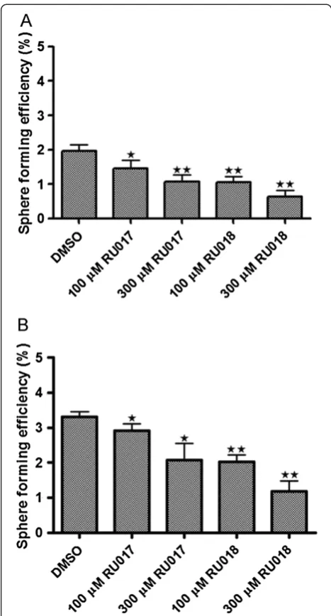
Treatment of second and third generation MCF-7 mammospheres with the algal compounds RU017 and RU018 causes a dose-dependent decrease in sphere forming efficiency
The inhibitory effect on MCF-7 mammosphere formation by the halogenated monoterpenes RU017 and RU018 was further assessed by screening these compounds against second and third generation mammospheres. Untreated mammospheres were dissociated, reseeded under anchorage-independent conditions and treated with either 100μM or 300μM of RU017 or RU018. In both cases, although the compounds were unable to com-pletely prevent MCF-7 mammosphere formation as was the case for primary mammospheres, treatment upon seeding of second and third generation mammospheres resulted in a dose-dependent decrease in sphere forming efficiency (Figure 5A and B). However, the decrease in SFE was greater for both compounds in the second vs. the third generation mammospheres. The inhibitory effect on mammosphere formation was not equal between the stereoisomers, with RU018 being more effective than RU017 in inhibiting both second and third gener-ation mammosphere formgener-ation (Figure 5A and B). However, neither of the compounds had a significant effect on cell viability in either second or third gener-ation mammosphere assays (data not shown).
Discussion
Figure 2The polyhalogenated monoterpenes RU017 and RU018 inhibit the formation but not development of MCF-7 mammospheres.
A) Mammosphere cultures were treated with either the DMSO vehicle control (a), 300μM of either of the halogenated monoterpene
stereoisomers RU017 (b) or RU018 (c) or10μM fucoxanthin (FXN) (d). The latter concentrations were derived from the IC50values in MDA-MB-231 or MCF7 breast cancer and MCF12A non-cancerous cells grown in anchorage-dependent conditions in the case of FXN. Images were captured under a light microscope at either 400× or 100× magnification using a Nikon Coolpix 990 camera on Day 1 (i), Day 3 (ii) and Day 6 (ii) after treatment with the relevant compounds upon seeding and on Day 6 for mammospheres treated with compounds four days after seeding (iv). Images were representative of at least three randomly selected fields for each treatment. Analysis of images and calculation of scale was carried out using ImageJ (NIH freeware). Scale bars representative of 0.1 mm.B) Sphere forming efficiency (SFE) andC) mammosphere viability on Day 8 as determined by WST-1 assay.D) Untreated mammospheres were dissociated and seeded as a single cell suspension into regular culture media containing either the vehicle control or 300μM of RU017 or RU018, cultured under adherent conditions and assessed for viability by means of an MTT assay. The percentage viability inC) andD) was calculated relative to the DMSO vehicle-treated control (taken as 100%) when an equal numbers of cells are seeded and treated for the same period. ForB),C) andD) error bars indicate the standard error of the mean where n = 3 (B) or 5 (CandD). Statistical significance was calculated using a Studentst-test (**p < 0.01, *p < 0.05).
de la Mareet al. Cancer Cell International2013,13:39 Page 7 of 14
only a specific subset of the bulk MCF-7 population, displaying unique functional and phenotypic character-istics, was capable of propagation in suspension and validated the mammosphere assay employed [7,19]. While the addition of the test compounds at the stage of seeding of MCF-7 cells into anchorage-independent
culture conditions assessed the effect on mammosphere formation, dosing with the compounds to existing mammospheres on Day 4 measured the ability to remove the mammospheres or alter further development. In addition, pre-treatment with compounds prior to seeding into mammosphere conditions provided preliminary insight
as to the mechanism of action, in particular the ability to remove CSCs from the bulk population when grown under adherent conditions.
The commonly used chemotherapeutic agent, Ptx, was unable to prevent mammosphere formation when added at a concentration which was toxic to both breast cancer and non-transformed breast epithelial cells under adher-ent conditions, nor was the drug able to reduce mammosphere viability to below 84%. This is consistent with previous reports which demonstrated that 10 ng/L (12 pM) of Ptx had no effect on MDA-MB-231 mammosphere survival [20]. In addition, when added at a concentration of roughly half the IC50value of MCF-7
cells, Ptx appeared to promote mammosphere formation and pre-treatment of MCF-7 cells prior to seeding pro-duced larger, more defined mammospheres. This data suggests that traditional chemotherapy regimens such as Ptx may be ineffective against cancer cells growing in suspension (as occurs in the bloodstream during metas-tasis) or that the cancer stem cell sub-population may be resistant to these agents, as has been suggested in the literature [21]. However, significant further work is required in order to substantiate these hypotheses.
The carotenoid compound, fucoxanthin (FXN), was shown to be toxic to MDA-MB-231 and MCF-7 breast cancer cells and, to a lesser extent, non-cancerous MCF12A breast epithelial cells. However, the com-pound was unable to prevent the formation of MCF-7 mammospheres when added either upon seeding or at Day 4, or when used to pre-treat MCF-7 cells prior to seeding into anchorage-independent mammosphere condi-tions. The inability of FXN to inhibit mammosphere forma-tion and development revealed that the anti-mammosphere activity was not a feature shared by all compounds of marine algal origin.
In the case of the halogenated monoterpene stereoiso-mers RU017 and RU018, both of which were shown to have no effect on the survival and proliferation of either breast cancer cells or non-transformed breast epithelial cells in vitro, the compounds were able to completely
inhibit mammosphere formation when added to MCF-7 cells upon seeding in the mammosphere assay. Since mammosphere formation is reportedly dependent on the presence of CSC [9] and isolated mammospheres in this study showed an increase in cells expressing the CD44
+
/CD24- breast cancer stem cell (BCSC) phenotype, the ability of RU017 and RU018 to inhibit mammosphere formation suggested that these compounds have putative anti-CSC activity and may inhibit a signal transduction pathway which is essential in mammosphere formation. Compounds RU017 and RU018 were not, however, able to remove existing mammospheres when added on Day 4. This could be attributed either to the inability of the compounds to penetrate the mammospheres or could indicate that they were only inhibitory during the early stages of mammosphere formation. This was supported by the fact that pre-treatment of MCF-7 cells with either RU017 or RU018 prior to seeding into anchorage-independent conditions had no effect on the ability of the cells to form mammospheres under these conditions. A possible explanation for this is that the effect of the compounds on the MCF-7 cells may be specific to their initial propagation under anchorage-independent serum-free growth conditions. This is supported by the fact that dissociated untreated mammospheres seeded into regu-lar anchorage-dependent conditions and then treated with the algal compounds experience only a minor decrease in viability as opposed to the large decrease in viability seen when cells are treated with these compounds upon seeding into anchorage-independent conditions.
To further assess the efficacy of the algal compounds, RU017 and RU018, their effect on sphere forming effi-ciency was assessed for second and third generation mammospheres. In both cases, although the compounds were unable to prevent second or third generation mammosphere formation as was the case for primary mammospheres, they did result in a dose-dependent decrease in sphere forming efficiency. This reduced ability of the algal compounds to inhibit mammosphere formation in subsequent generations may be due to
(See figure on previous page).
Figure 3The inhibitory effect of RU017 and RU018 on MCF-7 mammosphere formation is dose-dependent. A) Photographs of mammospheres formed after six days incubation in anchorage-independent serum-free conditions. MCF-7 cells were seeded as a single cell suspension into media containing either (a) DMSO vehicle control or a range of concentrations of the halogenated monoterpene stereoisomers (i) RU017 or (ii) RU018 [(b)25, (c)50, (d)100 or (e)300μM], (iii) fucoxanthin [(b)5, (c)10 or (d)15μM] or (iv) paclitaxel [(b)50 or (c)100 nM]. The latter concentrations of compounds tested was informed by the cytotoxicity values observed under adherent conditions. Cells were photographed under a light microscope at 100× magnification. All images are representative of at least three randomly selected fields and were set to the same scale using ImageJ (NIH freeware). Scale bars representative of 0.1 mm.B) Quantification of the mammospheres generated in terms of sphere forming efficiency (SFE) after six days for the various treatments. SFE was calculated as the number of spheres (average diameter = 100μm) formed in 96 wells plated with a single cell divided by the original number of single cells seeded and expressed as a percentage.C) Cell viability in treated mammosphere samples as assessed by WST-1 assay. The percentage viability after each of the treatments inC) was calculated relative to the DMSO-treated negative control (taken as 100%) after 8 days growth when an equal numbers of cells are seeded. Error bars indicate standard deviation where n = 3 (B) or 5 (C). Statistical significance of the differences in SFE and percentage survival relative to the DMSO control were calculated using a Studentst-test (**p < 0.01, *p < 0.05).
de la Mareet al. Cancer Cell International2013,13:39 Page 9 of 14
accumulated changes in growth characteristics of the cells as a consequence of prolonged culturing under anchorage-independent serum-free conditions, resulting in a more aggressive phenotype. This could also account for the increase in sphere forming efficiency in the ve-hicle control for the third generation as compared to that of the second generation. Interestingly, RU018 appeared more effective than RU017 in inhibiting second and third generation mammosphere formation, a finding which was consistent with the trend observed in terms of first generation mammosphere cell viability. This data implies that the stereochemistry of the monoterpene compounds plays a role in their mechanism of action against MCF-7 mammospheres.
The identification of CSCs within breast tumours has resulted in a major shift in focus in terms of the devel-opment of novel therapies to treat this disease [22]. It is now thought that complete eradication and preven-tion of relapse requires the removal of the stem cell subpopulation within a tumour [23]. Traditional treat-ments, such as chemotherapy and radiation, were ori-ginally developed to kill the rapidly dividing bulk population of cells within the tumour [22]. However, while these therapies are able to shrink the tumour, the effects are often transient and recurrence remains a reality for a substantial proportion of sufferers [22,24]. This has been attributed to the resistance of the CSC subpopulation to traditional therapies, such that these highly tumourigenic cells remain behind after chemo-therapy or radiation treatment [23,24]. Therefore, there is a need to find agents which are able to specific-ally inhibit CSCs.
Conclusions
The body of work described herein reports the first screening of marine algal compounds in a mammosphere assay. In particular, screening of the compounds FXN, RU017 and RU018 revealed that the latter monoterpene stereoisomers inhibited MCF-7 mammosphere formation. In contrast, the commonly used chemotherapeutic drug Ptx appeared to enhance both the formation and matur-ation of early mammospheres. More work is required to determine the specific molecular mechanisms mediating the mammosphere inhibitory activity of the halogenated monoterpenes as well as their respective cellular targets. In addition, the concentration of RU017 and RU018 re-quired for such activity falls outside the druggable range and rational chemical modification is needed to improve their efficacy. Since the compounds form part of a struc-tural series isolated from Plocamium corallorhiza and
Plocamium cornutum[17], they could find application as tool compounds to study CSC selectivity in halogenated monoterpenes.
Figure 5The halogenated monoterpenes RU017 and RU018 have a dose-dependent inhibitory effect on second and third generation mammosphere formation.Untreated mammospheres of first and second generation were dissociated and reseeded into anchorage-independent conditions in serum-free culture media containing the compounds of interest in order to assess second (A) and third generation (B) sphere forming efficiency (SFE), respectively. In both cases, mammosphere-derived MCF-7 cells were seeded as a single cell suspension into media containing either the DMSO vehicle control or either 100 or 300μM of the halogenated monoterpene stereoisomers RU017 or RU018. SFE was calculated as the number of spheres (average diameter = 100μm) formed after six days in 96 wells plated with a single cell divided by the original number of single cells seeded and expressed as a percentage. Error bars indicate the standard error of the mean where n = 5. Statistical significance of the differences in SFE relative to the DMSO control were calculated using a Studentst-test (**p < 0.01, *p < 0.05).
de la Mareet al. Cancer Cell International2013,13:39 Page 11 of 14
Methods Reagents
Dulbecco’s Modified Eagle Medium (DMEM) containing Glutamax™, Ham’s F-10 Medium containing Glutamax™, foetal calf serum (FCS), B-27 supplement and penicillin-streptomycin-amphotericin (PSA) were obtained from Gibco (Invitrogen). Epidermal growth factor (EGF), basic fibroblast growth factor (bFGF), hydrocortisone, heparin, Accutase®, paclitaxel and fucoxanthin were from Sigma-Aldrich. Insulin was obtained from NovoRapid (Novo Nordisk Pharmaceuticals). The MTT and WST-1 Cell Proliferation kits were from Roche. Hoescht 33342 dye for flow cytometric analyses was obtained from Invitrogen, while the allophycocyanin (APC)-conjugated mouse anti-human CD24 and fluorescein isothiocyanate (FITC)-conjugated rat anti-human CD44 antibodies were from e-Biosciences, as were the isotype controls. The mouse anti-human Oct3/4 antibody was obtained from Santa Cruz, while the goat mouse and goat anti-rabbit antibodies were from Sigma. The anti-rabbit anti-histone H3 antibody was from Cell Signalling Technologies.
Cell lines and culture conditions
The metastatic breast cancer cell line, MDA-MB-231 (ATCC: HTB-26) was maintained in culture in Dulbecco’s Modified Eagle Medium (DMEM) containing Glutamax™ and supplemented with 5% (v/v) heat-inactivated FCS, 100 U/ml penicillin, 100μg/ml streptomycin and amphotericin (PSA) at 37ºC in a humidified 9% CO2incubator. The
im-mortalized, non-transformed breast epithelial MCF-12A cells (ATCC: CRL-10782) were maintained using a 1:1 ratio of Ham’s F10 and DMEM containing Glutamax™ and supplemented with 10% (v/v) heat-inactivated FCS, PSA (as for MDA-MB-231 cells), 20 ng/ml EGF, 500 ng/ml hydro-cortisone and 10μg/ml insulin. The breast cancer cell line MCF-7 (ATCC: HTB-22) was maintained in culture either in anchorage-dependent or anchorage-independent condi-tions. In the former, cells were cultured as for the MDA-MB-231 line in regular 96-well plates.
Mammosphere assay
Anchorage-independent growth was assessed by mammosphere assay, modified from that previously described [11]. Briefly, cells were lifted with 0.25% (v/v) trypsin in 0.61% (w/v) ethylenediaminetetracetic acid (EDTA), washed with phosphate-buffered saline (PBS) (137 mM NaCl, 2.7 mM KCl, 10 mM Na2HPO4,
2 mM KH2PO4 pH 7.4) and passed through a 40 μM
cell strainer (BD Biosciences) to achieve a single cell suspension. Cell were seeded at a density of 1000 cells per well in ultralow attachment 96-well plates containing DMEM with Glutamax™supplemented with 1% (v/v) PSA, 2% (v/v) B-27 supplement, 20 ng/ml EGF and bFGF, 4 ng/ml heparin and 10μg/ml insulin.
Treatment with either 0.61% (v/v) dimethyl sulphoxide (DMSO) vehicle control or paclitaxel (Sigma-Aldrich), fucoxanthin (Sigma-Aldrich), RU017 or RU018 was carried out either upon seeding or at Day 4 for quintu-plicate samples. The cells were fed every 48 hours by the addition of fresh medium to existing culture vol-ume and the resultant mammospheres photographed in triplicate using a Nikon camera (Coolpix 990) attached to a light microscope at 100 × magnification. Images were analyzed and scale bars calculated using ImageJ (NIH). Quantification in terms of Sphere Forming Efficiency (SFE) was carried out by counting of mammospheres under a light microscope at 10× magnification and reported as the number of mammospheres/spheres (average diameter = 100μm) formed in 96 wells divided by the original number of single cells seeded and expressed as a percentage. Statistical significance for the various treatments was assessed using a Student’st-test in GraphPad Prism (GraphPad Inc.).
Dissociation of mammospheres for cell viability as-says and screening of second and third generation mammospheres was achieved using Accutase® solution (Sigma). Briefly, the mammospheres were collected by centrifugation, resuspended in 200 uL of 1 × Accutase® solution and incubated for 15 minutes before passing through a 40 μM cell strainer (BD Biosciences) to achieve a single cell suspension. Mammosphere-derived cells were seeded at a density of 6000 cells per well under regular anchorage-dependent conditions for cell viability analysis by MTT assay and at 1000 cells per well under anchorage-independent mammosphere culture con-ditions for second and third generation screening assays.
Cell surface marker analysis by flow cytometry
were recorded for each sample. Flow cytometry data was analysed using FlowJo software (Tree Star Inc.). Isotype controls were used to establish fluorescence threshold gates with the gates set on the isotype control to exclude the major population of cells. Gates were then copied onto the respective samples in order to determine the CD44/CD24 expression profile of the sample. Two-colour dot plots were then constructed showing FITC (CD24) fluorescence on the y-axis and APC (CD44) fluorescence on the x-axis.
Confocal microscopy analysis of anchorage-independent MCF-7 breast cancer cells
Anchorage-independent cells were seeded into 8-well chambered coverslips overnight. Cells were fixed by flash incubation in ice-cold methanol and left to air-dry before incubation in a blocking solution [1% (w/v) bovine serum albumin (BSA) in Tris-buffered saline (TBS)] at room temperature for 30 minutes. A primary antibody solution containing anti-CD44 conjugated to APC in 0.1% (w/v) BSA/TBS was added to the cells and incubated overnight at 4ºC. Cells were washed twice with agitation in blocking solution, after which the cells were washed in distilled water containing 1 μg/ml of DNA binding dye Hoechst 33342. The coverslips were mounted onto glass slides using Dako mounting media (Dako). Differential contrast (DIC) and fluorescent images were taken using a Zeiss LSM510 meta confocal microscope.
Western blot analysis
MCF-7 cells were seeded at a density of 4000 cells per well in anchorage independent mammosphere conditions and mammospheres allowed to develop over 8 days. In addition MCF-7 cells were cultured in anchorage-dependent regular culture conditions and lifted using 0.25% (v/v) trypsin. Whole cell lysates were prepared from adher-ent and mammosphere cultures. Briefly, cells were collected by centrifugation at 2000 rpm for 3 minutes and resuspended in 100μM SDS sample buffer (125 mM Tris– HCl pH 6.8, 2% (w/v) SDS, 20% (v/v) glycerol, 0.2% (w/v) bromophenol blue) before boiling for 5 minutes. Thereafter, 30 ug of protein from each lysate was loaded onto a 12% SDS polyacrylamide gels, separated by electrophoresis and transferred onto a nitrocellulose membrane, before probing with mouse anti-Oct3/4 (1 in 2000) at 4ºC overnight. The HRP-conjugated anti-mouse secondary antibody was detected using the ECL Advanced Western Blotting Kit (GE Healthcare) and visualized using the Molecular Imager ChemiDoc XRS System (BioRad). Lysates were probed with rabbit anti-histone H3 antibody (1 in 2500) as a loading control.
Extraction of compounds from marine algae
Specimens of the red alga Plocamium cornutum were gathered off the south-east coast of South Africa and
the halogenated monoterpenes extracted as previously described [25].
Cell viability assays
Cell viability after treatment with the compounds was eval-uated by means of the MTT assay in the case of adherent cells and WST-1 assay in the case of mammospheres, according to manufacturer’s instructions. For both assays, percentage viability after treatment with compounds was calculated relative to the DMSO vehicle control (taken as 100% viability) when equal numbers of cells are seeded. For the MTT Cell Proliferation Kit 1, MDA-MB-231, MCF-7 or MCF-12A cells were seeded at a density of 6000 cells/ well in 96-well plates and assessed for viability as previously described [17]. In the case of the WST-1 kit, after treatment and growth of the mammospheres for 8 days, 10μL of a 5 mg/ml WST-1 reagent was added to each well and incu-bated for a further 4 hours before reading the absorbance at 450 nm. Viability assays were carried out in quintuplicate and statistical significance assessed using GraphPad Prism.
Abbreviations
APC:Allophycocyanin; BCSC: Breast cancer stem cell; BSA: Bovine serum albumin; CSC: Cancer stem cell; DMEM: Dulbecco’s Modified Eagle Medium; DMSO: Dimethyl sulphoxide; EGF: Epidermal growth factor; FCS: Fetal calf serum; FGF: Fibroblast growth factor; FITC: Fluorescein isothiocyanate; FXN: Fucoxanthin; PBS: Phosphate-buffered saline; PSA: Penicillin-streptomycin-amphotericin; Ptx: Paclitaxel; SFE: Sphere forming efficiency; TBS: Tris-buffered saline.
Competing interests
The authors declare that they have no competing interests.
Authors’contributions
ALE, JdlM and GLB were involved in the conception and design of the study. JdlM and ALE drafted the manuscript. JdlM carried out the MCF-7
mammosphere assays and screening of marine compounds. JNS carried out the flow cytometry and MGS the Oct4 Western analysis. MTC and DRB were responsible for the purification of the compounds from marine algae. All of the authors read and approved the manuscript.
Acknowledgements
This research was supported by grants from the Cancer Research Initiative of South Africa (CARISA), the Medical Research Foundation (MRC) and the South African National Research Foundation (NRF). JdlM and JNS were supported by postgraduate scholarships from Rhodes University (RU), the NRF and RU Henderson Foundation (JdlM). MGS was supported by postdoctoral funding from Rhodes University (RU).
Author details
1The Biomedical Biotechnology Research Unit (BioBRU), Department of
Biochemistry, Microbiology and Biotechnology, Rhodes University, P. O. Box 94, Grahamstown 6140, South Africa.2Division of Pharmaceutical Chemistry,
Faculty of Pharmacy, Rhodes University, Grahamstown, South Africa.3College
of Health and Biomedicine, Victoria University, Melbourne, Australia.
Received: 7 November 2012 Accepted: 23 April 2013 Published: 1 May 2013
References
1. Lawson JC, Blatch GL, Edkins AL:Cancer stem cells in breast cancer and metastasis.Br Canc Res Treat2009,118:241–254.
2. Visvader JE:Cells of origin in cancer.Nature2011,469:314–322. 3. Bonnet D, Dick JE:Human acute myeloid leukemia is organized as a hierarchy
that originates from a primitive hematopoietic cell.Nat Med1997,3(7):730–737.
de la Mareet al. Cancer Cell International2013,13:39 Page 13 of 14
4. Wicha MS, Liu S, Dontu G:Cancer stem cells: an old idea–a paradigm shift.Canc Res2006,66:1883–1890.
5. Al-Hajj M, Wicha MS, Benito-Hernandez A, Morison SJ, Clarke MF:
Prospective identification of tumourigenic breast cancer cells.Proc Nat Acad Sci2003,100(7):3983–3988.
6. Patrawala L, Calhoun T, Schneider-Broussard R, Zhou J, Claypool K, Tang DG:
Side population is enriched in tumourigenic, stem-like cancer cells, whereas ABCG2+and ABCG2-cancer cells are similarly tumourigenic.Canc Res2005,
14(65):6207–6219.
7. Ponti D, Costa A, Zaffaroni N, Pratesti G, Petrangolini G, Coradini D, Pilotti S, Pierotti MA, Daidone MG:Isolation and in vitro propagation of tumourigenic breast cancer cells with stem/progenitor cell properties.Canc Res2005,
13(65):5506–5511.
8. Conley SJ, Gheordunescu E, Kakarala P, Newman B, Korkaya H, Heath AN, Clouthier SG, Wicha MS:Antiangiogenic agents increase breast cancer stem cells via the generation of tumor hypoxia.Proc Nat Acad Sci2012,
109:2784–2789.
9. Gupta PB, Onder TT, Jiang G, Tao K, Kuperwasser C, Weinberg RA, Lander ES:
Identification of selective inhibitors of cancer stem cells by high-throughput screening.Cell2009,138:645–659.
10. Reynolds BA, Rietze RL:Neural stem cells and neurospheres–re-evaluating the relationship.Nat Meth2005,5(2):333–336.
11. Dontu G, Abdallah WM, Foley JM, Jackson KW, Clarke MF, Kawamura MJ, Wicha MS:In vitropropagation and transcriptional profiling of human mammary stem/progenitor cells.Genes Dev2003,17(10):1253–1270. 12. Yeom Y, Fuhrmann G, Ovitt CE, Brehm A, Ohbo K, Gross M, Hubner K, Scholer HR:Germline regulatory element of Oct 4 specific for the totipotent cycle of embryonal cells.Development1996,122:881–894. 13. Jha RK, Zi-rong X:Biomedical compounds from marine organisms.
Mar Drugs2004,2:123–146.
14. Carté BK:Biomedical potential of marine natural products.Biosci1996,
46(4):271–286.
15. Andrianasolo EH, France D, Cornell-Kennon S, Gerwick WH:DNA methyl transferase inhibiting halogenated monoterpenes from the Madagascar red marine algaPortieria hornemannii.J Nat Prod2006,69(4):576–579. 16. Antunes EM, Afolayan AF, Chiwakata MT, Fakee J, Knott MG, Whibley CE,
Hendricks DT, Bolton JJ, Beukes DR:Identification andin vitro anti-esophageal cancer activity of a series of halogenated monoterpenes isolated from the South African seaweedsPlocamium suhriiand
Plocamium cornutum.Phytochem2011,72:769–772.
17. De la Mare J, Lawson JC, Chiwakata MT, Beukes DR, Edkins AL, Blatch GL:
Quinones and Halogenated Monoterpenes of Algal Origin show Anti-proliferative Effects against Breast Cancer Cellsin vitro.Invest New Drugs
2012. doi:10.1007/s10637-011-9788-0.
18. Dwight ES, Lawrence WD, Carl C, Nanci LW, Hangming R, Gunter D: Paclitaxel-induced apoptosis in MCF-7 breast-cancer cells.Int J Canc1997,70:214–220. 19. Korkaya H, Paulson A, Charafe-Jauffret E, Ginestier C, Brown M, Dutcher J,
Clouthier SG, Wicha MS:Regulation of mammary stem/progenitor cells by PTEN/Akt/β-Catenin Signaling.PLoS Biol2009,7(6):1–14.
20. Fillmore CM, Kuperwasser C:Human breast cancer cell lines contain stem-like cells that self-renew, give rise to phenotypically diverse progeny and survive chemotherapy.Br Canc Res2008,10:R25.
21. Dean M:ABC transporters, drug resistance and cancer stem cells.
J Mammary Gland Biol Neoplasia2009,14:3–9.
22. Kakarala M, Wicha MS:Implications of the cancer stem-cell hypothesis for breast cancer prevention and therapy.J Clin Oncol2008,26:2813–2820. 23. Massard C, Deutsch E, Soria J-C:Tumour stem cell-targeted treatment:
elimination or differentiation.Ann Oncol2006,17:1620–1624. 24. Marotta LLC, Polyak K:Cancer stem cells: a model in the making.
Curr Opin Genet Dev2009,19:1–7.
25. Afolayan AF, Mann MGA, Lategan CA, Smith PJ, Bolton JJ, Beukes DR:
Antiplasmodial halogenated monoterpenes from the marine red alga
Plocamium cornutum.Phytochem2009,70:597–600. doi:10.1186/1475-2867-13-39
Cite this article as:de la Mareet al.:Assessment of potential anti-cancer stem cell activity of marine algal compounds using anin vitro mammosphere assay.Cancer Cell International201313:39.
Submit your next manuscript to BioMed Central and take full advantage of:
• Convenient online submission
• Thorough peer review
• No space constraints or color figure charges
• Immediate publication on acceptance
• Inclusion in PubMed, CAS, Scopus and Google Scholar
• Research which is freely available for redistribution