ORIGINAL RESEARCH
Comparison of Scalpel versus Soft Tissue Diode Laser for
Biopsy of Oral Lesions
Sandeep Kashyap1, V V N Pavan Kumar2, Piyush Prakash3, Naqoosh Haidry4, Narahari Ranganath5, D S J D’Souza6
ABSTRACT
Background: Biopsy is a critical procedure in the diagnosis and treatment plan of surgical cases and is also highly tech-nique-sensitive. Various biopsy techniques are convention-ally used with each technique having its own pros and cons. A study was carried out to compare scalpel versus diode laser for biopsy of oral lesions under various parameters vital to his-topathological diagnosis.
Materials and Methods: A total of 40 patients (20 in each group) satisfying the inclusion criteria were randomly allocated using odd-even scheme into scalpel and laser groups by a blinded operator. The patients were evaluated intraoperatively for patient comfort, bleeding, time taken for excision, and post-operatively for pain and healing. The excised samples were sent for histopathological analysis as follows: Loss of archi-tecture in epithelium, loss of archiarchi-tecture in connective tissue, charring, and artifacts.
Results: The mean time taken for excision was statistically similar in both the groups. Distribution of patient comfort and pain was statistically not significant. Healing was uneventful in both the groups. In the histological parameters, there was loss of architecture in epithelium in 75% of cases in the laser group. In the same group, there was loss of architecture in 85% of the samples, and there was charring in 85% of the specimens.
Conclusion: Both techniques seem to be equally effective in performing excisional biopsies of oral lesions. Laser has the advantage of maintaining a bloodless field and avoidance of suturing as well. However, due to the associated thermal dam-age caused, there may be minor loss of histological architecture.
1,2Assistant Professor, 3Consulatant Oral and Maxillofacial
Surgeon, 4,5Senior Lecturer, 6Professor
1Department of Dental Surgery, Sikkim Manipal Institute of
Medical Sciences, Gangtok, East Sikkim, India,
2Department of Dental Surgery, Kasturi Medical College and
Hospital, Guntur, Andhra Pradesh, India,
3Consulatant Oral and Maxillofacial Surgeon, New Delhi, India,
4Department of Oral and Maxillofacial Surgery, Narsinbhai
Patel Dental College and Hospital, Visnagar, Gujarat, India,
5Department ofOral and Maxillofacial Surgery, Rajarajeswari
Dental College and Hospital, Bengaluru, Karnataka, India,
6Department of Dental Surgery, Maharastra Institute of Medical
Education and Research, Pune, Maharashtra, India
Corresponding Author: Dr. V V N Pavan Kumar, Assistant Professor, Department of Dental Surgery, Kasturi Medical College and Hospital, Guntur, Andhra Pradesh, India. Phone: +91-8555045006. e-mail: pavankumarv.v.n@gmail.com
Keywords: Excisional biopsy, Histopathology, Laser biopsy, Scalpel biopsy.
How to cite this article: Kashyap S, Kumar VVNP, Prakash P, Haidry N, Ranganath N, D’Souza DSJ. Comparison of Scalpel versus Soft Tissue Diode Laser for Biopsy of Oral Lesions. Int J Oral Care Res 2018;6(2):50-55.
Source of support: Nil
Conflict of interest: None
INTRODUCTION
Biopsy is one of the most vital procedures in the diag-nosis and treatment planning for any surgical specialty. The surgeon desires a quick and simple procedure, the histopathologist needs a large, truly representative, and undamaged specimen, and the patient would like a quick, painless operation with minimum post-oper-ative discomfort.[1] Various biopsy techniques used in excision of oral lesions are scalpel biopsy, punch biopsy, electrosurgery, and laser biopsy. The humble scalpel is still routinely employed because of economy, ease of use, accuracy, and minimal damage to adjacent tissues. However, it does not provide good hemostasis, which is critical when operating on highly vascular tissues or other lesions of the maxillofacial region.
It is crucial that the entire biopsy specimen be intact to make an unequivocal histological diagnosis. Hence, any surgical device that creates thermal or mechani-cal damage in the tissue to be examined is not recom-mended for performing biopsies. Due to these potential problems, the feasibility of routinely carrying out laser biopsies has been debatable. Recently, there have been reports of injection laser or diode laser, as a surgical aid to soft tissue surgery in the maxillofacial region. The advantages of diode lasers are minimal post-operative swelling and scarring, decreased post-operative pain, and maintaining bloodless field.[2] A study was carried out at a postgraduate dental institution to compare the efficacy of soft tissue diode laser and scalpel, to perform oral biopsies, using an array of clinical and histological parameters.
MATERIALS AND METHODS
Scalpel vesrus laser in oral biopsies
IJOCR
laser versus scalpel as a surgical aid in performing biopsy of oral lesions, to evaluate the healing process after scalpel and laser biopsy, to evaluate patient com-fort following laser and scalpel surgery, and to analyze the histological parameters of the specimen obtained with each of these techniques.
Patients presenting with benign intraoral lesions requiring biopsy were selected for the study. Appropriate clearance for carrying out the study was obtained prior from the institutional ethics committee. Each participant in the study was counseled by the primary investigator and written informed consent was obtained from each of the participants. Based on review of similar published data as well as looking at previous statistical data of our institute, a figure of 40 patients was arrived at as being statistically adequate and realistically available within the duration of the study.
A total of 40 patients (20 in each group) satisfying the inclusion criteria were included in the study and ran-domly allocated using an odd-even scheme by a blinded operator into scalpel and laser groups. The principal investigator had no role to play in the allotment of the participants of the trial to avoid any bias. Group I included biopsy taken from oral lesions with scalpel. Group II included biopsy taken from oral lesions with soft tissue diode laser.
Excisional biopsy following standard protocols was performed under local anesthesia for each case by the principal investigator each time. 20 patients under-went biopsy using scalpel and sutures were placed. In 20 patients, excision of the lesion was done with 970 nm diode laser. For all the lesions biopsied, a 0.5 mm of safety margin was considered in both the groups.
Efficacy of the technique was evaluated by visual assessment of intraoperative bleeding in both the groups and recording of time taken. Post-operative patient comfort was also recorded. Pain assessment was done after 24 h using a visual analog scale (VAS). Healing was assessed after 1 week. All of the above parameters were assessed by an independent senior fac-ulty who was blinded to the outcomes and parameters of the study. The excised samples were sent for histo-pathological analysis and various parameters such as loss of architecture in epithelium, loss of architecture in connective tissue, charring, and presence of artifacts were recorded. The data collected were tabulated and statistically analyzed.
RESULTS
Statistical Methods
The data recorded were subjected to descriptive and infer-ential statistical analysis as per the originally assigned
groups for proper understanding of the results. Results on continuous measurements are presented as mean ± standard deviation (minimum-maximum) and results on categorical measurements are presented as number (%). Significance was assessed at 5% level of significance.
Student’s t-test (two-tailed, independent) was used to find the significance of study parameters on contin-uous scale between two groups (intergroup analysis) on metric parameters. Chi-square/Fisher exact test was used to find the significance of study parameters on cat-egorical scale between two groups.
The comparative results for effectiveness of scalpel versus soft tissue diode laser for excisional biopsies of benign intraoral lesions are as follows:
Clinical Parameters
Intraoperative parameters [Figure 1]:
1. Patient comfort - In Group I, 40% of patients had a VAS score of 2, 40% of patients with VAS score of 3, 15 % of patients with VAS score of 4, and 5% with VAS score of 1. In Group II, 55% of patients had a VAS score of 2, 30% of patients had VAS score of 3, 15% of patients with VAS score of 3, and none had score 1 (P = 0.759)
2. Incidence of bleeding - 100% of patients in Group I had bleeding during the procedure, whereas none of the patients had bleeding in Group II (P < 0.001) 3. Time taken - In Group I, time taken for the
proce-dure in 55% of patients ranged from 6 to 11 min, in 30% of patients time taken ranged from 11 to 15 min, in 10% of patients ranged from 1 to 5 min, and in 5% of patients ranged from 16 to 20 min. The mean time taken in Group I was 9.15 min. In Group II, time taken for the procedure in 60% of patients ranged from 6 to 10 min, in 15% of patients ranged from 11 to 15 min, in 15% from 1 to 5 min, and in 10% from 16 to 20 min. The mean time taken in Group II was 9.30 min (P = 0.900).
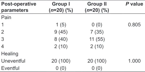
Post-operative parameters [Table 1]:
1. Pain - After 1 day, in Group I, 45% of the patients had a VAS for pain of 2, 40% had pain score of 3, 10% had pain score of 4, and 5% had pain score of 1. After 1 day, in Group II, 55% of patients had a pain score of 3, 35% had a pain score of 2, 10% had pain score of 4, and none had pain score of 1 (P = 0.805)
2. Healing - Healing was uneventful after 1 week in all the 40 patients (P = 1.000).
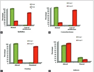
Histopathological parameters [Figure 2]:
2. Connective tissue - In Group I, there was no distor-tion of connective tissue in any of the specimens. In Group II, there was loss of architecture in the con-nective tissue in 85% of the specimens (P < 0.001) 3. Charring - In Group I, there was no charring in any
of the specimens. In Group II, there was charring in 85% of the specimens (P < 0.001)
4. Artifacts - In Group I, artifacts were present in 20% of the specimens. In Group II, artifacts were present in 10% of the specimens (P = 0.376).
DISCUSSION
A comparative study of carrying out excisional biopsy of oral lesions using scalpel versus soft tissue diode
Figure 1: (a) Distribution of patient comfort in the two groups studied, (b) bleeding in the two groups studied, (c) time taken in the two groups studied
a b
c
Figure 2: (a) Histological loss of architecture in epithelium, (b) Histological loss of architecture in connective tissue, (c) Degree of char-ring, (d) Histological artifacts in the two groups studied
a
c
b
Scalpel vesrus laser in oral biopsies
IJOCR
laser was done on 40 patients. Group I consisted of 20 patients in which scalpel was used and 20 patients in Group II that is in laser group. Patients aged 18–60 years were included in both the groups. The mean age of patient in Group I was 36.55 and that in Group II was 43.10. The mean age in both the groups was statistically similar. There were 12 males and 8 females in Group I and 13 males and 7 females in Group II.
The lesions treated included 28 fibromas (14 in Group I and 14 in Group II), 8 mucoceles (5 in Group I and 3 in Group II), pyogenic granuloma (none in Group I and two in Group II), papilloma (one in Group I and none in Group II), and lipoma (none in Group I and one in Group II). Distribution of diagnosis was statistically similar in both groups with P = 0.489
Patient comfort was measured using visual analog scale. There was no statistical difference in the intraop-erative discomfort in both groups. Yet, patients in the scalpel group complained a sense of pricking during suturing. Post-operative discomfort was less in laser as compared to conventional surgical procedures[3] which is also similar to findings reported in a study by Dhabekar et al., in 2010.[4]
Bleeding was observed only in scalpel group. Diode laser has an excellent cutting and coagulation ability with a tolerable damage zone, and hence, there is no post-operative bleeding.[5] Diode laser has also been reported to be more effective than conventional surgery in the reduction of intraoperative bleeding and post-op-erative pain.[6] Vessels up to 500 µm in diameter that supplies capillary and small venous vascular lesions are coagulated, allowing for en bloc excision of vascular lesions and also laser is highly desirable in patients who have coagulation disorders due to decreased potential blood loss compared with scalpel surgery.[7]
The mean time taken in the two groups was com-pared. Measurement of time taken measurement was recorded only after the effect of local anesthesia was firmed. The first incision with scalpel and the first con-tact with laser were taken as the starting time. Complete suturing with scalpel and completion of excision with laser were the end timings. Since there was no bleeding
in the laser group, suturing was not necessary in this group. The cutting efficiency of scalpel was quicker than laser, but some additional time was required for suturing. The mean time taken for the procedure in both the groups was statistically not significant (P = 0.009). Yagüe-García et al. found the total treatment time with laser to be less in comparison to scalpel which required a meticulous technique and also suturing at the end.[8] According to Kafas et al., the disadvantage of diode laser is the time required for excision as compared to scalpel blade.[9]
Pain was measured on the 1st post-operative day in all the patients. Pain was measured using visual ana-log scale. The experience in pain in the two groups was statistically insignificant with P = 0.805. Although the mechanism of analgesic effects of laser therapy is not well understood, an increased pain threshold through the alteration of neuronal stimulation and firing pattern, and the inhibition of the medullary reflexes is thought to be involved.[10] In addition, the laser effect is seen on prostaglandin synthesis, resulting in increased con-version of prostaglandin G2 and prostaglandin H2 into prostaglandin I2 (prostacyclin).[11] In a previous study by Baiju et al. comparing laser and scalpel biopsies, pain observed at the laser treated site as per VAS scale after 24 h was minimal compared to moderate pain in the scalpel-treated group. The same was noted in our study. This can be attributed to the fact that the cellular disintegration caused at the impact site does not allow for the release of inflammatory mediators which causes minimal or no pain in laser wounds.[12] The thin denat-uralized collagen layer observed on the surface of sur-gical wounds following laser surgery acts as a natural barrier to isolate the surgical wound from oral fluids. This further reduces the pain and maintains sterility of the wound.[4]
Postoperatively, the healing process was monitored based on clinical signs and symptoms. Pain, induration, or pus discharge, etc., from biopsy site, were taken into consideration. Scab formation was seen on the scalpel biopsy wounds, and the laser biopsy site was covered by a coagulum which served as a natural barrier from infection. There were no signs of any infection and wound dehiscence in any of the cases. Healing was uneventful in all the cases with P = 1.000. Following laser excision, the associated lymphatics and blood vessels are sealed which results in insignificant extrav-asation of fluids and limited inflammatory reaction. However, in the wound following scalpel excision, there is continued extravasation of blood and lymph fluid, which is manifested as a greater degree of swelling and inflammatory reaction. This is the reason for the longer resolution period.[7] The laser biopsy wounds exhibit a
Table 1: Post-operative parameters in the two groups studied
Post‑operative
parameters (nGroup I =20) (%) (nGroup II =20) (%) P value
Pain
1 1 (5) 0 (0) 0.805
2 9 (45) 7 (35)
3 8 (40) 11 (55)
4 2 (10) 2 (10)
Healing
Uneventful 20 (100) 20 (100) 1.000
growth is seen to commence at the edges and gradually covers the entire wound. The newly formed epithelium in laser biopsy specimens is seen to be more thin and parakeratotic in nature when compared with the epithe-lium formed after scalpel excision. Post-laser wounds show appreciably lesser quantity of myofibroblasts. This results clinically in lesser degree of wound contraction and scarring and shows improved post-operative func-tion, especially in critical areas of the tongue, floor of the mouth, soft palate, and buccal mucosa.[11] The only drawback is that healing occurs at a slower pace which needs to be explained to the patient. Laser wounds thus may heal completely after 2 or 3 weeks when compared to scalpel wounds, which generally heal in a week to 10 days.[13]
Histological analysis of the epithelium, connective tissue, charring, and presence of artifacts was under-taken. It is critical that the right type of laser setting be prudently employed owing to the fact that distinctive thermal effects are seen in biological tissue. This is the only way to ensure optimal clinical effectiveness, while ensuring that there is no destruction to the irradiated tissue. Dark substances such as hemoglobin show a greater degree of absorption and the depth of propaga-tion is also correlated to the wavelength and the absorp-tion coefficient of the irradiated tissue. The acabsorp-tion of most dental lasers occurs through photonic absorption which results in increased temperature (often by more than 100°C) within the tissue being focused on by the laser beam. This is the reason for irreversible or perma-nent damage in the surrounding tissues if the laser is not used judiciously. With this aim in mind, an effort was made to analyze the thermal effects, particularly at the marginal areas of the excised specimen in our study.[14]
A detailed search of the literature showed that diode laser often caused significant tissue damage such as tis-sue necrosis and sloughing and charring of the tistis-sue margin. However, in our study, since the excision was done using relatively low power setting (2.0 W, 810 nm), the tissue sections excised by diode laser, showed lim-ited degree of thermal effects at the margins of the lesion, epithelium, and underlying connective tissue. Instead, coagulation effects were seen in specific areas at the margins, related to epithelium and connective tissue. Despite this, there was no hindrance in carrying out histological diagnosis of the lesions. It is critical to note, however, that in the case of neoplastic or dysplas-tic lesions even these thermal effects, at the margins may cause difficulties in determining the extent of lesion as well as subsequent diagnosis. Hence, it is recommended to keep the incision slightly beyond the margins of the
logical picture.
The histological picture of the epithelium was nor-mal in all the samples of Group I, whereas there was some degree of loss of architecture in 15 samples in Group II. This may be as a result of thermal effects of the laser beam. This loss of architecture in epithelium was statistically significant with P < 0.001. There is no discernable epidermal destruction in histopathologic specimens following scalpel biopsy.[15] The energy transmitted by the laser beam results in warming, weld-ing, coagulation, protein denaturation, dryweld-ing, vapor-ization, and carbonization of the cellular tissue causing the histological changes such as intracellular vacuoliza-tion, cellular hyperchromatism, and loss of intracellular structure, with some degree of charring of tissues. These are described as the cytological artifacts which make the situation difficult to interpret the histological findings, especially in cases where the nature of the lesion is ques-tionable.[16]
The connective tissues were normal in all the sam-ples of Group I, but loss of architecture was present in 17 cases of Group II. This again correlates to the tissue injury caused by the thermal effects of laser. The loss of architecture in connective tissue was statistically sig-nificant with P < 0.001. Pogrel in his study of 23 exci-sional biopsies noted that the greatest degree of thermal effects was seen in dense connective tissue and mucosal epithelium with a lesser amount in loose connective tis-sue. A variable zone of thermal changes that are revers-ible was seen close to the area affected by the laser. It is possible and recommended to control and reduce these thermal effects by judicious choice of power, pulse dura-tion, and pulse repetition rate in the laser settings.[17]
Charring was understandably absent in all cases of Group I but was present in 17 samples of Group II which was statistically significant value (P < 0.001). The singular artifact seen subsequent to laser biopsies is the presence of marginal charring of tissue; however, as long as an adequate depth was maintained; this did not hamper the diagnosis.[4] In the event of excessively fibrotic lesions which are difficult to excise and need higher power settings, the entire marginal region may show significant charring.[18] This was also the experi-ence in our study as well.
Scalpel vesrus laser in oral biopsies
IJOCR
fixative. Artifacts may be produced secondary to crush-ing, hemorrhage, splitting of the tissue, or fragmenta-tion of the specimen. Laser biopsies result in relatively lesser need of handling of tissue when compared to scalpel excisions. This reduces the chances of producing artifacts within the specimen and subsequent better his-topathological diagnosis.[1]
CONCLUSION
It appears that either technique seems to be equally effective in carrying out excisional biopsies of benign oral lesions under the study parameters. Further stud-ies with larger sample size may be carried out in future to corroborate these results. Lasers have the advantage in maintaining bloodless field and avoidance of sutur-ing as well as better post-operative sequelae. However, due to the dangers of associated thermal damage, there may be some loss of histological architecture which can be minimized by utilizing the minimum power settings that are needed for atraumatic excision.
REFERENCES
1. Moule I, Parsons PA, Irvine GH. Avoiding artefacts in oral biopsies: The punch biopsy versus the incisional biopsy. Br J Oral Maxillofac Surg 1995;33:244-7.
2. Tamarit-Borrás M, Delgado-Molina E, Berini-Aytés L, Gay-Escoda C. Removal of hyperplastic lesions of the oral cavity. A retrospective study of 128 cases. Med Oral Patol Oral Cir Bucal 2005;10:151-62.
3. Shalawe WS, Ibrahim ZA, Sulaiman AD. Clinical compar-ison between diode laser and scalpel incisions in oral soft tissue biopsy. Al–Rafidain Dent J 2012;12:337-43.
4. Dhabekar GS, Dandekar RC. Laser as an alternative to scal-pel biopsy ? J Dent Lasers 2010;4:24-7.
5. Goharkhay K, Moritz A, Wilder-Smith P, Schoop U, Kluger W, Jakolitsch S, et al. Effects on oral soft tissue pro-duced by a diode laser in vitro. Lasers Surg Med 1999;25:401-6. 6. Pogrel MA, Yen CK, Hansen LS. A comparison of car-bon dioxide laser, liquid nitrogen cryosurgery, and
scalpel wounds in healing. Oral Surg Oral Med Oral Pathol 1990;69:269-73.
7. Pirnat S. Versatility of an 810nm diode laser in dentistry: An overview. J Laser Health Acad 2007;4:1-9.
8. Yagüe-García J, España-Tost AJ, Berini-Aytés L, Gay-Escoda C. Treatment of oral mucocele-scalpel versus CO2 laser. Med Oral Patol Oral Cir Bucal 2009;14:e469-74. 9. Kafas P, Stavrianos C, Jerjes W, Upile T, Vourvachis M,
Theodoridis M, et al. Upper-lip laser frenectomy without infiltrated anaesthesia in a paediatric patient: A case report. Cases J 2009;2:7138.
10. Tam G. Low power laser therapy and analgesic action. J Clin Laser Med Surg 1999;17:29-33.
11. Wlodawsky RN, Strauss RA. Intraoral laser surgery. Oral Maxillofac Surg Clin North Am 2004;16:149-63.
12. Baiju CS, Khashu H, Machanda S. Comparative clinical eval-uation of gingival depigmentation using scalpel and diode laser: Case series. J Dental lasers 2011;5:11-17.
13. D’Arcangelo C, Di Nardo Di Maio F, Prosperi GD, Conte E, Baldi M, Caputi S. A preliminary study of healing of diode laser versus scalpel incisions in rat oral tissue: A compar-ison of clinical, histological, and immunohistochemical results. Oral Surg Oral Med Oral Pathol Oral Radiol Endod 2007;103:764-73.
14. Munisekhar MS, Reddy M, Ahmed SA, Surl C, Priyadarshini E. Conventional scalpel versus laser biopsy: A comparative pilot study. Int J Laser Dent 2011;1:41-4. 15. Liboon J, Funkhouser W, Terris DJ. A comparison of
muco-sal incisions made by scalpel, CO2 laser, electrocautery, and constant-voltage electrocautery. Otolaryngol Head Neck Surg 1997;116:379-85.
16. Kende P, Gaikwad R, Yuwanati M, Jain B. Application of diode laser in oral biopsy: Removal of white patch over tongue. A case report. J Ind Dent Assoc 2011;9:985-7. 17. Suter VG, Altermatt HJ, Sendi P, Mettraux G, Bornstein MM.
CO2 and diode laser for excisional biopsies of oral muco-sal lesions. A pilot study evaluating clinical and histo-pathological parameters. Schweiz Monatsschr Zahnmed 2010;120:664-71.