COMPARISON OF PATHOGENESIS OF P. BERGHEI
INFECTION IN MOUSE AND RAT MODELS
Chin VK1, Chong WC2, Nordin N3, Lee TY4, Zakaria ZA2, Hassan H2, Basir R2.
1 School of Biosciences, Taylor’s University, No 1, Jalan Taylor’s, 47500 Subang Jaya, Selangor, Malaysia
2 Department of Human Anatomy, Faculty of Medicine and Health Sciences, Universiti Putra Malaysia, 43400 Serdang, Selangor, Malaysia
3 Department of Biomedical Sciences, Faculty of Medicine and Health Sciences, Universiti Putra Malaysia, 43400 Serdang,
Selangor, Malaysia
4 School of Foundation Studies, Perdana University, 43400 Serdang, Selangor, Malaysia
Correspondence:
Associate Professor Dr Rusliza Basir Department of Human Anatomy, Faculty of Medicine and Health Sciences, Universiti Putra Malaysia,
43400 Serdang, Selangor, Malaysia Email: rusliza@upm.edu.my
Abstract
Background: The cytokine cascade in the immunopathogenesis of malaria infection had been widely studied. However, their specific association with survival and severe infection remained obscure.
Methods: Thestudy investigated the cytokine profiles and histopathological features of malaria in the severe infection and survival models by using male ICR mice and male Sprague Dawley rats respectively.
Results: The severe model, the infected ICR mice, exhibited a high parasitemia with 100% mortality after peak parasitemia at day 5 post-infection. The survival model, the infected Sprague Dawley rats, showed mild parasitemia with full recovery by day 14 of infection. Both severe and survival models showed similar histopathological severity during peak parasitemia. The severe model produced highly elevated levels of pro-inflammatory cytokines, TNF-α and IL-1α, and low levels of the anti-pro-inflammatory cytokine, IL-4; while the survival model showed low levels of TNF-α and IL-1α with high levels of IL-4.
Conclusion: There were differences in the pathogenesis of the severe and survival models of malaria infection. These could be a basis for immunotherapy of malaria in the future.
Keywords:Malaria, Survival and Severe Models, Cytokines, Immunopathogenesis of Malaria
Introduction
With more than 219 million cases and an estimated 435000 deaths recorded in 2017, malaria is one of the most devastating parasitic diseases worldwide (1). It is caused by the Plasmodium spp. and is transmitted through
the infected female Anopheles mosquitoes. There are five
main Plasmodium (P) species causing malaria in man P. vivax, P. falciparum, P. malariae, P. ovale, and P. knowlesi
(2-3). Up to 99% of deaths are caused by P. falciparum with 70% of deaths in children under 5 years of age. Malaria occurs mostly in the WHO African regions, followed the WHO Southeast Asia regions and the WHO Eastern Mediterranean regions (1).
The pathogenesis of the malaria lies in the ability of the parasite to invade host cells, especially hepatocytes and erythrocytes, and multiply and proliferate, causing severe anaemia and cerebral malaria (CM) in man (4). CM is a
life-threatening encephalopathy, from Plasmodium falciparum
infection characterized by the sequestration of parasitized red blood cells (pRBC), in the deep cerebral vascular beds, together with the release of elevated levels of pro-inflammatory cytokines (5-6).The underlying mechanisms of CM remain poorly understood as even an autopsy can only provide an endpoint of the infection. There is a need
of eight mice and eight rats each, namely a malaria-infected group and control group. The malaria groups were injected intraperitoneally (i.p) with 2x10⁷ pRBCs. The control groups
received normal RBCs (8). Parasitemia development was monitored through Leishman’s stained thin blood smears prepared daily by a drop of blood collected from the vein in the tail of the animals. The blood smears were viewed under a light microscope at 1000 X magnification in which five different microscopic fields of approximately 200 cells each were counted for a total of ~1000 cells. The average parasitemia from the five different fields was determined. The number of animals that succumbed to death on each day was also documented to determine the survival rate. Parasite density was calculated using the formula, as shown below.
Parasite density
(parasitemia) = total number of red blood cells countnumber of infected red blood cells x 100
Plasma cytokine analysis for mice and rats
Sixteen male ICR mice were assigned into two groups of eight mice, one group served as the uninfected control, and the other served as the malaria-infected group. Sixteen Sprague Dawley rats were also used and subjected to the same grouping system. Blood from the animals was collected by cardiac puncture on day 5 of peak parasitemia for the mice and day 8 of peak parasitemia for the rat. The plasma samples were prepared by centrifugation and were then stored at -70˚C before analysis.
Plasma concentrations of TNFα, IL-1α, IFNγ, IL-10, IL-13 and IL-4 were measured using commercial ELISA Quantikine kits (R&D Systems, US). For the rat, theELISA kit employed solid-phase quantitative sandwich enzyme immunoassay designed to measure rat cytokine levels in serum. The assays were all performed according to the manufacturer’s protocols.
Histopathological analysis
The morphology and pathological changes of major organs including brain, liver, spleen, kidney and lungs in both survival and severe models were assessed by light microscopy. Organs were harvested on the day when parasitemia level peaked, on day 5 for mice and day 8 for rats. Tissues were excised and fixed, and sectioned and stained with haematoxylin-eosin (H&E). Tissue morphology in both survival and severe models were assessed via light microscopy under 400x magnification.
Statistical analysis
All statistical analyses were carried out using GraphPad Prism (version 5.0). Results were appropriately expressed as mean ± SEM (standard error of the mean). Group data were compared using unpaired Student’s t-test and the p-value of <0.05 was considered statistically significant.
We had demonstrated that Plasmodium berghei ANKA (PbA) in ICR mice could be used to study malaria immunopathogenesis (8). The mice developed a severe infection with parasitic sequestration in the microcirculation of major organs that resembled those in human CM. Several studies documented the use of Sprague-Dawley rats (NIMR) in P. berghei infections (9-10). The juvenile rats were susceptible to P. berghei infection with high parasitemia, severe anaemia, and massive splenomegaly, although the adult rats were resistant (11-13).
The balance between Th1 and Th2 cytokines released during the immune response against malaria strongly determined
the outcome of the infection. High levels of systemic inflammatory Th1 cytokines such as TNF-α, IFN-γ and IL-1α were extensively implicated in the severe pathogenesis of CM in man (14), with anti-inflammatory Th2 cytokines
including IL-10 (15), IL-4 and IL-13 (16-17) to counter the pro-inflammatory responses and contributes to parasitic clearance (18). The specific association of the cytokine cascade with the survival and severe infection required further study.
This study was undertaken to determine the differences in disease presentation between the severe and the survival models of malaria infection, with P. berghei ANKA infection (PbA) in ICR mice as the model of a severe lethal infection,
and P. berghei ANKA infection (PbA) in Sprague Dawley rats as the model of survival in the infection. Measurement and observation of several important parameters and investigation of cytokine profiles in the two models were
carried out to understand the immunopathogenesis of the
infection further.
Materials and Methods
Animals and malarial infection
Plasmodium berghei ANKA polyclonal line of malaria parasite used in this study was initiallyobtained from the Institute for Medical Research (IMR) Malaysia. Male ICR mice, 2–3 weeks old, weighing between 20-25 g and male Sprague Dawley rats 3–4 weeks old, weighing between 70-80 g were used throughout the study. Malaria infection was established by inoculating male ICR mice and male Sprague Dawley rats intraperitoneally (i.p) with 2 x 10⁷ parasitized
red blood cells (pRBCs) obtained from a homologue donor mouse/rat pre-infected with the parasites. The control group received an equal volume and dilution of normal RBC obtained from an uninfected donor mouse/rat (8). Ethical clearance for the experimental protocols used in this study was obtained from the Animal Care and Use Committee of University Putra Malaysia (approval no: UPM/FPSK/PADS/ BR-UUH/00307).
Animals and malarial infection
Results
Survival rate in the severe and survival malarial models
The survival rate in survival and severe model of malarial infection was shown in Figure 1. The survival model showed no mortality throughout the infection with full recovery after 14 days following infection. The severe model showed 100% mortality by day 6 of infection after the peak parasitemia.
Cytokines profile in the severe and survival models Cytokines profile in the survival and severe model was shown in Figure 3. Both models showed a very significant difference in the level of TNFα, IL-1α and IL-4 release. The survival model showed low levels of TNFα and IL-1α but high levels of IL-4 during the peak parasitemia. This is in contrast with the severe model which showed high levels of TNFα and IL-1α release but low levels of IL-4 during peak parasitemia. Both models however showed almost the same level of IFNγ. The severe model also showed relatively higher levels of IL-10 and IL-13 as compared to the survival model.
Figure 3: Cytokines release profile in the survival and severe models by malaria infection status. Plasma concentration of TNF-α, IL-1α, IFN-γ, IL-10 IL-13 and IL-4 cytokines in survival and severe models (day 8 and day 5 post infection, respectively), were compared between malaria and control group using unpaired Student’s t-test, * indicates P<0.05, ** indicates P<0.005, *** indicates P<0.0005
Histopathology of survival and severe models
Brain
The histopathological changes in brain in severe and survival models were shown in Figure 4. Haemorrhages were observed in the white matter of the brain. The absence of pRBCs around the haemorrhagic vessels would indicate the cytoadherence of pRBCs to the endothelium without free bleeding into the parenchyma. This result was similar to the other reports of tissue oedema and congestion of infected erythrocytes and mononuclear cells in the capillaries in the brain of PbA-infected mice (19-20). Both necrotic and pyknotic neurons were observed in the brain of malaria-infected rats suggestive of neurodegeneration. In contrast, the capillaries of control rats did not show anomalies of pRBCs and leukocytes sequestration or inflammation.
Liver
The histopathological changes in the liver of severe and survival models were shown in Figure 5. Healthy livers obtained from both control mice and rats demonstrated
Figure 1: Percentage survival of survival and severe models between malaria and control groups. Control groups (severe and survival) showed 100% survival rate throughout the study. Malaria-infected mice in severe group showed 0% survival at day 6 post infection while malaria infected rat in survival group showed 100% survival throughout the study. Data were analyzed using Kaplan-Meier survival estimator (N=8)
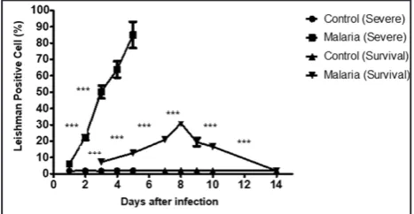
Figure 2: Parasitemia levels in the survival and severe models by malaria infection status. At specific time intervals post infection, parasitemia levels from mice and rats were measured. Results represent the mean +SEM of eight mice per time intervals. The parasitemia levels in survival and severe models were compared between malaria and
Parasitemia levels in the severe and survival malarial models
Figure 4: Light micrographs of brain tissues in control mice (A), malarial mice (B), control rats (C) and malaria-infected rats (D, E & F). Arrows indicate sequestration of pRBCs in brain microvessels (B2) and perivascular space enlargement (B1) in malarial mice. Arrows indicate the presence of pRBCs (D1), enlargement of perivascular space (D2) and lymphocytes infiltration (D1), necrotic neuron (E1) and pyknotic neuron (E2), petechia haemorrhage (F2) and perivascular oedema (F1) in malarial rats (H & E stain, x 400)
Figure 5: Light micrographs of liver tissues in control mice (A) and malarial mice (B); control rats (C) and malarial rats (D). Arrows indicate sinusoids dilatation (B1) and congestion with malarial pigments (B2) and hypertrophy and hyperplasia of Kupffer cells (B3) in malarial mice. Arrows indicate the presence of pRBCs (D1) and macrophage engulfing elements in portal veins and inflammatory infiltrate in portal tract (D2) in malarial rats (H & E stain, x 400)
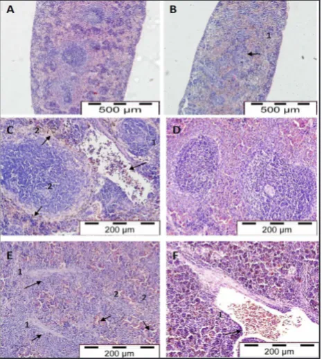
Figure 6: Light micrographs of spleen tissues in control mice (A), malaria-infected mice (B & C), control rats (D) and malaria-infected rats (E & F). Arrows indicate expanded red and white pulp (B1), the presence of pRBCs in splenic vessel (C1), pigment deposition in sinuses and splenic cord (C2) in malarial mice. Arrows indicate thickened trabeculae (E1), congested sinuses and splenic cord (E2), the presence of pRBCs and lymphocytes in microvessel (F1) in malarial rats (A and B: H & E, x 100; C, D, E and F: H & E, x 200) Figure 5
1
1
1
1 2
2
2
2
1 3
2 2
1
2
Figure 6
1
1 2
2
1 1
2 2
1
cells within sinusoids. Malarial livers harvested from mice and rats were enlarged and demonstrated a dark red to slate grey colour with a dense texture. Dilatation in hepatic sinusoids and accumulation of hypertrophied Kupffer cells in association with hyperplasia were observed. Cell clusters were noted around the dilated sinusoids packed with heavily-pigmented Kupffer cells. Reticuloendothelial changes of the malarial rat liver were associated with pRBCs, haemozoin pigments and macrophage-engulfing elements in portal veins. There were inflammatory infiltrates in the portal tract and an increase of macrophages in the sinusoids.
Spleen
were filled with pRBCs and macrophage engulfing elements and lymphocytes, as describedby Franke-Faryard, B, Janse CJ, Cunha-Rodrigues M et al. (21) who noted that in malaria the spleen was one of the major organs for accumulation of schizonts.
Kidney
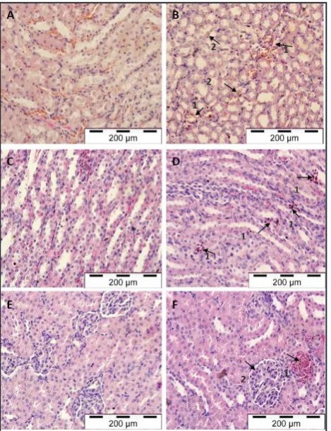
The histopathological changes in the kidney in severe and survival models were shown in Figure 7. The cortex and medulla of the murine kidney had extensive deposition of pigments and parasitized erythrocytes in microvessel and interstitial tissues. There was less cytoadherence and pigmentation in renal tissue than in the liver, with partial engorgement. Macroscopic examination on kidneys of malarial rats did not exhibit colour or size changes. Pigment-laden macrophages and pRBCs were occasionally seen on the medullary and interstitial blood vessels along with cortical haemorrhages during peak parasitemia on day 8. Endothelial changes characterized by enlarged and increased cellularity of the glomerulus were observed in association with infiltration of inflammatory cells suggestive of glomerulonephritis.
Lungs
The histopathological changes in lungs in severe and survival models were shown in Figure 8. The lung tissues of normal mice were characterized by thin septal walls with numerous capillaries. All PbA-infected mice generally showed thickened septal walls along with congestion of pRBCs, RBCs, macrophage-engulfing elements. Malarial pigments within the alveoli and microvessels of alveolar septa were observed. No pulmonary oedema was noted. The lung of uninfected rats showed a normal endothelium. Malaria-infected rats had thickened alveolar septa, patchy alveolar and septal haemorrhages along with hyaline membrane formation. The capillaries in the alveolar wall were congested with pRBCs, normal RBCs and inflammatory infiltrates. In malaria-induced lung injury the presence of pink oedema fluid in the lung interstitium was indicative of pulmonary oedema and focal oedema. In addition, the alveoli and septal capillaries were occluded with pRBCs, mononuclear cells and pigment-containing macrophages.
Figure 7: Light micrographs of kidney medullary tissue in control mice (A) and malarial mice (B), control rats (C & E) and malaria-infected rats (D &F). Arrows indicate pRBCs presence in microvessel (B1) and pigment deposition in interstitial tissue (B2) in malarial mice. Arrows indicate the presence of pRBCs in microvessels (D1), increased in glomerular cellularity (F2), glomerulus surrounded by inflammatory infiltrates and cortical haemorrhage (F1) in
Figure 8: Light micrographs of lung tissues in control mice (A), malarial mice (B), control rats (C) and malarial rats (D – F). Arrows indicate the presence of pRBCs and macrophage engulfing elements in microvessel (B1), alveolar septa congested with haemozoin pigment (B2) in malarial mice. Arrows indicate alveolar and septal haemorrhages and interalveolar capillaries congested with pRBCs, RBCs and inflammatory infiltrates (D) in malarial rats. Arrows indicate alveolar and interstitial tissue filled with pink oedema fluid (E) and the presence of pigment-laden macrophage, pRBCs and mononuclear lymphocytes (F) in malarial rats. (A, C, and D- H & E stain, x 200; B, E, and F – H & E stain, x400) Figure 7
1 2
2 1
1 2 1
1 1
1
Figure 8
1 1
2
Discussion
Due to the clinical significance of malarial infection in humans, it was important to understand the interaction and disruption of host cell function, and the pathogenesis and pathophysiology of the infection in disease progression. This study utilized Plasmodium berghei ANKA (PbA) infection in ICR mice as a model of severe infection, and
Plasmodium berghei ANKA (PbA) infection in Sprague Dawley rats as a model of survival.
The study confirmed the susceptibility of ICR mice to P. berghei ANKA (PbA) infection with a 100% fatality within six days. A high burden of infected erythrocytes was seen in the circulatory system with a peak parasitemia of 80–90% around day 5 to day 6 post-infection before death. The hyperparasitemia caused severe hemolysis with a depletion of the total RBCs resulting in severe malarial anaemia (22). The high level of the parasitemia was associated with the increased production of pro-inflammatory cytokines of mainly IL-6, TNF-α, IFN-γ and an anti-inflammatory cytokine, IL-10 (23-24). The Sprague dawley rat model of
PbA infection was distinctively different from PbA-infected ICR mice. Rats were generally less susceptible to P. berghei
parasites with peak parasitemia of only 30-40% on day 8 post-infection. The recovery rate was 100% partly due to parasite clearance during acute phase of infection (25). Since pathogenesis of malaria infection depended highly on the inflammatory response and the expressions of cytokines. It was therefore important to determine the release of both pro-inflammatory and the anti-inflammatory mediators. The study demonstrated different cytokine profiles in the severe and the survival models of malaria. In the severe model, high levels of circulating TNFα, IL-1α, IFNγ, IL-10 and IL-13 with an exceptionally low level of circulating IL-4 were observed. In contrast, the survival model showed high levels of circulating IFNγ, IL-10, IL-13 and IL-4 with exceptionally low levels of circulating TNFα and IL-1α.
Past studies attributed the severity of the malaria infection to the excess production of Th1 inflammatory mediators such as TNF-α, IFNγ and IL-1 both in human (26-27) and murine malaria (28, 29). Overproduction of Th1 type
cytokines, particularly TNF-α and IFNγ postulated to exacerbate the inflammatory response and worsen malarial pathology (30).
Past studies attributed the severity of the malaria infection to the excess production of Th1 inflammatory mediators such as TNF-α, IFNγ and IL-1 both in human (26-27) and murine malaria (28-29). Overproduction of Th1 type
cytokines particularly TNF-α and IFNγ was postulated to exacerbate the inflammatory response and worsen malarial pathology (30). In murine P.berghei ANKA infection high levels of TNF-α accelerated the development of irreversible hypothermia and the rapid onset of cerebral pathology (31). The excessive production of TNF-α could also increase the binding capacity of PRBCs to vascular endothelium and disturb the blood-brain barrier permeability increasing disease severity (32).
IL-1α is an endogenous pyrogen that acted synergistically with TNF-α to promote an acute inflammatory response against invading pathogens. Sustained levels of IL-1α had been reported to correlate with severe malarial anaemia (33). Rockett KA, Awburn MM, Rockett EJ et al. (34) proposed that the positive feedback mechanism between IL-1α and TNF-α enhanced the secretion of nitric oxide and IFNγ leading to hypoglycaemia in P. vinckei models of
murine malaria (34). Also, post mortem brain analysis of cerebral malaria patients revealed significant expression of TNF-α and IL-1α (35-36).
IL-4 is a cytokine with anti-inflammatory responses which ameliorated the parasitic infections through interference with the maturation of Th1 cytokines (37). In this study the circulating IL-4 concentration was lower in the severe model than in the survival model. Plasma IL-4 concentrations in patients who died of malaria infection were lower than the levels in malaria patients who survived (38). Saeftel M, Krueger A, Arriens S et al. (39) also reported that low levels of IL-4 could reduce the resistance against murine
P. berghei infection.
The survival model of malarial infection exhibited significantly high concentrations of IL-4. During the erythrocytic stage, high levels of IL-4 in the circulatory system of mice were found to inhibit the recrudescence of parasitemia and temper the inflammatory response. IL-4 was also crucial in the humoral immune response to human malaria through the enhancement of the production of
Plasmodium-specific IgG1 and IgG3 antibodies (40-41). Additionally, IL-4 was likely involved in the development
of CD8+ T cell and the memory response against the
Plasmodium parasites multiplying in the liver (42). The significance of IL-10 and IL-13 release in this study was unclear. Both models produced relatively high levels of the two cytokines during peak parasitemia with the severe model of infection showing slightly higher levels. IL-13 is a Th2 cytokine and is a potent mediator similar as
IL-4. Both IL-4 and IL-13 shares the IL-4 receptor α chain, with overlapping functions has been reported between these cytokines (43-44). IL-13 could inhibit the production
of Th1 cytokine and nitric oxide by activated macrophage (45). Lately it was identified as a negative predictor of haemoglobin (Hb) levels for anaemia of malaria in children (46). As high levels of IL-13 were detected in both models, further studies could determine its exacerbation or amelioration of malaria infection. Understanding the relationship between IL-13 and the pathogenesis of malaria could lead to the development of new malaria immunotherapeutic approaches.
had been extensively studied (50-51), in this study, the high level of IL-10 expression in the severe model was insufficient to counter the high levels of circulating TNF-α, IFNγ and IL-1α, which resulted in high mortality in the mice. The histopathological features of the survival model showed a similar degree of severity as those observed in the severe infection model with widespread pRBCs sequestration, haemozoin deposition and inflammation leading to the occlusion of the microvasculature of the major organs, particularly the brain, liver and lungs. Van der Heyde HC, Batchelder , Sandor M et al. (52) proposed that the microcirculatory dysfunction was the result of a cascade of inflammatory events that began with platelet activation mediated by pRBC antigens an inflammatory response activation with an increased expression of endothelial cell adhesion molecules such as ICAM-1 and
a haemostatic dysregulation from local pro-coagulant
activation. The malarial rats with only a mild parasitemia were able to recover fully from the infection and survive completely, despite the widespread severe pathological features, that were similar to the fatally ill infected mice with high parasitemia. It was possible therefore that the severe histopathology had only an indirect relationship with the parasitemia level and was not a contributing factor towards death.
The high TNFα and IL-1 could be key contributing factors towards death, as the murine severe model released high levels of both cytokines during the peak parasitemia and suffered a 100% mortality but not the rats who had low levels of the cytokines and survived. These two cytokines were again not directly related to the severe pathological conditions seen in both the rats and mice. High level of IL-4 in the rat survival model and its low level in the murine severe model during peak parasitemia could also point to a crucial role for IL-4 as a contributing factor for survival during malaria. Since IL-4, was a Th2-type cytokine, survival in rats could largely be due to a predominantly
Th2-type response during the critical stage of the infection, in contrast with the murine severe model that showed a
predominantly Th1-type response during the late critical stage of the infection.
Lastly, the high survival rate in the rat survival model could be due to the probable immunization against lethal
P. berghei infection with a complete resolution of its mild parasitemia without any recrudescence. The plasma IgG titers of rats could have protective activity against malaria (53). There was a significant and rapid upregulation of cytophilic IgG2 and production of cytosolic IgG1 production during the early and late phases of PbA infection in the survival from PbA infection while fatality in the susceptible murine strain could be due to a slower and declining IgG2 response and insufficient IgG1 production. The surrogate marker of protection from the erythrocytic stage of malaria in the rat was provided by the sequential upregulation of parasite-specific IgG response, with the resolution of parasitemia correlated to an intense IgG1 and IgM
an assumption could be made that the protective antibody response was driven by Th2 cells through B cell production induced by Th2 cytokines, mainly IL-4 and IL-13 (56).
Conclusion
This was a study on the pathogenesis of malaria infection. Two models of malaria infection were used, a model of a severe infection and a model of survival. Severe histopathology was demonstrated in both. With the severe model, the ICR, were unable to survive the malaria infection possibly due to the production of high levels of IL-α and TNF-α. With the survival model, the Sprague Dawley rats, could survive the infection with the production of high levels of IL-4 and low levels of TNFα during peak parasitemia. These differences in the pathogenesis of the severe and survival models of malaria infection could be a basis for immunotherapy of malaria in the future.
Acknowledgement
We would like to thank Universiti Putra Malaysia and The Ministry of Science, Technology and Innovation (MOSTI) for providing financial and infrastructure support for the conduct of the research study. The research work was supported by E-Science funding from The Ministry of Science, Technology and Innovation (MOSTI) (Grant number: 02-01-04-SF1313).
Competing interests
The authors declare that they have no competing interests.
References
1. WHO: World Malaria Report 2018; World Health Organization: Geneva, Switzerland 2018.
2. Singh B, Sung LK, Matusop A, Radhakrishnan A, Shamsul SS, Cox-Singh J, Thomas A, Conway DJ.
A large focus of naturally acquired Plasmodium knowlesi infections in human beings. Lancet. 2004;363:1017-24.
3. Singh B, Daneshvar C. Human infections and
detection of Plasmodium knowlesi. Clin Microbiol
Rev. 2013;26:165-84.
4. Miller LH, Good MF, Milon G. Malaria pathogenesis. Science. 1994;264:1878-83.
5. Clark IA, Rockett KA. The cytokine theory of human cerebral malaria. Parasitol Today. 1994;10:410-2. 6. Artavanis-Tsakonas K, Riley EM. Innate immune
response to malaria: rapid induction of IFN-γ from human NK cells by live Plasmodium falciparum
-infected erythrocytes. J Immunol. 2002;169:2956-63. 7. De Souza JB, Riley EM. Cerebral malaria: the
contribution of studies in animal models to our understanding of immunopathogenesis. Microbes Infect. 2002;4(3):291-300.
in ICR mice as a model of cerebral malaria. Iran J Parasitol. 2012;7:62-74.
9. Brown IN, Phillips RS. Immunity to Plasmodium berghei in rats: passive serum transfer and role of the spleen. Infect Immun. 1974;10:1213-8.
10. Orago AS, Solomon JB. Antibody-dependent and-independent cytotoxic activity of spleen cells for
Plasmodium berghei from susceptible and resistant rats. Immunology. 1986;59:283-8.
11. Singer I, Hadfield R, Lakonen M. The influence of age on the intensity of infection with Plasmodium berghei
in the rat. J Infect Dis. 1955;97:15-21.
12. Smalley ME. The nature of age immunity to
Plasmodium berghei in the rat. Parasitology. 1975;71:337-47.
13. Solomon, J.B. Natural cytotoxicity for Plasmodium
berghei in vitro by spleen cells from susceptible and resistant rats. Immunology. 1986;59(2); 277-81. 14. Schofield L, Grau GE. Immunological processes
in malaria pathogenesis. Nat Rev Immunol. 2005;5(9):722.
15. Day NP, Hien TT, Schollaardt T, Loc PP, Chuong LV, Hong Chau TT, et al. The prognostic and pathophysiologic
role of pro- and anti- inflammatory cytokines in
severe malaria. J Infect.Dis. 1999;180:1288-97. 16. Riley EM, Wahl S, Perkins DJ, Schofield L. Regulating
immunity to malaria. Parasite Immunol. 2006;28:35-49.
17. Foulds KE, Wu CY, Seder RA. Th1 memory: implications for vaccine development. Immunol Rev. 2006;211:58-66.
18. Yanez DM, Manning DD, Cooley AJ, Weidanz WP, Van Der Heyde HC. Participation of lymphocyte subpopulations in the pathogenesis of experimental murine cerebral malaria. J Immunol. 1996;157:1620-4.
19. Bagot S, Boubou MI, Campino S, Behrschmidt C, Gorgette O, Guénet JL, et al. Susceptibility to experimental cerebral malaria induced by
Plasmodium berghei ANKA in inbred mouse strains recently derived from wild stock. Infect Immun. 2002;70:2049-56.
20. White VA. Malaria in Malawi: inside a research autopsy study of pediatric cerebral malaria. Arch Pathol Lab Med. 2011;135:220-6.
21. Franke-Fayard B, Janse CJ, Cunha-Rodrigues M, Ramesar J, Büscher P, Que I, et al. Murine malaria parasite sequestration: CD36 is the major receptor, but cerebral pathology is unlinked to sequestration. Proc Natl Acad Sci. 2005;102:11468-73.
22. Perkins DJ, Were T, Davenport GC, Kempaiah P, Hittner JB, Ong’echa JM. Severe malarial anemia: innate immunity and pathogenesis. Int J Biol Sci. 2011;7:1427-42.
23. Clark IA, Alleva LM, Vissel B. The roles of TNF in brain dysfunction and disease. Pharmacol Ther. 2010;128:519-48.
24. Urquhart AD. Putative pathophysiological interactions of cytokines and phagocytic cells in severe human falciparum malaria. Clin Infect Dis. 1994;19:117-31. 25. Hernandez-Valladares M, Naessens J, Nagda S,
Musoke AJ, Rihet P, Iraqi FA. Comparison of pathology in susceptible A/J and resistant C57BL/6J mice after infection with different sub-strains of Plasmodium chabaudi. Exp Parasitol. 2004;108:134-41.
26. Ringwald P, Peyron F, Vuillez JP, Touze JE, Le Bras J, Deloron P. Levels of cytokines in plasma during
Plasmodium falciparum malaria attacks. J Clin Microbiol. 1991;29:2076-8.
27. Depinay N, Franetich JF, Grüner AC, Mauduit M, Chavatte JM, Luty AJ, et al. Inhibitory effect of TNF-α on malaria pre-erythrocytic stage development: influence of host hepatocyte/parasite combinations. PLoS One. 2011;6:e17464.
28. Grau GE, Fajardo LF, Piguet PF, Allet B, Lambert PH, Vassalli P. Tumor necrosis factor (cachectin) as an essential mediator in murine cerebral malaria. Science .1987;237:1210-2.
29. Amani V, Vigário AM, Belnoue E, Marussig M, Fonseca L, Mazier D, et al. Involvement of IFN-γ receptor-mediated signaling in pathology and anti-malarial immunity induced by Plasmodium berghei infection. Eur J Immunol. 2000;30:1646-55.
30. Jennings VM, Actor JK, Lal AA, Hunter RL. Cytokine profile suggesting that murine cerebral malaria is an encephalitis. Infect Immun. 1997;65:4883-7. 31. Curfs JH, Van der Meer JW, Sauerwein RW, Eling WM.
Low dosages of interleukin 1 protect mice against lethal cerebral malaria. J Exp Med. 1990;172:1287-91.
32. Lou J, Lucas R, Grau GE. Pathogenesis of cerebral malaria: recent experimental data and possible applications for humans. Clin Microbiol Rev. 2001;14:810-20.
33. Ouma C, Davenport GC, Awandare GA, Keller CC, Were T, Otieno MF, et al. Polymorphic variability in the interleukin (IL)-1β promoter conditions susceptibility to severe malarial anemia and functional changes in IL-1β production. J Infect Dis. 2008;198:1219-26. 34. Rockett KA, Awburn MM, Rockett EJ, Clark IA. Tumor
necrosis factor and interleukin-1 synergy in the context of malaria pathology. Am J Trop Med Hyg. 1994;50:735-42.
35. Kwiatkowski D, Sambou I, Twumasi P, Greenwood BM, Hill AV, Manogue KR, et al. TNF concentration in fatal cerebral, non-fatal cerebral, and uncomplicated
Plasmodium falciparum malaria. Lancet. 1990;336:1201-4.
36. Krishna S, Waller DW, Kuile FT, Kwiatkowski D, Crawley J, Craddock CF, et al. Lactic acidosis and hypoglycaemia in children with severe malaria: pathophysiological and prognostic significance. Trans R Soc Trop Med Hyg. 1994;88:67-73.37.
38. Cabantous S, Poudiougou B, Oumar AA, Traore A, Barry A, Vitte J, et al. Genetic evidence for the
aggravation of Plasmodium falciparum malaria by
interleukin 4. J Infect Dis. 2009;200:1530-9.
39. Saeftel M, Krueger A, Arriens S, Heussler V, Racz P, Fleischer B, et al. Mice deficient in interleukin-4 (IL-4) or IL-4 receptor α have higher resistance to sporozoite
infection with Plasmodium berghei (ANKA) than do
naive wild-type mice. Infect Immun. 2004;72:322-31. 40. Winkler S, Willheim M, Baier K, Schmid D, Aichelburg
A, Graninger W, et al. Frequency of cytokine-producing T cells in patients of different age groups with Plasmodium falciparum malaria. J Infect Dis. 1999;179:209-16.
41. Arévalo-Herrera M, Soto L, Perlaza BL, Céspedes N, Vera O, Lenis AM, Bonelo A, et al.
Antibody-mediated and cellular immune responses induced
in naive volunteers by vaccination with long
synthetic peptides derived from the Plasmodium
vivax circumsporozoite protein. Am J Trop Med Hyg. 2011;84(2-suppl):35-42.
42. Morrot A, Hafalla JC, Cockburn IA, Carvalho LH, Zavala F. IL-4 receptor expression on CD8+ T cells is required for the development of protective memory responses against liver stages of malaria parasites. J Exp Med. 2005;202:551-60.
43. Muchamuel T, Menon S, Pisacane P, Howard MC, Cockayne DA. IL-13 protects mice from lipopolysaccharide-induced lethal endotoxemia: correlation with down-modulation of TNF-alpha, IFN-gamma, and IL-12 production. J Immunol. 1997;158:2898-903.
44. Lai YH, Heslan JM, Poppema S, Elliott JF, Mosmann TR. Continuous administration of Il-13 to mice induces extramedullary hemopoiesis and monocytosis. J Immunol. 1996;156:3166-73.
45. Wood N, Whitters MJ, Jacobson BA, Witek J, Sypek JP, Kasaian M, et al. Enhanced interleukin (IL)-13 responses in mice lacking IL-13 receptor α 2. J Exp Med. 2003;197:703-9.
46. Ong’echa JM, Davenport GC, Vulule JM, Hittner JB, Perkins DJ. Identification of inflammatory biomarkers for pediatric malarial anemia severity using novel statistical methods. Infection and Immunity. 2011;79:4674-80.
47. Niikura M, Inoue SI, Kobayashi F. Role of interleukin-10 in malaria: focusing on coinfection with lethal and nonlethal murine malaria parasites. J BioMed Biotechnol. 2011;38:3962.
48. Hugosson E, Montgomery SM, Premji Z, Troye-Blomberg M, Björkman A. Higher IL-10 levels are associated with less effective clearance of
Plasmodium falciparum parasites. Parasite Immunol. 2004;26:111-7.
49. Fiorentino DF, Zlotnik A, Mosmann TR, Howard M, O’Garra A. IL-10 inhibits cytokine production by activated macrophages. J Immunol. 1991;147:3815-22.
50. Del Prete G, De Carli M, Almerigogna F, Giudizi MG, Biagiotti R, Romagnani S. Human IL-10 is produced by both type 1 helper (Th1) and type 2 helper (Th2) T cell clones and inhibits their antigen-specific proliferation and cytokine production. J Immunol.1993;150:353-60.
51. Groux H, Bigler M, De Vries JE, Roncarolo MG. Interleukin-10 induces a long-term antigen-specific anergic state in human CD4+ T cells. J Exp Med. 1996;184:19-29.
52. van der Heyde HC, Batchelder JM, Sandor M, Weidanz WP. Splenic γδ T cells regulated by CD4+ T cells are
required to control chronic Plasmodium chabaudi
malaria in the B-cell-deficient mouse. Infect Immun. 2006;74:2717-25.
53. Jarra W, Hills LA, March JC, Brown KN. Protective immunity to malaria. Studies with cloned lines of
Plasmodium chabaudi chabaudi and P. berghei in
CBA/Ca mice. II. The effectiveness and inter-or intra-species specificity of the passive transfer of immunity with serum. Parasite Immunol. 1986;8:239-54. 54. Smith EC, Taylor-Robinson AW. Parasite-specific
immunoglobulin isotypes during lethal and non-lethal murine malaria infections. Parasitology Research. 2003;89:26-33.
55. Couper KN, Phillips RS, Brombacher F, Alexander J. Parasite-specific IgM plays a significant role in the protective immune response to asexual erythrocytic
stage Plasmodium chabaudi AS infection. Parasite Immunol. 2005;27:171-80.
56. Taylor-Robinson AW, Phillips RS. Reconstitution of B-cell-depleted mice with B cells restores Th2-type