Nihal Bakirkalay Aydin
1, A–c, f, Turgut Teke
1, A, d–f, Hatice Toy
2, B, c, f, Kursat Uzun
1, A, c–fThe Effect of Theophylline on the Prevention
of Mechanical Ventilation-Induced
Diaphragm Atrophy in Rats
1 department of chest diseases, Necmettin Erbakan University, Meram Medical faculty, Konya, Turkey 2 department of Pathology, Necmettin Erbakan University, Meram Medical faculty, Konya, Turkey
A – research concept and design; B – collection and/or assembly of data; C – data analysis and interpretation;
D – writing the article; E – critical revision of the article; F – final approval of article; G – other
Abstract
Background. Movement disorders and atrophy occur in the diaphragm, the most important muscle of respiration, because of mechanical ventilation (MV).
Objectives. In this animal model, we aimed to evaluate the effect of intravenous theophylline administration on the prevention of mechanical ventilation-induced diaphragmatic atrophy.
Material and Methods. In our study, 30 healthy male Sprague-dawley rats were used. They were divided into 3 equal groups. Group 1: the control group (no MV); group 2: the placebo group that received MV; Group 3: the theophylline group composed of rats that received both MV and theophylline therapy. In all 3 groups, the dia-phragmatic atrophy was evaluated histopathologically.
Results. In the histopathological examination, no macroscopic thickening and microscopic atrophy were observed in the diaphragm in the control group. In the placebo group (group 2), macroscopically definite thickening was observed in all rats, and microscopically, heavy (+++) atrophy was observed. In the theophylline group (group 3), there was no atrophy in one rat. In 8 rats, light (+), and in 1 rat medium (++) atrophy was observed.
Conclusions. In our study, it was shown that atrophy occurred in the diaphragms of rats after MV, and the atrophy was decreased after theophylline administration (Adv Clin Exp Med 2014, 23, 1, 33–38).
Key words: mechanical ventilation, diaphragmatic atrophy, theophylline, rat.
Adv clin Exp Med 2014, 23, 1, 33–38 ISSN 1899–5276
ORIGINAL PAPERS
© copyright by Wroclaw Medical University
Mechanical ventilation (MV) is a lifesaving method that is used to sustain the blood gas balance of patients who are unable to maintain alveolar ven-tilation. However, 25% of the patients are in need of prolonged ventilation support and this causes a rise in infection rates, length of stay in the intensive care unit, morbidity and mortality [1]. Prolonged venti-lation causes a decrease in the contraction strength of diaphragmatic muscles and leads to atrophy. In most of the studies, it is stated clearly that MV causes diaphragmatic atrophy and loss of isometric strength production, diaphragmatic mass and the protein content of diaphragmatic muscle [2–4].
Therefore, in order to maintain the mass and function of respiratory muscles during ventila-tion, strategies have been developed. These are: MV modes that help the diaphragm contribute to
Material and Methods
Animals
The rat was chosen as the experimental animal because of its being favored for biomedical studies, its small dimensions and its maintainability. for this purpose, four-month-old male Sprague-daw-ley 30 rats with an average weight of 315–375 g were housed at the Selcuk University Experimen-tal Medicine Research and Practices facility and fed rat pellet feed and water ad libitum and main-tained on a 12 : 12 h light-dark cycle for 3 weeks before initiation of this study. This study was ap-proved by the University of Selcuk Experimental Animals Ethics committee.
Experimental Design
The rats were randomly assigned to one of 3 ex-perimental groups. Group 1 (control group): Intrap-eritoneal anesthesia was applied as acutely. Group 2 (MV group):After reaching a surgical plane of in-traperitoneal anesthesia, the animals were tracheos-tomized and mechanically ventilated and monitored for 24 h. Group 3 (MV + Theophylline Group):After reaching a surgical plane of intraperitoneal anesthe-sia, animals were tracheostomized and mechanical-ly ventilated and monitored for 24 hand theophyl-line infusion was given (15 mg/kg as a loading dose; 0.05 mg/kg/h infusion as maintenance).
Protocol for Control Animals
The animals in the control group were not me-chanically ventilated or exposed to long-term an-esthesia before study. These animals were weighed and then sodium pentobarbital was given intraperi-toneally (50 mg/kg). After a surgical plane of anes-thesia was achieved, the diaphragm was rapidly re-moved, and its weight was measured on a precision scale. The largest dimensions of the diaphragm and membranous region were measured and segments from the costal region were fixed in a buffered 10% formalin solution for histopathological evaluation.Mechanical Ventilation Protocol
All surgical procedures were performed us-ing aseptic techniques. After weighing and reach-ing a surgical plane of anesthesia (sodium pentobar-bital, 50 mg/kg, intraperitoneally), the animals were tracheostomized and mechanically ventilated with a volume-controlled small animal ventilator (SAR- -830, IITc Life Science, USA).The tidal volume was 1 mL/100 g of body weight, with a respiratory rate of 80 breaths/min.This respiratory rate was selected tomimic the breathing frequency of adult rats at rest. Additionally, positive end-expiratory pressure (PEEP) of 1 cm H2O was used throughout the protocol.
An arterial catheter was placed in the carotid artery to permit the continuous measurement of blood pressure.A venous catheter was inserted in-to the femoral vein epigastria branch for continu-ous infusion of isotonic saline. A surgical plane of anesthesia was maintained with sodium pentobar-bital infusion (10 mg/kg/h) over the entire period of MV.The level of anesthesia was monitored with methods such as heart rate, blood pressure and
corneal-lid reflexes.Body temperature was
main-tained at 37°c using a heating blanket.In addition to this, the heart rate and electrical activity of the heart were monitored placing subcutaneous elec-trodes through an EKG. The fluid balance of the body was maintained with the continuous applica-tion of 2 mL/kg/h IV isotonic saline. continuous care during the MV protocol included expressing the bladder, removing airway mucus, moistening the eyes, passive movements of the limbs and rotat-ing the animal. This care was maintained through-out the experimental period at hourly intervals.
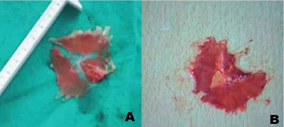
On completion of MV, the body weight of the animals was reweighed and the diaphragm was re-moved under abdominal surgery. The existence of macroscopic abnormalities (color change, thick-ening, and hemorrhage) was checked and seg-ments of the costal diaphragm were fixed in a buff-ered 10% formalin solution for histopathological evaluation.
Histopathological
Evaluation
costal diaphragm samples 1 cm in length and 0.5 cm in width were taken. These samples were stained with hematoxylin eosin and were exam-ined with a light microscope.
Histopathological evaluations were done by a pathologist who had no previous knowledge of the protocol and procedures of this study. Accord-ing to these histopathological evaluations, the at-rophy of the diaphragm muscle, neutrophil in-filtration, fibrosis and steatosis were analyzed. diaphragmatic atrophy was scored that 0–33% was light (+), 34–66% was medium (++) and more than 67% was heavy (+++).
Statistical Analysis
groups and a Mann-Whitney U test was used for binary comparisons if a significant difference was observed among the groups. A value of p < 0.05 was consideredsignificant.
Results
None of the animals were eliminated due to in-fection. There was no significantly difference be-tween initial and final the body weight of the ani-mals between the experimental groups (p > 0.05). These results confirm that our program of nutri-tion and hydranutri-tion was adequate. To determine whether our MV protocol was successful at sus-taining homeostasis, we monitored blood pressure and heart rate during the MV period. It was main-tained within the physiological range.
The measurements done on the diaphrag-matic tissue of the control, placebo (MV only) and theophylline groups are summarized in Ta-ble 1. compared with the control, the applica-tion of MV resulted in a statistically significant
decrease in diaphragm weight, large diameter of the diaphragm (fig. 1A), large diameter of the membranous portion (fig. 1B) and the ratio of diaphragm weight to body weight (respectively; p < 0.01, p < 0.01, p < 0.01 and p = 0.05). Theo- phylline administration restored these values to the control values. compared with the place-bo, the administration of theophylline resulted in a statistically significant increase in diaphragm weight (p < 0.05), large diameter of the diaphragm (p < 0.05), large diameter of the membranous por-tion (p < 0.01) and the ratio of diaphragm weight to body weight (p = 0.05).
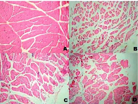
The histopathological evaluation results are shown in Table 2. As a result of this histopathologi-cal evaluation, it is seen that macroscopic thickening and microscopic atrophy were observed in the dia-phragm of rats in the control group and all are nor-mal (fig. 2A). Macroscopically evident thickening and heavy (+++) atrophy microscopically were ob-served in the diaphragm of the rats in the MV ap-plied placebo group (Group 2). An increase in the distance between the muscle fibers was detected and
Table 1. Measurements of diaphragm tissue in control, placebo and theophylline groups
Control
(mean ± SD) MV(mean ± SD) MV plus Teophyphilline(mean ± SD)
Weight 122.20 ± 1.92 116.50 ± 4.71a 119.90 ± 3.03c
Large diameter 4.90 ± 0.15 4.44 ± 0.21a 4.73 ± 0.14c
Large diameter of the membranous portion 2.42 ± 0.08 2.19 ± 0.11a 2.36 ± 0.08d
dW/BW 0.34 ± 0.00 0.33 ± 0.01b 0.34 ± 0.01e
MV – Mechanical ventilation, dW/BW – ratio of diaphragmatic weight to body weight,ap < 0.01 – compared with the
con-trol group, bp = 0.05 – compared with the control group, cp < 0.05 – compared with the MV group, dp < 0.01 – compared
with the MV group, dp = 0.05 – compared with the MV group.
muscle fiber diameters were variable (fig. 2B). In the theophylline group, no atrophy was observed and in only one animal it was completely normal. Eight ani-mals had light (+) atrophy, the distance between mus-cle fiber increased minimally and the diameters were similar to each other (fig. 2c). One of the animals in the theophylline group had medium (++) atrophy and the distance between the muscle fiber increased moderately (fig. 2d). Histopathologically, no neutro-phil infiltration and fibrosis was observed in the dia-phragms of both the placebo and theophylline groups, and there was a similarity in both in terms of fat.
Discussion
As far as we know, this study is the first histo-pathological experiment on the effects of intrave-nous theophylline administration on MV-induced diaphragmatic atrophy. As a result of this study, it is seen that MV causes atrophy in the diaphragm muscle of rats in as little as 24 h and theophylline therapy inhibits the formation of atrophy and the reduction in diaphragm mass.
In the literature, there are many studies in-vestigating the dysfunction of respiratory mus-cles after MV. Le Bourdelles et al. [3]studied the
Fig. 2. Histopathological evaluation (HE, 10 × 10), A; the normal appearance of the diaphragm in the control group, B; diaphragm + + + atrophy in group 2, c; + atrophy of diaphragm in group 3, d; + + atrophy of the diaphragm in group 3
Table 2. Histopathological evaluation results
MV
(n) MV plus Teophyphilline (n)
0 + ++ +++ 0 + ++ +++
Atrophy – – – 10 1 8 1 –
Neutrophil infiltration – – – – – – – –
fibrosis – – – – – – – –
Steatosis 8 2 – – 9 1 – –
effects of 48 h MV on both atrophy and contrac-tion strength of rat diaphragms. As a result, a sig-nificant decrease in isometric strength production, diaphragm mass and protein content were report-ed. Shanely et al. [4] showed clearly that short term, controlled MV causes diaphragmatic atro-phy rapidly. 18 h MV causes a 7% decrease in cos-tal diaphragm mass and tocos-tal protein destruction accelerates after MV [4].Powers et al. [2] showed in a similar study that MV causes a significant de-crease in diaphragm specific strength of the rat. Powers et al. [2] presented that diaphragm dys-function develops fast at the beginning (12 h) and this dysfunction proceeds during the first 24 h of MV in the ventilator. Therefore, the controlled MV period is identified as 24 h in our experimen-tal study.
Theophylline has been used for many years in the treatment of chronic obstructive pulmonary disease. Many studies have shown that theophyl-line increases the contraction strength of the di-aphragm, decreases the weakness and has a pro-tective effect against fatigue [6, 8, 14–18]. In vitro
studies have shown that theophylline strengthens muscle contraction, revealed by direct and indi-rect electrical stimuli in isolated preparations [9]. The same effect was seen in the diaphragm prep-arations including isolated phrenic nerve semi-in-cision in rats [10]. Studies on humans show that aminophylline regulates the diaphragm contrac-tion funccontrac-tion of tired people significantly [11]. Aubier et al. [13] demonstrated in their study on animals that theophylline increases diaphragm contraction strength and respiratory minute ven-tilation. Apart from the studies indicating that theophylline increases the production of strength and decreases the weakness, there are some stud-ies claiming that theophylline has little or no effect on diaphragm contraction strength and weakness in the diaphragm [19, 20].
There are several mechanisms mentioned in the literature about the regulatory effect of theoph-ylline on diaphragmatic function. The first of these is the increase of diaphragmatic perfusion by the-ophylline. Theophylline leads to this by increasing
cardiac output and by providing vasodilation in diaphragmatic arterioles [21]. The second mecha-nism [22] is the adjustment of diaphragm muscle contraction even during the fatigue period when a shortening is seen in muscle fibers [7, 11, 23]. The third mechanism is by increasing the sensi-tivity of the respiratory center to the diaphragm motor neuron [24], and the last one is the inotro-pic effect on diaphragm muscle fibers [7, 11, 23, 25]. However, none of these mechanisms are clear and this subject is still controversial. danialou et al. [22] showed that theophylline causes a signif-icant vasodilatation on diaphragm arterioles. On the contrary, the study by Mayock et al. [26] on animals reported that there was no change in di-aphragmatic blood flow after aminophylline infu-sion. However, methylxantine-derived drugs are reported to weaken the intensity of diaphragmatic weakness or can delay the start. In fact, it is known that theophylline has vasodilator effects [27]. The-ophylline might increase the blood flow of the dia-phragm as a result of systematic vasodilatation and might improve diaphragmatic strength. One of the diaphragm function-adjustment mechanisms may be the prevention of diaphragmatic atrophy which we revealed histopathologically for the first time in our study on animals.
We chose the rat model for several reasons for this experimental study. first, adult rats are of suf-ficient size for surgical procedures. Second, and most importantly, human and rat diaphragms are similar in anatomical features, function and mus-cle fiber-type composition [28, 29].
In conclusion, although studies on both ani-mals and humans indicate that theophylline clear-ly increases diaphragm contraction strength, the mechanism of this increase is not clearly explained. In our study, the preventive effect of theophylline was observed when compared to the placebo in rats with MV-induced diaphragm atrophy. The re-sults obtained lead us to think that an increase in diaphragm contraction strength by using theoph-ylline can be achieved, preventing muscle atrophy. However, a great number of animal and human studies are needed on the molecular level.
References
[1] Esteban A, Frutos F, Tobin MJ, Alia I, Solsona JF, Valverdu I: A comparison of four methods of weaning patients from mechanical ventilation. N Engl J Med 1995, 332, 345–350.
[2] Powers SK, Shanely RA, Coombes JS, Koesterer TJ, McKenzie M, Van Gammeren D: Mechanical ventilation results in progressive contractile dysfunction in the diaphragm. J Appl Physiol 2002, 92, 1851–1858.
[3] Le Bourdelles G, Viires N, Boczkowski J, Seta N, Pavlovic D, Aubier M: Effects of mechanical ventilation dia-phragmatic contractile properties in rats. Am J Respir crit care Med 1994, 149, 1539–1544.
[4] Shanely RA, Zergeroglu MA, Lennon SL, Sugiura T, Yimlamai T, Enns D: Mechanical ventilation-induced dia-phragmatic atrophy is associated with oxidative injury and increased proteolytic activity. Am J Respir crit care Med 2002, 166, 1369–1374.
[6] Aubier M, De Troyer A, Sampson M, Macklem PT, Roussos C: Aminophylline improves diaphragmatic contrac-tility. N Engl J Med 1981, 305, 249–252.
[7] Murciano D, Aubier M, Lecocguic V, Pariente R: Effects of theophylline on diaphragmatic strength and fatigue in patients with chronic obstructive lung disease. N Engl J Med 1984, 311, 349–353.
[8] Sigrist S, Thomas D, Howell S, Roussos CH: The effect of aminophylline on inspiratory muscle contractility. Am Rev Respir dis 1982, 126, 46–50.
[9] Jones DA, Howell S, Roussos C, Edwars RHT: Low-frequency fatigue in isolated skeletal muscles and the effects of methylxanthines. clin Sci 1982, 63, 161–167.
[10] Kentera D, Varagic VM: The effects of cyclic N-2-0-dibutyryl-adenosine 3’, 5’-monophosphate, adrenaline and aminophylline on the isometric contractility of the isolated hemidiaphragm of the rat. Br J Pharmacol 1975, 54, 375–381.
[11] Wanke T, Merkle M, Zifko U, Formanek D, Lahrmann H, Grisold W: The effect of aminophylline on the force-length characteristics of the diaphragm. Am J Respir crit care Med 1994, 149, 1545–1549.
[12] Aubier M: Effect of theophylline on diaphragmatic and other skeletal muscle function. J Allergy clin Immunol 1986, 78, 787–792.
[13] Aubier M, Murciano D, Viires N, Lecocguic Y, Palacios S, Pariente R: Increased ventilation due to improved diaphragmatic efficiency during aminophylline infusion. Am Rev Respir dis 1983, 127, 148–154.
[14] Supinski GS, Deal ED, Kelsen SG: comparative effects of theophylline and adenosine on respiratory skeletal and smooth muscle. Am Rev Respir dis 1986, 133, 809–813.
[15] Viires N, Aubier M, Murciano D, Marty C, Pariente R: Effects of theophylline on isolated diaphragmatic fibers: a model for pharmacologic studies on diaphragmatic contractility. Am Rev Respir dis 1986, 133, 1060–1064.
[16] Kolbeck RC, Speir WA: diltiazem, verapamil and nifedipine inhibit theophylline-enhanced diaphragmatic con-tractility. Am Rev Respir dis 1989, 139, 139–145.
[17] Kuei JH, Sieck GC: chronic aminophylline administration: effect of diaphragm contractility and fatigue resistance in vitro. Am Rev Respir dis 1991, 144, 121–125.
[18] Sassoon CS. Zhu E, Caiozzo VJ: Assist-control mechanical ventilation attenuates ventilator-induced diaphrag-matic dysfunction. Am J Respir crit care Med 2004, 170, 626–632.
[19] Derom E, Janssens S, De Bock V, Decramer M: Theophylline minimally alters contractile properties of canine diaphragm in vitro. J Appl Physiol 1990, 69, 1390–1396.
[20] Janssens S, Derom E, Reid MB, Tjandramaga TB, Decramer M: Effects of theophylline on canine diaphragmatic contractility and fatigue. Am Rev Respir dis 1991, 144, 1250–1255.
[21] Dow-Edwards D, DeCrescito V, Tomasula JJ: Effect of aminophylline and isoproterenol on spinal cord blood flow after impact injury. J Neurosurg 1980, 53, 385–390.
[22] Danialou G, Vicaut E, Aubier M, Boczkowski J: Theophylline dilates rat diaphragm arterioles via the prostaglan-dins pathway. Br J Pharmacol 1998, 124, 1355–1362.
[23] Sherman MS, Lang DM, Matityahu A, Campbell D: Theophylline improves measurements of respiratory muscle efficiency. chest 1996, 110, 1437–1442.
[24] Bhatia J: current options in the management of apnea of prematurity. clin Pediatr 2000, 39, 327–336.
[25] Eldridge FL, Millhorn DE, Waldrop TG, Kiley JP: Mechanism of respiratory effects of methylxanthines. Respir Physiol 1983, 53, 239 –261.
[26] Mayock DE, Standaert TA, Woodrum DE: Effect of methylxanthines on diaphragmatic fatigue in the piglet. Pediatr Res 1992, 32, 580–584.
[27] Rall TW: drugs used in the treatment of asthma. The methylxanthines, cromolyn sodium and other agents. In: The Pharmacological Basis of Therapeutics. Eds.: Goodman LS, Gilman A, MacMillan co., New York 1990, 618–630.
[28] Mizuno M: Human respiratory muscles: fibre morphology and capillary supply. Eur Respir J 1991, 4, 587–601.
[29] Powers S, Demirel H, Coombes J, Fletcher L, Calliaud C, Vrabas I, Prezant D: Myosin phenotype and bioener-getic characteristics of rat respiratory muscle. Med Sci Sports Exerc 1997, 29, 1573–1579.
Address for correspondence:
Turgut Teke
Necmettin Erbakan Universitesi Meram Tip fakultesi Hastanesi Gogus Hastaliklari Anabilim dali
42080 Meram-Konya Turkey
Tel.: +90 332 2236218 E-mail: [email protected] conflict of interest: None declared Received: 10.08.2012