Zbigniew Hruby
1, 6, A–D, Maria Stanek-Piotrowska
2, B, C, G, Jakub Turek
3, 6, B, C, G,
Wojciech Witkiewicz
3, 6, A, E, F, Anna Jonkisz
4, G, Andrzej Konieczny
1, 6, B, C, G,
Mariusz Kosiński
5, 6, B, C, G, Tadeusz Dobosz
4, E–GThe Clinicopathological Determinants
of Native Arteriovenous Fistula Failure
in Patients on Maintenance Hemodialysis*
Uwarunkowania kliniczne i histopatologiczne niewydolności przetoki
z naczyń własnych pacjenta u chorych przewlekle hemodializowanych
1 Department of Nephrology, Regional Specialist Hospital Center for Research & Development, Wrocław, Poland 2 Fresenius Nephrocare Dialysis Center, Wrocław, Poland
3 Department of Vascular Surgery, Regional Specialist Hospital Center for Research & Development,
Wrocław, Poland
4 Department of Molecular Biology, Wroclaw Medical University, Wrocław, Poland
5 Department of Pathology, Regional Specialist Hospital Center for Research & Development, Wrocław, Poland 6 WroVasc Integrated Cardiovalscular Center, Regional Specialist Hospital Center for Research & Development,
Wrocław, Poland
A – research concept and design; B – collection and/or assembly of data; C – data analysis and interpretation;
D – writing the article; E – critical revision of the article; F – final approval of article; G – other
Abstract
Background. Progressive narrowing of the venous part of dialysis fistulae is caused by hemodynamic and inflam-matory factors.
Objectives. The pathogenic and clinical determinants of deterioration of the functioning of arteriovenous fistulae in chronically hemodialyzed patients were evaluated.
Material and Methods. The hemodynamic parameters and the activity of inflammatory growth factors in the vessel wall of newly implanted fistulae were assessed and correlated with the clinical course of 34 hemodialyzed patients. Measurements taken at the time of implanting the fistulae included blood flow in the venous part of the anastomosis and its widest diameter by ultrasound Power Doppler, a histopathologic examination of fistula wall samples and measurements of mRNA expression for growth factors PDGFβ1 and TGFβ in the fistula wall. The results were correlated with clinical data from 36 months’ observation: duration of fistula maturation, adequacy of dialysis treatment (eKt/V), the patient’s survival, morbidity linked with vascular access problems and general cardiovascular morbidity.
Results. The mean duration of fistula maturation was 44.9 days (N = 43, SD = 38.6), whereas the average dura-tion of fistula usage as dialysis access was 795.9 ± 480.6 days. Fistula blood flow at the time of implantadura-tion, aver-aged 1782.2 ± 1735.3 ml/min. The mean number of hospitalization days due to vascular access morbidity was 9.9 ± 15.6 days and it correlated positively with the fistula blood flow (R = 0.596, P = 0.004). There was a negative correlation between the expression of PDGFβ1 mRNA and fistula blood flow (R = –0.673, P = 0.011), as well as between TGFβ expression and patient survival (R = –0.722, P = 0.002).
Conclusions. Inflammatory activity of the vessel wall growth factors PDGFβ1 and TGFβ implies impairment of fistula function and the patient’s cardiovascular morbidity (Adv Clin Exp Med 2013, 22, 4, 495–500).
Key words: maintenance hemodialysis, dialysis fistula, hemodynamic parameters, growth factors, morbidity, patient’s survival.
Adv Clin Exp Med 2013, 22, 4, 495–500 ISSN 1899–5276
oRIGINAl PAPERS
© Copyright by Wroclaw Medical University
Vascular access problems limit dialysis deliv-ery, compromising the efficacy of treatment and resulting in increased patent morbidity and mor-tality [1]. The most frequent abnormality is throm-bosis caused by local stenosis with subsequent ces-sation of fistula blood flow [2]. The condition of the vascular endothelium lining the inner sur-face of arteriovenous anastomoses is influenced by many factors, such as the hemodynamic character-istics of the fistula blood flow and the local inflam-matory environment [3].
In this study, the hemodynamic and histopath-ological determinants of failing native fistulae in chronically hemodialyzed patients were analyzed, and possible correlations between these parameters and the clinical course in these patients were in-vestigated. The main positive correlates of patents’ vascular access morbidity were the fistula hemo-dynamic characteristics, while fistula wall expres-sion of genes for the inflammatory growth factors PDGFβ1 and TGFβ showed a significant negative correlation with fistula blood flow and patient sur-vival during the three-years follow-up. The authors infer from these results that inflammatory condi-tions that already affect vascular walls in the pre-dialysis period may have a negative impact on pa-tients’ survival in the early course of maintenance dialysis therapy.
Material and Methods
Design of the Study
Patients were included in study group upon the creation of an arteriovenous fistula as vascu-lar access for chronic hemodialysis treatment. At the time of enrollment a sample of the venous part of anastomosis was obtained to assess its his-topathological characteristics and the expression of mRNA for PDGFβ1 and TGFβ. Doppler Ul-trasound (DU) examinations analyzing the fistu-la blood flow and anastomosis diameter were per-formed four weeks after the first dialysis and then at three month intervals or whenever fistula failure was suspected during physical examination and observation of the dialysis parameters. Patients were observed prospectively for the 36 subsequent months, noting the following clinical parame-ters every three months: the adequacy of dialysis (eKT/V); the number of dialysis days with inad-equate, reduced blood flow; the number of days with the use of a temporary catheter to substitute for a failing fistula; the patient’s survival time and morbidity (number of hospitalization days) due to vascular access problems; and overall cardiovas-cular morbidity. The results of the histopatholog-ical assessments of the fistulae and the measure-ments of mRNA for growth factors that were done at the start of the study were then correlated with the clinical parameters collected during the obser-vation period.
Streszczenie
Wprowadzenie. Postępujące zwężenie żylnej części przetoki dializacyjnej jest spowodowane czynnikami hemody-namicznymi i zapalnymi.
Cel pracy. ocena wyznaczników patogenetycznych i klinicznych dysfunkcji przetoki tętniczo-żylnej z naczyń włas-nych pacjenta u chorych przewlekle hemodializowawłas-nych.
Materiał i metody. oceniano wskaźniki hemodynamiczne i aktywność prozapalnych czynników wzrostu w ścia-nie naczynia zespolenia tętniczo-żylnego po jego implantacji, po czym badano istotność korelacji tych czynników z parametrami klinicznymi u 34 hemodializowanych pacjentów obserwowanych w ciągu 36 miesięcy. Bezpośrednio po implantacji zespolenia badano szybkość przepływu krwi w jego żylnej części oraz jej największą średnicę za pomocą ultradźwiękowej techniki dopplerowskiej, oceniano histopatologicznie preparaty wycinków ściany naczy-nia, mierząc przy tym ekspresję mRNA dla czynników wzrostu PDGFβ1 i TGFβ. Wyniki korelowano z danymi klinicznymi zebranymi w czasie 36-miesięcznej obserwacji tych pacjentów: czasem „dojrzewania” zespolenia, ade-kwatnością dializoterapii (eKt/V), czasem przeżycia pacjentów, ich chorobowością związaną z dostępem naczynio-wym i ogólną chorobowością z przyczyn sercowo-naczyniowych.
Wyniki. Średnia liczba dni hospitalizacji z powodu powikłań dostępu naczyniowego w czasie obserwacji wyniosła 9,9 ± 15,6 dnia i była dodatnio skorelowana z przepływem krwi w żylnej części przetoki (R = 0,596; p = 0,004). Ekspresja mRNA dla PDGFβ1 wykazywała znamienną negatywną korelację z szybkością przepływu krwi przez przetokę (R = –0,673; p = 0,011), a ekspresja mRNA dla TGFβ korelowała negatywnie z okresem przeżycia pacjen-tów w ciągu 36-miesięcznej obserwacji (R = –0,722; p = 0,002).
Wnioski. Wzmożona aktywność genów dla prozapalnych czynników wzrostu PDGFβ1 i TGFβ w ścianie zespolenia zapowiada dysfunkcję przetoki i skrócone przeżycie pacjentów hemodializowanych (Adv Clin Exp Med 2013, 22, 4, 495–500).
Study Subjects
The patients enrolled were treated with main-tenance hemodialysis for end-stage renal disease at the Fresenius Nephrocare Dialysis Center and the Dialysis Center of the Regional Specialist Hospital in Wroclaw, Poland. The study group consisted of 34 participants (17 males and 17 females), mean age 66.4 years (27–86 years). The reasons for re-nal failure in these patients included diabetic ne-phropathy (29.4%), hypertensive nene-phropathy (23.5%), glomerulonephritis (17.6%), chronic py-elonephritis (5.9%) and systemic vasculitis (5.9%). The causes in the remaining 17.7% were not deter-mined. The control group for the real-time
poly-merase chain reaction (RT-PCR) studies included
10 samples of vein wall collected during reparative surgery for traumatic hand injury.
Each study subject gave his or her informed consent for participation. The protocol of the study was prepared in accordance with the Helsinki Dec-laration and was approved by the Bioethical Com-mittee of Wroclaw Medical University.
Histopathological Study
Slices of the venous part of an anastomosis (1.5 mm thick) were collected during the creation of a dialysis fistula, fixed in Methacarn solution (Sigma), dehydrated and embedded in paraffin. Subsequently the paraffin blocks were cut into slices, rehydrated, stained with
hematoxylin-eo-sin and inspected under light microscope
magni-fication (x400). Each time the thickness of the ves-sel wall was measured and the mean value of the shortest and longest diameter was calculated (ten preparations per individual). The results were ex-pressed as mean ± SD for 10 preparations.
Expression of mRNA for
PDGFβ1 and TGFβ
Assessment of mRNA activity for growth fac-tors PDGFβ1 and TGFβ in the venous part of the fistula walls was performed on slices (1.5 mm) col-lected during creation of anastomosis and pre-served in RNAlater solution (Sigma Aldrich). Iso-lation of the total RNA was accomplished with the EZNA Total RNA Kit (omega Bio-Tek), fol-lowing the protocol provided by the manufactur-er. After isolation, the concentration of tRNA in each sample was checked with a NanoDrop spec-trophotometer (Thermo Scientific) at the 260 nm wavelength. The RT-PCR reaction was then per-formed using a High Capacity cDNA Reverse Transcription Kit with RNAse Inhibitor (Applied
Biosystems). Expression of the following genes was assessed: PDGFβ1, PDGFβ, TGFβ and GADPH as a “housekeeping gene” (positive control). An ABI PRISM 7900 HT Fast RT-PCR System (Applied Biosystems) thermocycler was used with 45 cycles
at 95oC. Gene expression was calculated from the
standard curve of fluorescence of the reporter dye versus time (cycle number).
Duplex Doppler Ultrasound
Examination
A BK Pro Focus device (BK Medical) with
a 12 MHz vascular head was used for the DU, ex-amining a segment of the venous part of fistula 20 cm from the anastomosis. Each time the widest diameter of the vessel and blood flow were mea-sured. The latter was calculated from the formula:
Fb = π (D/2)2V, where Fb= blood flow, D = vessel
di-ameter and V = mean velocity of blood flow. Mea-surements were taken routinely every three months, or at any clinical symptom of a failing fistula.
Clinical Data
For the purposes of the study the medical his-tories and dialysis reports of all the participants were analyzed. The patients were enrolled between November 1, 2006, and November 30, 2010. In each case the following data were noted: the dura-tion of fistula maturadura-tion (the time from creadura-tion to first dialysis), the number of inadequate dialy-ses (reduced blood flow due to fistula insufficiency, single needle dialyses), the number of days using a temporary catheter due to a failing fistula, patient morbidity (the number of hospitalization days) caused by vascular access problems, general car-diovascular morbidity and patient mortality (the number of days from fistula creation to death).
Statistical Analysis
Results
During the study period (2006–2010), 451 na-tive arteriovenous anastomoses were created as vascular access for the maintenance dialysis pro-grams of patients at the Region Specialist Hospital Department of Nephrology and the Fresenius Di-alysis Center, both in Wroclaw, Poland. out of this number 34 patients were enrolled in the study af-ter having Cimino Brescia fistulae created on their wrists (21 patients) or cubital fossa (13 patients).
The mean thickness of the venous part of fis-tula walls measured in the histopathological prep-arations (N = 34) collected during fistula creation averaged 8.03 ± 4.05 mm.
The average velocity of blood flow in the fis-tula at 24 months after creation of the anastomosis was 1782.18 ± 1735.26 ml/min; at 48 months it was
2825.36 ± 3968.64 ml/min. Similarly, the longest fistula diameter increased from 7.87 ± 3.37 mm at 24 months to 9.57 ± 4.54 mm at 48 months.
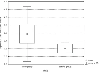
Measurements of mRNA expression for growth factors in the vascular wall (RT-PCR, samples col-lected during the creation of the anastomosis) yielded the following results: PDGFβ1 3.57 ± 0.7 (N = 34). The mean value of the study group was significantly higher than that of control group (3.20 ± 0.16, N = 10, U Mann Whitney’s test Z = 2.157, P = 0.031, Figure 1).
The mean values of mRNA for PDGFβ2 and TGFβ in the study group did not significantly dif-fer from controls (PDGFβ2: 4.51 ± 1.14, N = 34, controls: 4.39 ± 1.13, N = 10; TGFβ: 8.72 ± 0.96, N = 34, controls: 8.49 ± 0.21, N = 10).
The most relevant correlations between study parameters (Spearman test) are listed in Table 2.
Table 1. Principal clinical data on fistula maturation and use during 36 months of observation
Tabela 1. Najistotniejsze dane kliniczne dotyczące dojrzewania i użycia przetoki w ciągu 36-miesięcznej obserwacji
Duration of maturation – days from implantation to first use
(Czas dojrzewania – dni od implantacji do pierwszego użycia) 44.94 (N = 34, 4–154, SD = 38.6) Number of dialyses with reduced blood flow due to fistula insufficiency
(liczba zabiegów ze zmniejszonym przepływem krwi z powodu niewydolności przetoki) 18.71 ± 28.98 Number of hospitalization days due to fistula malfunction
(liczba dni hospitalizacji z powodu dysfunkcji przetoki) 9.95 ± 15.57
General cardiovascular morbidity – number of hospitalization days for cardiovascular reasons
(ogólna chorobowość sercowo-naczyniowa – liczba dni hospitalizacji) 16.4 ± 31.97 Number of days with temporary catheter due to fistula malfunction
(liczba dni z użyciem cewnika tymczasowego z powodu dysfunkcji przetoki) 48.76 ± 162.06 Duration of fistula use – days from implantation to patient death
(Czas używania przetoki – liczba dni od implantacji przetoki do śmierci pacjenta) 795.89 ± 480.6
Data are presented as mean ± standard deviation.
Dane przedstawiono jako średnią ± odchylenie standardowe.
Table 2. Selected significant correlations between study parameters (Spearman test)
Tabela 2. Wybrane znamienne statystycznie korelacje między parametrami badania (test Spearmana)
Correlations
(Korelacje) Spearman R P value
TGFβ mRNA and patient survival after fistula implantation
(TGFβ mRNA i przeżycie pacjentów) –0.722 0.0002
PDGFβ1 mRNA and fistula blood flow
(PDGFβ1 mRNA i przepływ krwi przez przetokę) –0.674 0.0011
longest diameter of fistula and cardiovascular morbidity
(Największa średnica przetoki i chorobowość sercowo-naczyniowa) –0.504 0.0199
Mean thickness of fistula wall and cardiovascular morbidity
(Średnia grubość ściany przetoki i chorobowość sercowo-naczyniowa) 0.449 0.0059 longest diameter of fistula and morbidity due to fistula insufficiency
Discussion
Functional abnormalities of dialysis fistulae ac-count for 15–24% of hospitalizations in chronically hemodialyzed patients, contributing to their mor-tality [1] and high healthcare costs. Failure of ar-teriovenous anastomoses may be caused by either abnormal maturation of newly planted fistulae or their progressive narrowing, initiated by thrombo-sis. Although strict definitions of maturation vary among vascular societies, there is a general consen-sus that fistula maturation time is the period be-tween its implantation and first use for routine he-modialysis [4]. For the patients in this study, the mean maturation time was 44.9 ± 38.6 days, which did not considerably differ from those reported by Ng et al. in a cohort of 4929 patients [5]. In the present study the duration of fistula maturation correlated with its longest diameter as assessed by Doppler ultrasound examination: Wider fistulas were ready for first use earlier than narrower ones (R = –0.59, P = 0.022). Surveillance of fistula mat-uration and subsequent use for hemodialysis was based on frequent physical examinations by med-ical personnel [2] backed by color Doppler ultra-sound assessment of fistula flow and possible re-circulation [6]. In the current study (N = 34), the
blood flow in fistulae 24 months after creation av-eraged 1782 ± 1735 ml/min and was comparable to reference values [7]. Nevertheless, the reliabil-ity of this comparison is questionable for two rea-sons: firstly because of high standard deviations in the measurements in the current study, and sec-ondly because the majority of the fistulae in this group were implanted at the wrist, as opposed to the cubital fossa in the cited study [7]. The mean duration of fistula use (the number of days from implantation to patient death) was also insignifi-cantly different from data reported by other au-thors for comparable fistulas [8]. The observa-tions in the current study indicated considerable morbidity in hemodialyzed patients caused by im-paired fistula patency [1]: The mean number of hospitalization days due to vascular access failure (9.95 ± 15.57) within the study period was only in-significantly lower than the number of hospitaliza-tion days caused by cardiovascular complicahospitaliza-tions (16.39 ± 31.97), which is a well-recognized reason for morbidity and mortality of patients receiving hemodialysis [9].
The main aim of the study was to assess pos-sible clinicopathological correlates of fistula fail-ure. Reduced fistula blood flow measured by Dop-pler ultrasound examination, a well-established
Fig. 1. Expression of mRNA for PDGFβ1 in vascular walls (mean values, standard errors and standard deviations) assessed in patients during the creation of anastomosis (study group) and in persons subjected to reparative vascular surgery due to trauma (control group). The mean value in the study group significantly exceeded that of the control subjects (P = 0.031)
Ryc. 1. Ekspresja mRNA dla PDGFβ1 w ścianie naczyń (wartości średnie, błąd standardowy oraz odchylenia stan-dardowe) oceniano u pacjentów podczas tworzenia zespolenia (grupa badana) oraz u osób poddanych reparacyjnej chirurgii naczyniowej z powodu urazu (grupa kontrolna). Średnie wartości w grupie badanej były istotnie większe niż te z grupy kontrolnej (p = 0,031)
mean mean ± SD
study group control group
group 2.8
3.0 3.2 3.4 3.6 3.8 4.0 4.2 4.4
method of access flow surveillance [10], was been found to be significantly negatively correlated on-ly with the expression of PDGFβ1 in the vascular wall (R = –0.674, P = 0.0011). No other parame-ter of the study significantly correlated with fistu-la blood flow, whether assessed by the Spearman test or by multiple regression analysis. The impor-tance of growth factor expression in the vascular wall in determining fistula patency is hardly sur-prising, since immune/inflammatory mechanisms, along with the suppression of nitric oxide genera-tion by the endothelium, mediate neointimal hy-perplasia leading to fistula failure [3].
In addition to demonstrating a negative corre-lation between fistula blood flow and expression of mRNA for PDGFβ1 in the fistula wall, the current study has shown that after the creation of an anas-tomosis the local activity of this gene significant-ly exceeded respective values in the control group. The exact cellular source of PDGF in this setting
is unknown, since the gene expression in vascular wall was determined quantitatively, without
his-tological PCR insitu studies, but it is conceivable
that local inflammatory mechanisms mediated by infiltrating macrophages could by implicated [3]. In fact, activity of proinflammatory cytokine Il-6 and growth factor IGF have been found in neointi-mal hypertrophy of stenotic fistulae [11, 12]. The current study demonstrated that the expression of gene for TGFβin fistula wall is predictive of patients’ survival during three years of main-tenance hemodialysis treatment. Since samples of fistula walls for assessment of PDGFβ1 and TGFβ expression were collected during the cre-ation of anastomoses in predialysis period, these results indicate that inflammatory conditions in patients being prepared for hemodialysis thera-py could influence both the patency of vascular access and patient survival during renal replace-ment treatreplace-ment.
Conflict of interest: None declared
Received: 30.08.2012 Revised: 25.02.2012 Accepted: 12.08.2013
References
[1] Hakim RM, Breyer J, Ismail N, Schulman G: Effects of dose of dialysis on morbidity and mortality. Am J Kidney Dis 1994, 23, 661–669.
[2] Besarab A, Asif A, Roy-Chadhury P, Spergel LM, Ravani P: The native arteriovenous fistula in 2007. Surveillance and monitoring. J Nephrol 2007, 20, 656–667.
[3] Roy-Chadhury P: Pathophysiology of fistula maturation and failure. ASN Renal Week 2009.
[4] Owens CD, Wake N, Kim JM, Hentschel D, Conte MS, Schanzer A: Endothelial function predicts positive arte-rial-venous fistula remodeling in subjects with stage IV and V chronic kidney disease. J Vasc Access 2010, 11, 329–334.
[5] Ng YY, Wu SC, Hung YN, Ko PJ: Effect of demographic characteristics and timing of vascular access maturation on patency in Chinese incident haemodialysis patients. Nephrol Dial Transplant 2009, 24, 3447–3453.
[6] Schwartz G, Mitterbauer C, Boczula M, Maca T, Funovics M, Heinze G, Lorenz M, Kovarik J, Oberbauer M:
Flow monitoring: performance characteristics of ultrasound dilution versus color Doppler ultrasound compared with fistulography. Am J Kidney Dis 2003, 42, 539–545.
[7] Begin V, Ethier J, Dumont M, Leblanc M: Prospective evaluation of intra-access flow of recently created native arteriovenous fistulae. Am J Kidney Dis 2002, 40, 1277–1282.
[8] Poch E, Martinez X, Rodrigo JA, Tovar JL: Prevalence and functional profile of unsuspected radial artery stenosis in native radiocephalic fistula dysfunction. Nephrologia 2006, 26, 581–586.
[9] Foley RN, Gilbertson DT, Murray T, Collins AJ: long interdialytic interval and mortality among patients receiv-ing hemodialysis. N Engl J Med 2011, 365, 1095–1107.
[10] Tessitore N, Bedogna V, Poli A, Mantovani W, Lipari G, Baggio E, Mansueto G, Lupo A: Adding access blood flow surveillance to clinical monitoring reduces thrombosis rates and costs and improves fistula patency in the short term: a controlled study. Nephrol Dial Transplant 2008, 23, 3578–3584.
[11] Marrone D, Pertosa G, Simone S, Loverre A, Capobianco C, Cifarelli M, Memoli B, Schena FP, Grandaliano G:
local activation of interleukin 6 signaling is associated with arteriovenous fistula stenosis in hemodialysis patients. Am J Kidney Dis 2007, 49, 664–673.
[12] Stracke S, Konner K, Kőstlin I, Schneider M, Herzog R, Jehle PM, Keller F, Friedl R: over-expression of IGF-related peptides in stenosis of native arteriovenous fistulas in hemodialysis patients. Growth Horm IGF Res 2007, 17, 297–306.
Address for correspondence:
Zbigniew Hruby
Department of Nephrology
Regional Specialist Hospital Center for Research & Development Kamieńskiego 73A
51-124 Wrocław Poland