Andrzej Myśliwiec
1, 2, A–D, F, Edward Saulicz
1, 3, A, C, E, F, Michał Kuszewski
1, A, C, E, F,
Przemysław Sładkowski
4, B–D, Tomasz Wolny
1, 3, E, F, Mariola Saulicz
1, 3, E, FAssessment of the Cervical Spine Range
of Motion After the Use of the Saunders Traction Device
in Different Positioning of the Upper Extremities
1 Department of Physiotherapy, The Jerzy Kukuczka Academy of Physical Education in Katowice, Poland 2 Department of Physiotherapy, College of Strategic Planning in Dąbrowa Górnicza, Poland
3 Academy of Business in Dąbrowa Górnicza, Poland
4 Graduate Studies, Department of Kinesiotherapy and Special Physiotherapy Methods, The Jerzy Kukuczka Academy of Physical Education in Katowice, Poland
A – research concept and design; B – collection and/or assembly of data; C – data analysis and interpretation;
D – writing the article; E – critical revision of the article; F – final approval of article; G – other
Abstract
Background. Among the procedures used in the therapy of spinal pain syndromes one of the most frequently recommended is the cervical traction. The methods of performing the traction are varied.
Objectives. It was decided to examine in the research whether the abduction and external rotation of the brachium, causing relaxation of the pectoral girdle muscles as well as the cervical spine, affects the quality and efficiency of traction in the patient’s subjective assessment and the changes in the cervical spine range of motion.
Material and Methods. Fifty subjects aged 20 to 42 were involved in the study, including 26 women and 24 men. The criterion of inclusion into the research project was the age between 20 and 40 years and the result of question-naire containing the NDI scale for evaluation of the degree of dysfunction of the cervical spine, ranging in value between 5 and 14 points. The admitted group of 50 subjects was randomly divided into two experimental groups. The traction of the cervical spine was performed twice in both groups by means of the Saunders device. In the first group used the traditional positing, with the upper extremities placed along the torso, was adapted as first whereas in the second group the modified position was applied, in which the upper extremities were in the external rotation in abduction and with flexion in the cubital articulation, in other words the patients placed their hands next to head. In both cases the patient was in the supine position on a therapeutic table with a support roll under the knees.
Results. The evaluation of the cervical spine range of motion pointed, that in both position all the obtained differ-ences proved statistical significance. The subjects taking part in the research claimed that the procedure performed by means of the traditional method was more pleasant.
Conclusions. No significant difference was discovered in the effectiveness of the suggested positioning of the arms during performing the traction procedure of the cervical spine. The modified position caused greater sensation of discomfort than position with traditional arrangement of the arms (Adv Clin Exp Med 2014, 23, 5, 769–774).
Key words: traction, neck, pain, range of motion.
Adv Clin Exp Med 2014, 23, 5, 769–774 ISSN 1899–5276
ORIGINAL PAPERS
© Copyright by Wroclaw Medical University
Alongside the progress of civilization, cervical spine pain complaints have become an increasing-ly large therapeutic and epidemiological problem. It is connected with a radical change in the level of physical activity and in lifestyle [1–5]. During the initial phase of these ailments, the feeling of over-load of the structures located directly in the nape area prevails. It is represented by increased tension of the muscles or even their “stiffness”, then pain
movement area and especially the upper part of the spiral and lateral line and the superficial back and arms lines [9]. Neglected pain of the cervical spine may lead to a developing cervical spondylitis which gradually affects the decrease in the range of motion in one segment, simultaneously overload-ing the neighboroverload-ing ones [10].
Among the procedures used in the therapy of spinal pain syndromes, one of the most frequent-ly recommended is cervical traction [11]. Thanks to a deloading of the spine, and as a consequence, the relaxing of the tensed paravertebral structures, the decompression of the irritated nerve roots as well as improvement in the circulation within the vertebral artery, and as a consequence, increased statokinetic efficiency, occur [12, 13]. The meth-ods of performing the traction are varied (Peake 2005, Myśliwiec 2010). There is no conclusive answer whether the best results are obtained by means of traction with the use of a static traction force [14], intermittent traction [15, 16], manual traction [17] or a vibratory traction device [10]. Diversity may be also noticed in reference to the suggested force of the traction [6, 15, 18] as well as the patient’s position. Some researchers indicate the possibility to perform the traction in a sitting position [12, 18, 19], whereas others claim that cer-vical traction should be always performed in the supine position [6, 11, 14, 15].
A difference in opinions may also be noted with regard to the suggested additional treatments, performed before or after the traction session. The types of procedures which prevail among these treatments are dynamic and/or isometric exercis-es, massage, thermotherapy and briefing, which focuses on maintaining the correct posture [13, 17, 20–22]. Other methods connected with traction are nonsteroidal anti-inflammatory medicines [18] or physiotherapy treatment procedures [21, 23]. There are also authors who suggest performing ex-clusively the manual traction treatment [24].
It was decided to examine in the research whether the abduction and external rotation of the brachium, causing relaxation of the pectoral gir-dle muscles as well as the cervical spine, affects the quality and efficiency of traction in the patient’s subjective assessment and changes in cervical spine range of motion.
Material and Methods
The research was carried out in accordance with the principles of the Helsinki Convention and based on the approval of Bioethics Commit-tee No. 12/2006 functioning by the Jerzy Kukuczka Academy of Physical Education in Katowice. The
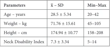
research procedures were carried out in the physio-therapy room in Rybnik and the Academy of Physi-cal Education in Katowice. Fifty subjects aged 20 to 42 were involved in the study, including 26 women and 24 men, who were inhabitants of Silesia. The subjects were selected intentionally on the basis of voluntary applications. The criterion for inclusion into the research project was the age between 20 and 42 years and the results of a questionnaire con-taining the NDI scale for evaluation of the degree of dysfunction of the cervical spine, ranging in val-ue between 5 and 14 points [25]. People with no or minimal pain complaints as well as those who in-dicated a value above 14 points were not admitted to the research. An additional criterion of exclu-sion was the occurrence of radiation of the symp-toms along the upper extremities. All the peo-ple qualified to take part in the research obtained a physician’s permission which, in a specific way, eliminated other than overload-induced causes of developing the condition such as neoplastic disease or hypermobility of the segment. The occurrence of vertebral artery occlusion was also eliminated. The biometric data as well as the values of the intensifi-cation of pain are presented in Table 1.
The group of 50 subjects admitted was ran-domly divided into 2 experimental groups. The na-ture of the groups was typically organizational and, after assigning the subjects a given group, a draw-ing was held in which it was indicated from which initial position the group would start the experi-ment. In both groups, the traction force was dosed in such a way that the patients perceived notice-able but painless traction [6, 11, 23]. The length of the traction was 15 min. After carrying out the first treatment session, a one week break took place, af-ter which the method of performing the traction was changed in both experimental groups.
In order to assess the range of motion of the cervical spine, the CROM (cervical range of mo-tion) instrument was used. It made it possible to perform the measurement to an accuracy of 2 de-grees in all 3 planes [26]. The measurements were carried out by a person who did not know the meth-od of the implemented traction, in a chair sitting
Table 1. Biometric data and intensification of pain com-plaints expressed in the NDI (neck of disability index) points of patients qualified to take part in the experiment
Parameters x– – SD Min–Max
Age – years 28.5 ± 5.34 20–42
Weight – kg 71.76 ± 15.61 45–105
Height – cm 174.94 ± 10.77 158–208
position with the upper extremities placed along the torso and back firmly supported at a right an-gle. Special attention was paid to prevent the pa-tient from deepening the movement by bending or torso rotation and to perform the movement in accordance with maintaining the plane. Each of the measurements was performed three times and so the result of the best test was used in further analysis. The movement of flexion and extension, side bends to the left and right as well as rotation movements in both sides were subjected to eval-uation. Another area of study was the evaluation of the difference in pain complaints occurring be-fore and after carrying out the traction procedure. In order to assess the pain, the analogue pain scale VAS (visual analogue scale) was used [27, 28]. The patients marked on a 10 cm line their current sen-sation of the pain occurring, assuming that on the left side of the line no pain occurs (0) whereas on the right side appears the pain sensation identi-fied as unbearable (10). After marking on the line, the person conducting the research performed the measurement by means of a centimeter scale rul-er. The results were noted down to an accuracy of 1 mm. The evaluation of feelings of comfort while performing the traction procedure, which was car-ried out immediately after completing the session, was implemented by means of a similar, modified instrument. The patient was to mark on the above-mentioned line, the point which corresponded to his/her sensation of discomfort and feeling of safe-ty during the session. The 0 value represented the feeling of complete safety and comfort whereas the feeling of anxiety and discomfort, connected with the process of the procedure, increased toward the right side of the scale. The sensation perceived at the level of 10 was such that it resulted in an ab-solute necessity to interrupt the treatment proce-dure. The result was noted down to an accuracy of 1 mm also in this test.
The last parameter which was evaluated was the traction force. The result was obtained from the reading of the measuring instrument of the traction device at the moment when the patient reported a sensation of a noticeable and painless traction, which was a sign to stop increasing the in-tensity of the procedure. The value was read off to an accuracy of 1 kG of force.
The results obtained were subjected to statis-tical analysis performed by means of the Statisti-ca 9.0 program. The arithmetiStatisti-cal mean values (x–) and standard deviation (SD) were calculated and the minimum and maximum values were indicat-ed. In the evaluation of difference significance (p) in the measurement of the range of motion in the pre-post sample, the Student’s t-test for dependent samples was used. The measurement of differences
between the groups was implemented on the basis of a Student’s t-test for independent samples. In view of the abnormal distribution, non-paramet-ric tests were used to indicate the statistical signifi-cance of differences in results concerning the sub-jective evaluation as well as the traction force. The
U Mann-Whitney test was used to evaluate the in-tergroup differences whereas the Wilcoxon signed-rank test was used to assess the differences in the pre-post sample. The critical level of significance was p < 0.05.
Results
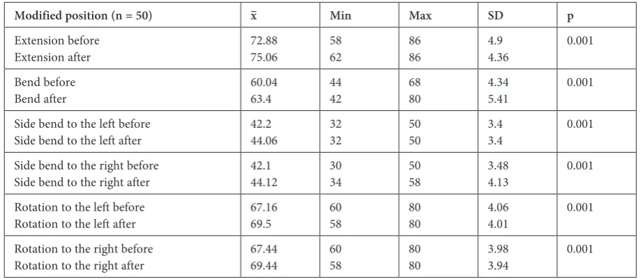
The evaluation of the cervical spine range of motion, implemented by means of a CROM in-strument, made it possible to state that in the treat-ment procedure performed with the traditional positioning of the upper extremities as well as with the modified positioning, all the differences ob-tained proved statistically significant. No signifi-cant differences were discovered between the stud-ied groups in the test preceding the traction. It is worth emphasizing that the range of motion after performing the traction in the modified position-ing obtained greater statistical significance. The obtained results are illustrated in Tables 2 and 3.
The analysis of the difference in pain com-plaint intensification level before and after the traction made it possible to state that the group that had the procedure performed with the tradi-tional positioning of the upper extremities demon-strated a greater reduction of the symptoms, which is presented in Table 4.
The subjects taking part in the research claimed that the procedure performed by means of the tra-ditional method was more pleasant than with the hands placed by the head. It was also observed that the force of traction carried out in the traditional positioning of the upper extremities made it pos-sible to obtain higher values than in the modified positioning. The results achieved in these observa-tions are presented in Table 5.
Discussion
be assumed that the traction of the cervical spine has an initial function, preparing for deep stabili-zation exercises and postural re-education [2, 20].
This type of algorithm should encourage the for-mulation of such methods which would allow the most effective influence of traction. The traction
Table 3. Mean value, standard deviation, minimum and maximum value of cervical spine motion after the procedure with the modified positioning of the upper extremities and the significance level in the Student’s t-test
Modified position (n = 50) x– Min Max SD p
Extension before
Extension after 72.8875.06 5862 8686 4.94.36 0.001
Bend before
Bend after 60.0463.4 4442 6880 4.345.41 0.001
Side bend to the left before
Side bend to the left after 42.244.06 3232 5050 3.43.4 0.001
Side bend to the right before
Side bend to the right after 42.144.12 3034 5058 3.484.13 0.001
Rotation to the left before
Rotation to the left after 67.1669.5 6058 8080 4.064.01 0.001
Rotation to the right before
Rotation to the right after 67.4469.44 6058 8080 3.983.94 0.001
Table 4. Mean value, standard deviation, minimum and maximum value of the pain intensity formulated by the VAS (visual analogy scale) scale and the level of signifi-cance in the Wilcoxon signed-rank test
Traditional Modified
x– ± SD
min–max x– ± SDmin–max
Point on VAS
scale before 1.09 ± 0.950–3.2 1.06 ± 0.830–3.2 Point on VAS
scale after 0.71 ± 0.810–3 0.88 ± 0.830–3
p 0.001 0.034
Table 5. Mean value, standard deviation, minimum and maximum value of the evaluation of discomfort and trac-tion force as well as the significance level in the U Mann-Whitney test
Traditional Modified p
x– ± SD
min–max x– ± SDmin–max
Discomfort (points on ana-logic scale)
2.12 ± 1.59
0–6 3.42 ± 2.110–8 0.003
Traction force
(kG) 9.65 ± 3.215–18 7.72 ± 2.684–16 0.002
Table 2. Mean value, standard deviation, minimum and maximum value of cervical spine motion after the procedure with the traditional positioning of the upper extremities and the significance level in the Student’s t-test
Traditional position (n = 50) x– Min Max SD p
Extension before
Extension after 73.6475.18 6068 8290 5.074.81 0.02
Bend before
Bend after 59.9861.76 4040 7070 5.075.01 0.002
Side bend to the left before
Side bend to the left after 41.7443.08 3234 5052 3.733.57 0.001
Side bend to the right before
Side bend to the right after 41.9843.08 2030 4852 4.334.02 0.003
Rotation to the left before
Rotation to the left after 66.5267.8 4050 8080 5.725.22 0.07
Rotation to the right before
preceded by physiotherapy procedures including electrotherapy and thermotherapy appears to be more effective in consideration of their analgetic and relaxing effect [13]. While observing the pro-cess of the traction procedure while carrying out the therapy with patients with cervical spine pain syndrome, it is difficult to resist the impression concerning the necessity of creating the most com-fortable position for the patient. The patients who are subjected to the therapy are frequently asked to relax, which often leads to their falling asleep [11, 23]. It is difficult here to agree with the methodolo-gy recommending performing the traction in a sit-ting position, in which the very act of maintaining the body’s upright position requires appropriate muscle tone [12, 18, 19].
The position suggested in the research per-formed was inspired, among others, by the fact that while resting, a lot of people adopt the po-sition in which the hands are placed under the head. Such positioning causes rising of the brachi-al girdle, which in consequence decreases the tone of muscles localized in the nape area by relaxing the back line and arms lines [9]. Adopting such a position is possible basically only in a situation when the radiation of symptoms does not occur. In this case, the suggested positioning might have caused intensification of the complaints and made
it impossible to continue the procedure [30, 31]. In the case of pain complaints, in which no irrita-tion of the brachial plexus as well as the peripheral nerves of the upper extremity occurred, adopting such a position did not cause any difficulties for the patients, consequently giving a full opportu-nity to carry out the observation. However, main-taining the position for a longer time resulted in greater discomfort than was in the case of the tra-ditional position. Also in this position, the feeling of sufficient force of traction was reported faster by the patients. This type of situation may prove that such a position decreases muscle protection, provoking faster tension in the area of the passive locomotor system, represented by ligaments. To a certain extent, this observation may explain the achieving of higher statistical significance in refer-ence to cervical spine mobility, especially in flex-ion and side bending. This advantage; however, is not convincing in relation to the minor reduction of pain complaints.
The authors concluded that no significant dif-ference was discovered in the effectiveness of the suggested positioning of the arms while perform-ing the traction procedure of the cervical spine. The modified position caused greater sensations of discomfort than the position with traditional ar-rangement of the arms.
References
[1] Falla D: Unravelling the complexity of muscle impairment in chronic neck pain. Man Ther 2004, 9, 125–133.
[2] Falla D, Jull G, Hodges P, Vicenzimo B: An endurance–strength training regime in effective in reducing myo-electric manifestation of cervical flexor muscle fatigue in females with chronic neck pain. Neurophysiol Clin 2006, 117, 828–837.
[3] Cote P, Cassidy JD, Carroll L: The epidemiology of neck pain: what we have learned from our population – based studies. JCCA 2003, 47, 284–290.
[4] Nowotny J, Nowotny-Czupryna O, Brzęk A, Kowalczyk A, Czupryna K: Body posture and syndromes of back pain. Ortop Traumatol Rehabil 2011, 13, 1, 59–71.
[5] Hill J, Levis M, Papageorgiou A, Dziedzic K, Croft P: Predicting persistent neck pain. Spine 2004, 29, 1648–1654.
[6] Saunders HD, Saunders R: Evaluation, Treatment and Prevention of Musculoskeletal Disorders. The Saunders Group, Minnesota 1995.
[7] Pascarelli EF, Hsu Yp: Understanding work-related upper extremity disorders; clinical findings in 485 computer users, musicians and others. Journal Occupy Rehab 2001, 11, 1–21.
[8] Ariens GA, van Mechelen W: Physical risk factors for neck pain. Scand J Work Environ Health 2000, 26, 7–19.
[9] Myers TW: Anatomy Trains. Myofascial Meridians for manual and movement therapists. Churchill Livingston, Elsevier, Wrocław 2009, 2nd ed.
[10] Gieremek K, Saulicz E, Piłat A, Molicka D: La eficacia del aparato vibratorio de extension cervical en el trata-miento de los pacientes con espondylosis cervical. Cuestiones Fisioterapia 2003, 21–28.
[11] Myśliwiec A, Saulicz E, Kuszewski M, Kokosz M, Wolny T: Assessment of the influence of Saunders traction and trancutaneous electrical nerve stimulation on hand grip force in patients with neck pain. Ortop Traumatol Rehabil 2011, 1960, 13, 37–44.
[12] Tsai CT, Chang WD, Kao MJ, Wang CJ, Lai PT: Changes in blood pressure and related autonomic function dur-ing cervical traction in healthy women. Orthopedics 2011, 34, 295–301.
[13] Peake N, Harte A: The effectiveness of cervical traction. Phys Ther Rev 2005, 10, 217–229.
[14] Klaber-Moffett JA, Hughes GI, Griffiths P: An investigation of the effects of cervical traction. Clinical effective-ness. Clin Rehabil 1990, 4, 205–211.
[15] Baker P, Marcoux BC: The effectiveness of home cervical traction on relief of neck pain and impaired cervical range of motion. Phys Ther 1999, 2, 145–151.
[17] Zylbergold RS, Piper MC: Cervical spine disorders. A comparison of three types of traction. Spine 1985, 10, 864–871.
[18] Constantoyannis C, Konstantinou D, Kourtopoulos H, Papadakis N: Intermittent cervical traction for cervical radioculopathy causes by large – volume herniated disks. J Manipulative Physiol Ther 2002, 25, 188–192.
[19] Pan PJ, Tsai PH, Tsai CC, Chou CL, Lo MT, Chiu JH: Clinical response and autonomic modulation as seen in heart rate variability in mechanical intermittent cervical traction: o pilot study. J Rehabil Med 2011, 44, 229–234.
[20] Shakoor M, Ahmed MS, Kibria G, Khan AA, Mian MAH, Hasan SA: Effects of cervical traction and exercise therapy in cervical spondylosis. Bangladesh MRC Bull 2002, 28, 61–69.
[21] Brewerton DA, Beardwell A, Blower PW, Brown MR, Campbell EDR, Cochrane GM: British association of physical medicine. Pain in the neck and arm: a multicentre trial of the effects of physiotherapy. BMJ 1996, 2, 253–258.
[22] Saal JS, Saal JA, Yurth F: Non-operative management of herniated cervical intervertebral disc with radiculopathy. Spine 1996, 21, 1877–1883.
[23] Myśliwiec A, Saulicz E, Kuszewski M, Kokosz M, Gnat R, Wolny T: Changes in the subjective sensation of pain in patients with cervical spine dysfunction treated by mean of Saunders traction and TENS. FP 2010, 3, 10, 211–221.
[24] Valtonen EJ, Kiuru E: Cervical traction as a therapeutic tool. Scand J Rehabil Med 1970, 2, 29–36.
[25] Vernon H, Mior S: The neck disability index: a study of reliability and validity. J Manipulative Physiol Ther 1991, 14, 409–415.
[26] Hole DE, Cook JM, Bolton JE: Reliability and concurrent validity of two instruments for measuring cervical range of motion: effects of age and gender. Man Ther 1995, 1, 36–42.
[27] Wewers ME, Lowe NK: A critical review of visual analogue scales in the measurement of clinical phenomena. Res Nurs Health 1990, 13, 227–236.
[28] Kilderso J, Wyon D, Skov T, Schneider T: Visual analogue scales for detecting changes in symptoms of sick build-ing syndrome in an intervention study. Scan J Work Environ Health 1999, 25, 361–367.
[29] Sipko T, Bieć E, Demczuk-Włodarczyk E, Ciesielska B: Mobility of cervical spine and postural equilibrium in patients with spinal overload syndrome. Ortop Traumatol Rehabil 2007, 2, 9, 141–148.
[30] Kleinresink GJ, Stoeckart R, Mulder PG, Hoek G, Broek T, Vleeming A, Snijders C: Upper limbs tension tests as tools in the diagnosis of nerve and plexus lesions. Anatomical and biomechanical aspects. Clin Biomech 2000, 15, 9–14.
[31] Shacklock M: Clinical neurodynamics. A new system of musculoskeletal treatment. Elsevier, Wrocław 2008.
Address for correspondence:
Andrzej Myśliwiec
The Jerzy Kukuczka Academy of Physical Education Department of Physiotherapy
Mikołowska 72a 40-065 Katowice Poland
Tel.: +48 32 207 53 54
E-mail: [email protected]
Conflict of interest: None declared