Ewa Zaczyńska
B, C, E, Maja Kocięba
B, E, Ewelina Śliwińska
B, Michał Zimecki
A, C–FBovine Lactoferrin Enhances Proliferation
of Human Peripheral Blood Lymphocytes
and Induces Cytokine Production
in Whole Blood Cultures
Laboratory of Immunobiology, Institute of Immunology and Experimental Therapy, Polish Academy of Science, Wrocław, Poland
A – research concept and design; B – collection and/or assembly of data; C – data analysis and interpretation;
D – writing the article; E – critical revision of the article; F – final approval of article; G – other
Abstract
Background. Lactoferrin belongs to the immunoregulatory milk proteins involved in iron metabolism as well in providing innate immunity to newborns. The protein has been the subject of numerous clinical studies.
Objectives. The aim of this investigation was to evaluate the effects of bovine lactoferrins (bLF), differing in source and iron content, on spontaneous proliferation of human peripheral blood mononuclear cells (PBMC) and cyto-kine production by human whole blood cultures.
Material and Methods. The following bLF preparations were used: partially iron saturated or devoid of iron bLF from milk and bLF from colostrum. The study was conducted on 12 healthy volunteers (men, 20–24 years old). The effects of bLFs on the proliferation of PBMC in four-day cultures was studied at 50–0.6 µg/mL concentration range and the rate of proliferation was determined using the MTT colorimetric method. TNF α and IL-6 levels, induced by the bLFs in 24 h whole blood cultures, were measured by ELISA.
Results. The lactoferrins stimulated autologous proliferation of human peripheral blood mononuclear cells (PBMC) in a dose-dependent manner, with a comparable efficacy. This stimulation occurred both in the constant presence of bLFs in the cultures and also upon preincubation of PBMC with bLFs with subsequent exhaustive wash of cells. Only bLF from colostrum induced production of tumor necrosis factor alpha (TNF-α) and interleukin-6 (IL-6) in cultures of whole blood cells. This phenomenon took place predominantly at concentration of 50 µg/mL.
Conclusions. The results showed potent stimulation of the proliferative response of PBMC by bovine lactoferrin, associated with the induction of proinflammatory cytokines only in the case of colostral bLF. This observation may be of importance when high doses of bLF are used in therapy and by designing diet supplementation with this protein (Adv Clin Exp Med 2014, 23, 6, 871–876).
Key words: IL-6, proliferation, lactoferrin, TNF-α, autologous PBMC culture.
Adv Clin Exp Med 2014, 23, 6, 871–876 ISSN 1899–5276
ORIGINAL PAPERS
© Copyright by Wroclaw Medical University
Lactoferrin is a protein abundant in colostrum and milk as well in the excretory fluids of mam-mals, constituting an important element of innate immunity [1]. The protein, which is also contained in secondary granules of neutrophils [2], plays a role in iron metabolism [3].
Lactoferrin affects the immune system by promotion of lymphocyte maturation [4, 5] and stimulation of mucosal and systemic immune re-sponse [6]. Lactoferrin interacts with a wide spec-trum of receptors, distributed in virtually all organs
and cell types [7]. They include, among others, mannose receptors [8], toll-like receptors [9], hep-aran sulfate [10], CD14 [11], nucleolin [12], int-electin [13], sialoadhesin [14] and CD22 [15].
are, in fact, immunoregulatory and depend on the individual immune reactivity of healthy volunteers and status of the patient [19–21].
Bovine lactoferrin has been the subject of nu-merous clinical studies (for review, see [22]) and is used as an ingredient in infant food [23]. It was, therefore, of importance to investigate the effect of lactoferrin on the proliferation of autologous hu-man lymphocytes, a model which has not been studied yet. In addition, the ability of bLF to in-duce production of tumor necrosis factor alpha (TNF-α) and interleukin-6 (IL-6) in whole blood cell cultures was determined. For the studies, par-tially iron saturated and iron-free milk bLFs and colostrum-derived bLF, were applied.
Material and Methods
Reagents
RPMI medium, Hanks’ medium with and without Ca+2 and Mg+2 and LSM Lymphoprep
sep-aration medium (1.077 g/mL) were from Cytogen GmbH (Germany). MTT-93-[4,5-dimethylthia-zol-2-yl]-2,5diphenyltetrazolium bromide derived from Sigma-Aldrich (USA). Fetal calf serum was purchased from Gibco (USA). The cytokines (TNF-α and IL-6) were measured using ELISA kits manufactured by RD Systems (USA & Canada).
Lactoferrins
The following forms of bovine lactoferrin were used: (1) bLF from Tatua Co-Operative Dairy Company, New Zealand; (2) Apolactoferrin (apo-bLF) prepared from bLF (Tatua) as originally de-scribed by Mansson; 1% bLF solution in distilled water (100 mg of protein/10 mL water), dialyzed against 20 × volume of 0.1 M citrate acid for 36 h, then dialyzed against distilled water for 3 days at 4oC (3 times in 20-fold water volume) and finally
lyophilized. (3) Colostral bovine lactoferrin (cbLF) from Sigma (no. L4765).
Preparation of Peripheral
Blood Mononuclear Cells
Venous blood from each of the donors was withdrawn into syringes with sodium citrate and diluted twice with PBS. PBMCs were isolated by centrifugation on a Lymphoprep gradient (density 1.077 g/mL) and centrifuged at 800 × g for 20 min at 4oC. Cells from the interphase, consisting of
lym-phocytes (20%) and monocytes (80%), were then washed 3 times with Hanks’ medium and re-sus-pended in a culture medium, referred to below as
the culture medium, consisting of RPMI-1640 sup-plemented with 10% fetal calf serum, L-glutamine, sodium pyruvate, 2-mercaptoethanol and antibiot-ics, at a density of 2 × 10⁶ cells/mL.
Preincubation of PBMC
with Lactoferrins
PBMCs resuspended in the culture medium, were transferred to a 6-well culture plate (3 × 10⁶ cell/
/mL) and the respective concentrations of the stud-ied lactoferrins were added (50 µg/mL). The cells were incubated for 1 h at 37oC in a cell culture
incu-bator. After the incubation, the cells were harvested in cold Hanks’ medium devoid of Ca++ and Mg++.
Next, the cells were resuspended in Hanks’ contain-ing Ca+2, Mg+2 and washed 5 times (centrifuged for
10 min at 1.200 rpm at 4oC). Then, the cells were
resuspended in the culture medium at a density of 2 × 10⁶ cells/mL and transferred to a 96-well plate.
Determination of Autologous
Cell Proliferation
The PBMCs were distributed into 96-well flat-bottom plates in 100 µL aliquots (2 × 10⁵ cells/
/well). The lactoferrins were added at a concen-tration range of 50–0.6 µg/mL. Alternatively, the cells were pulsed with lactoferrin and washed as described above. After a 4-day incubation in a cell culture incubator, the proliferative response of the cells was determined using the colorimetric MTT method. The data is presented as a mean OD value from quadruplicate wells ± SE.
Induction of Cytokines
in the Human Blood Culture
Human whole blood from healthy males, 20–24 years old, was diluted 5 × with RPMI 1640 medium and distributed to 24-well culture plates in 1 mL aliquots. After an overnight incubation, the supernatants were harvested and frozen at –80oC until cytokine determination.
Colorimetric MTT Assay
for Cell Growth and Kill
Statistical Analysis
The results are presented as mean values ± stan-dard error (SE). Brown-Forsyth’s test was used to determine the homogeneity of variance between groups. When the variance was homogenous, anal-ysis of variance (one-way ANOVA) was applied, followed by post hoc comparisons with the Tukey’s test to estimate the significance of the difference between groups. Nonparametric data was evaluat-ed with the Kruskal-Wallis analysis of variance, as indicated in the text. Significance was determined at p < 0.05. Statistical analysis was performed using STATISTICA 7 for Windows.
Results
Effects of Lactoferrins
on Spontaneous Proliferation
of Human PBMC
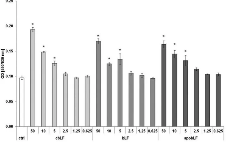
The lactoferrins were added at a concentration range of 50–0.6 μg/mL to the PBMC cultures, in-cubated for 4 days and the rate of proliferation was measured. The results, presented in Fig. 1, show a representative example of 12 determinations in individual subjects with very similar results. All bLFs displayed similar, dose-dependant stimulato-ry effects on the proliferation of cells in an autolo-gous system. The strongest effect was observed at 50 µg/mL whereas 2.5 µg/mL was not stimulatory.
In order to check whether pulsing of PMBC with bLFs is sufficient to induce the stimulatory
effect on cell proliferation, PBMCs were incubat-ed for 1 h with 50 µg of lactoferrins and washincubat-ed 5 times. The results (Fig. 2) indicate that only in the case of colostral bLF was the 1 h exposure of PBMC to bLFs sufficient to exert the stimulatory effects on cell proliferation. However, the stimula-tory effects were lower in comparison to cultures with constant presence of lactoferrins (Fig. 1).
Effects of Lactoferrins on
Production of TNF-α and IL-6
in Whole Blood Cell Cultures
BLF has been shown in the past to induce cy-tokine production in vivo and in vitro [19, 24, 25]. Here we determined the effects of colostrum-de-rived lactoferrin on the production of TNF-α and IL-6 in the whole blood cell cultures of 4 represen-tative individuals (Table 1). The results show that cbLF, at 50 μg/mL induced production of TNF-α
Fig. 1. Stimulatory effects of lactoferrins on proliferation of peripheral blood mononuclear cells (*, p < 0.05, when compared with control cultures)
Table 1. Induction of TNF-α and IL-6 by cbLF in human whole blood cell cultures
Donor TNF-α (pg/mL) IL-6 (pg/mL) cbLF (µg/mL) cbLF (µg/mL)
50 10 50 10
1 404 – 1761 99
2 154 – 896 30
3 90 – 653 8
and IL-6 to various degrees. However, at the con-centration of 10 μg/mL only some level of IL-6 was induced. Other lactoferrins were not effective in cytokine induction (not shown).
Discussion
In the present investigation, we showed that bovine lactoferrins, irrespectively of origin and iron saturation, were capable of significantly stim-ulating the proliferation of PMBCs in autologous cultures. This action was distinctly uniform (stim-ulatory) in all tested individuals in contrast to the phytohemagglutinin A-induced proliferation of PBMC [20], where the effects of bLF were differ-ential and strongly depended on the individual re-activity of blood donors. The responding cell in the autologous mixed lymphocyte reaction is the CD4+
T cell [26]. Therefore, all effects exerted by lacto-ferrin on T cells, mainly described as phenotypic changes and induction of signaling pathways, are relevant in this phenomenon. These effects include induction of the CD4 antigen [4] and lymphocyte adhesion molecule-1 [27], which may have signif-icance in interaction with ICAM-1 receptors on accessory cells. Lactoferrin is also able to increase expression of the zeta chain in the CD3 complex, responsible for transmission of the stimulatory signal in T cells [28]. More importantly, lactofer-rin was shown to activate mitogen-activated pro-tein kinase in Jurkat cells [29], the action reveal-ing a triggerreveal-ing of the signalreveal-ing pathway leadreveal-ing to activation of T cells. In addition, other actions of lactoferrin described may contribute to increased cell proliferation, involving inhibition of apop-tosis [30] and the property of scavenging harm-ful hydroxyl radicals [31]. Effective stimulation of
cell proliferation in the case of pulsing of the cell with cbLF (Fig. 2) supports the notion that a short time signal delivered by lactoferrin may be suffi-cient for T cells to initiate proliferation. It also ap-peared that the presence of iron in the lactoferrin molecule did not affect the degree of cell prolifer-ation, indicating that the conformation of the lac-toferrin molecule was without effect on the in-teraction of a bLF ligand with the respective cell receptor. In this particular case, the receptor for bLF may bear a resemblance to that present on the surface and inside Jurkat and stimulated human lymphocytes [32].
The results also confirmed our previous find-ings on the induction of TNF-α and IL-6 in whole blood cultures [25]. The induction of TNF-α could represent an additional stimulus for cell prolifer-ation as reported elsewhere [33] and account for a stronger induction of cell proliferation. Of inter-est, the induction of cytokines was only observed in the case of colostrum-derived lactoferrin. This phenomenon could be associated with a differ-ent pattern of glycosylation in cbLF as compared with milk-derived lactoferrin. Generally, colos-tral proteins are highly sialylated as compared to milk-derived proteins [34]. Lactoferrin isolated from bovine colostrums is a glycoprotein carry-ing complex and high-mannose type glycans [35]. Therefore, a putative candidate for the interaction of bLF is siglec7, present on monocytes. This re-ceptor is responsible for activation of cells for cy-tokine production, including TNF-α and IL-6, by ligands terminated by sialic acid. Nevertheless, it seems that the signals leading to cell proliferation and cytokine production are separate since milk- -derived lactoferrins, not able to induce cytokine production, were equally effective in inducing cell proliferation.
Fig. 2. Effects of preincuba-tion with lactoferrins on lymphocyte proliferation in autologous culture
In summary, this is the first demonstration of a potent stimulation of the proliferative re-sponse of PBMC in autologous culture by bovine lactoferrin. This observation should be taken in-to account when high doses of bLF are used in
therapy and by designing diet supplementation with this protein. The results of this study may also explain the strong stimulatory effects of oral bLF on the response of the gut-associated lym-phoid system.
References
[1] Vogel HJ: Lactoferrin, a bird’s eye view. Biochem Cell Biol 2012, 90, 233–244.
[2] Masson PL, Heremans JF, Schonne E: Lactoferrin, an iron-binding protein in neutrophilic leukocytes. J Exp Med 1969, 130, 643–658.
[3] Johnson EE, Wessling-Resnick M: Iron metabolism and the innate immune response to infection. Microbes Infect 2012, 14, 207–216.
[4] Zimecki M, Mazurier J, Machnicki M, Wieczorek Z, Montreuil J, Spik G: Immunostimulatory activity of lacto-transferrin and maturation of CD4- CD8-murine thymocytes. Immunol Lett 1991, 30, 119–123.
[5] Zimecki M, Mazurier J, Spik G, Kapp JA: Human lactoferrin induces phenotypic and functional changes in murine splenic B cells. Immunology 1995, 86, 122–127.
[6] Debbabi H, Dubarry M, Rautureau M, Tome D: Bovine lactoferrin induces both mucosal and systemic immune response in mice. J Dairy Res 1998, 65, 283–293.
[7] Suzuki YA, Lopez V, Lonnerdal B: Mammalian lactoferrin receptors: structure and function. Cell Mol Life Sci 2005, 62, 2560–2575.
[8] Zimecki M, Kocieba M, Kruzel M: Immunoregulatory activities of lactoferrin in the delayed type hypersensitivity in mice are mediated by a receptor with affinity to mannose. Immunobiology 2002, 205, 120–131.
[9] Curran CS, Demick KP, Mansfield JM: Lactoferrin activates macrophages via TLR4-dependent and -independent signaling pathways. Cell Immunol 2006, 242, 23–30.
[10] Chien YJ, Chen WJ, Hsu WL, Chiou SS: Bovine lactoferrin inhibits Japanese encephalitis virus by binding to heparan sulfate and receptor for low density lipoprotein. Virology 2008, 379, 143–151.
[11] Baveye S, Elass E, Fernig DG, Blanquart C, Mazurier J, Legrand D: Human lactoferrin interacts with soluble CD14 and inhibits expression of endothelial adhesion molecules, E-selectin and ICAM-1, induced by the CD14-lipopolysaccharide complex. Infect Immun 2000, 68, 6519–6525.
[12] Legrand D, Vigie K, Said EA, Elass E, Masson M, Slomianny MC, Carpentier M, Briand JP, Mazurier J, Hovanessian AG: Surface nucleolin participates in both the binding and endocytosis of lactoferrin in target cells. Eur J Biochem 2004, 271, 303–317.
[13] Shin K, Wakabayashi H, Yamauchi K, Yaeshima T, Iwatsuki K: Recombinant human intelectin binds bovine lactoferrin and its peptides. Biol Pharm Bull 2008, 31, 1605–1608.
[14] Choi BK, Actor JK, Rios S, d’Anjou M, Stadheim TA, Warburton S, Giaccone E, Cukan M, Li H, Kull A, Sharkey N, Gollnick P, Kocieba M, Artym J, Zimecki M, Kruzel ML, Wildt S: Recombinant human lactoferrin expressed in glycoengineered Pichia pastoris: effect of terminal N-acetylneuraminic acid on in vitro secondary humoral immune response. Glycoconj J 2008, 25, 581–593.
[15] Zimecki M, Artym J, Kocięba M, Duk M, Kruzel ML: The effect of carbohydrate moiety structure on the immu-noregulatory activity of lactoferrin in vitro. Cell Mol Biol Lett 2014, 19, 284–296.
[16] Djeha A, Brock JH: Effect of transferrin, lactotransferrin and chelated iron on human T-lymphocytes. Br J Haematol 1992, 80, 235–241.
[17] Miyauchi H, Kaino A, Shinoda I, Fukuwatari Y, Hayasawa H: Immunomodulatory effect of bovine lactoferrin pepsin hydrolysate on murine splenocytes and Peyer’s patch cells. J Dairy Sci 1997, 80, 2330–2339.
[18] Slater K, Fletcher J: Lactoferrin derived from neutrophils inhibits the mixed lymphocyte reaction: Blood 1987, 69, 1328–1333.
[19] Zimecki M, Właszczyk A, Cheneau P, Brunel AS, Mazurier J, Spik G, Kübler A: Immunoregulatory effects of a nutritional preparation containing bovine lactoferrin taken orally by healthy individuals. Arch Immunol Ther Exp 1998, 46, 231–240.
[20] Zimecki M, Stępniak D, Szynol A, Kruzel ML: Lactoferrin Regulates Proliferative Response of Human Peripheral Blood Mononuclear Cells to Phytohemagglutinin and Mixed Lymphocyte Reaction. Arch Immunol Ther Exp 2001, 49, 147–154.
[21] Zimecki M, Właszczyk A, Wojciechowski R, Dawiskiba J, Kruzel M: Lactoferrin regulates the immune responses in post-surgical patients. Arch Immunol Ther Exp 2001, 49, 325–333.
[22] Zimecki M, Kruzel ML: Milk-derived proteins and peptides of potential therapeutic and nutritive value. J Exp Ther Oncol 2007, 6, 89–106.
[23] Artym J, Zimecki M: The role of lactoferrin in the proper development of newborns. Postepy Hig Med Dosw (Online) 2005, 59, 421–432.
[24] Machnicki M, Zimecki M, Zagulski T: Lactoferrin regulates the release of tumour necrosis factor alpha and inter-leukin 6 in vivo. Int J Exp Pathol 1993, 74, 433–439.
[26] Smolen JS, Luger TA, Chused TM, Steinberg AD: Responder cells in the human autologous mixed lymphocyte reaction. J Clin Invest 1981, 68, 1601–1604.
[27] Zimecki M, Międzybrodzki R, Mazurier J, Spik G: Regulatory Effects of Lactoferrin and Lipopolysaccharide on LFA-1 Expression on Human Peripheral Blood Mononuclear Cells. Arch Immunol Ther Exp 1999, 47, 257–264.
[28] Frydecka I, Zimecki M, Boćko D, Kosmaczewska A, Teodorowska R, Ciszak L, Kruzel M, Włodarska-Polinsk J, Kuliczkowski K, Kornafel J: Lactoferrin-induced up-regulation of zeta (zeta) chain expression in peripheral blood T lymphocytes from cervical cancer patients. Anticancer Res 2002, 22, 1897–1901.
[29] Duthille I, Masson M, Spik G, Mazurier J: Lactoferrin stimulates the mitogen-activated protein kinase in the human lymphoblastic T Jurkat cell line. Adv Exp Med Biol 1998, 443, 257–260.
[30] Hou JM, Chen EY, Wei SC, Lin F, Lin QM, Lan XH, Xue Y, Wu M: Lactoferrin inhibits apoptosis through insulin-like growth factor I in primary rat osteoblasts. Acta Pharmacol Sin 2014, 35, 523–530.
[31] Ogasawara Y, Imase M, Oda H, Wakabayashi H, Ishii K: Lactoferrin directly scavenges hydroxyl radicals and undergoes oxidative self-degradation: a possible role in protection against oxidative DNA damage. Int J Mol Sci 2014, 15, 1003–1013.
[32] Bi BY, Leveugle B, Liu JL, Collard A, Coppe P, Roche AC, Nillesse N, Capron M, Spik G, Mazurier J:
Immunolocalization of the lactotransferrin receptor on the human T lymphoblastic cell line Jurkat. Eur J Cell Biol 1994, 65, 164–171.
[33] Yokota S, Geppert TD, Lipsky PE: Enhancement of antigen- and mitogen-induced human T lymphocyte prolif-eration by tumor necrosis factor-alpha. J Immunol 1988, 140, 531–536.
[34] Takimori S, Shimaoka H, Furukawa J, Yamashita T, Amano M, Fujitani N, Takegawa Y, Hammarström L, Kacskovics I, Shinohara Y, Nishimura S: Alteration of the N-glycome of bovine milk glycoproteins during early lactation. FEBS J 2011, 278, 3769–3781.
[35] Le Parc A, Dallas DC, Duaut S, Leonil J, Martin P, Barile D: Characterization of goat milk lactoferrin N-glycans and comparison with the N-glycomes of human and bovine milk. Electrophoresis 2014, 35, 1560–1570.
Address for correspondence:
Michał Zimecki
Laboratory of Immunobiology
Institute of Immunology and Experimental Therapy Polish Academy of Science
R. Weigla 12 53-114 Wrocław Poland
E-mail: [email protected]
Conflict of interest: None declared