Original Article
Detection of Spiked
Fasciola
hepatica
Eggs in Stool Specimens
Using LAMP Technique
Sahar GHODSIAN 1, *Soheila ROUHANI 1, Shirzad FALLAHI 2, Seyyed Javad
SEYYED-TABAEI 1, Niloofar TAGHIPOUR 1
1. Department of Parasitology and Mycology, School of Medicine, Shahid Beheshti University of Medical Sciences, Tehran, Iran
2. Department of Parasitology and Mycology, School of Medicine, Lorestan University of Medical Sciences, Khorramabad, Iran
Received 19 May 2018
Accepted 11 Aug 2018 Abstract Background: Fascioliasis is one of the most important food-borne worm disease caused by Fasciola sp. Parasitological diagnosis is more difficult due to the low para-site burden and a few eggs shedding of helminths. Therefore, it will be valuable to development of simple, fast and reliable diagnostic tests for detection of human and animal fascioliasis.
Methods: Infected liver collected from abattoir in Tehran, Iran in 2017. F. hepatica
eggs were detached from the uterus of worms under a stereo microscope. Various numbers of eggs were spiked to 200 mgr. of negative feces samples. DNA was ex-tracted and then target regions (nuclear IGS) were amplified by LAMP assay using six primers. Fecal specimens without egg and DNA of other helminths were used as negative controls. F. hepatica sample which confirmed by morphologic criteria and PCR- RFLP was used as positive control.
Results: LAMP products by using SYBR Green I could detect even a single egg in
fecal samples which was visible by change of color from orange to green. There was no cross amplification by other helminths including; Taenia saginata, Dicrosolium dendriticum and F. gigantica.
Conclusion: LAMP seems a rapid, sensitive, cost-effective technique for detection
of human fascioliasis.
Keywords:
LAMP; Fasciola hepatica; Spiked egg
*Correspondence
Email:
Introduction
ascioliasis is a zoonotic and serious disease caused by infection with the digenetic trematodes of the genus
Fasciola. Fascioliasis is important in medicine
and veterinary medicine. Parasite causes medi-cal and economic losses in livestock products
F
Iranian Society of Parasitology http://isp.tums.ac.ir
Iran J Parasitol
Open access Journal at http://ijpa.tums.ac.ir Tehran University of Medical
(1-3). This disease is more commonly found in America, Europe, Africa and Asia (1, 3-5). These parasites inhabit in the biliary system of the affected hosts. F. hepatica and F. gigantica typified based on morphological features (4, 6-9). F. hepatica, is a cosmopolitan parasite of cattle, sheep, buffaloes, goats as an intermedi-ate host and human are accidental host (10).
Today, many diagnostic techniques have been used to detect F. hepatica infection in ru-minants, including microscopic observation of worm eggs in feces, detection of antibodies in sera (11), coproantigen ELISA (cELISA) (12) in feces and several biochemical markers in the blood (13). In human, parasitological di-agnosis is more difficult due to the low site burden and a few eggs shedding of para-site.
Recent molecular techniques such as PCR and loop mediated isothermal amplification are more rapid and accurate for detection of
Fasciola sp. (14). PCR method has some
limita-tions such as needs thermocycler and special detection devices. LAMP technique is a rela-tively new technology in which the reaction can run at a steady temperature and allows amplification of target nucleic acids with high specificity, sensitivity, rapidity, and precision (15, 16).
This assay uses a DNA polymerase, named Bst polymerase stranded replacement activity (17). This technique is highly specific due to a set of six primers, which binds to six distinct regions in the specific target DNA (17, 18). Simple visual monitoring of DNA amplifica-tion with the naked eye under a sunlight and UV light in the presence of SYBR Green I is one of the advantages of LAMP technique (16). The sequence selected in this study was the ribosomal intergenic spacer (IGS) se-quence of F. hepatica. IGS sese-quence is located between the 3′ end of the 28S rRNA gene and the 5′ end of the 18S rRNA gene.
The aim of this study was to develop and evaluate rapid, sensitive, specific, and useful LAMP assay based on the IGS sequences for
detection of spiked F. hepatica eggs in human feces.
Materials and Methods
Parasite and Spiking Protocol
F. hepatica samples were collected from the
Maysam slaughterhouses, Tehran, Iran in 2017. The eggs were detached from the uterus of worms under a dissecting microscope. Eggs were accurately counted and variable number [1,1-5,10-15, 20-30, 50-60] of F. hepatica eggs were spiked to 200 mgr. of negative fecal samples were taken from under one-year-old kid (confirmed to be free of F. hepatica egg by the formalin-ether technique). Fecal speci-mens without egg and DNA of other hel-minths, including T. saginata, F. gigantica and D.
dendriticum were used as a negative control. F. hepatica specimens previously confirmed by the
authors of this paper by molecular methods and morphological standard were used as pos-itive control (2, 3).
PCR and LAMP assay
an-nealing (30 sec at 52 °C), and extension (30 sec at 72 °C), and final extension for 7 min at 72 °C.
The LAMP assay was conducted in a 25 µL volume with 2.5 µL of 10x Buff, 1.4 mM of dNTP, 8mM of Mgso4, 0.8 mM of Betaine, 40 pmol of each FIP and BIP primers, 20 pmol of each LF and LB primers, 5pmol of each F3 and B3 primers, 1 µL of target DNA, 8 Units of Bst DNA polymerase (New England.
Bi-olabs), 4.5 µL of double distilled water. The reaction was incubated at 62 °C in a water bath for 1 hour. Amplifications were visually detected by adding 2 μl of 1:10 diluted 10000x concentration SYBR Green I dye (Invitrogen). We observed positive solution’s color changed from orange to green. Additionally, the LAMP products were monitored using 1.5% agarose gel electrophoresis stained with safe stain.
Table 1: LAMP primers sequences used for detection F. hepatica targeting the IGS region
Primer Length
(bp) Sequence (5
/-3/)
F3 20 CATTACCGACTCAGCTTGCA
B3 20 ACCAAACGTTCGGTTAAGGT
FIP 42 GCCGAATCAACCAGCCCTGAAAATGACGGTCCGGTATAGGTC
BIP 40 AGCGGATTCCAACTTCCATGGCACGCGACGCTCATGAGAT
FLP 20 GATGGCGCTGGAGCGTCGGA
BLP 18 CACCGTCCTGCTGTCTGG
Results
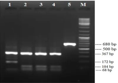
The F. hepatica samples, previously con-firmed by the authors of this paper were used as positive control (Fig. 1).
Fig. 1: PCR-RFLP pattern of Fasciola, Lane1: ITS1 product of F. gigantica after digestion with RsaI enzyme, Lane2-4: ITS1 product of F. hepatica after digestion with enzyme, Lane 5: ITS1 product of Fasciola before digestion, Lane M: 100 bp DNA ladder
The specificity of the LAMP primers for the identification and differentiation of F. hepatica was tested with other helminthes (control negative) (Fig. 2).
Primers amplified a fragment of 212 bp; af-ter sequencing, our results showed a 100% Identification, 99% Query coverage with the
sequence (Fig. 3), consensus partial sequence of the 28S ribosomal RNA gene of Fasciola spp.
Fig. 3: Sequence alignment of PCR band obtained in present study in comparison with database FhCM1-F. hepatica (GU903890)
In the following, the LAMP method was highly appropriate for the differentiation of the species. There was no cross-amplification by other helminths, including T. saginata, D.
dendriticum and F. gigantic (Fig.4).
Fig. 4: LAMP products under sunlight after the addition of SYBR Green I, a) F. hepatica, b) D.
dendiriticum c) T. saginata, d) Negative control
To instate the detection limit of the Fasciola by the LAMP method, various numbers of
Fasciola hepatica egg were tested. Results
showed high sensitivity of LAMP, even in the low egg numbers. The Green fluorescent of the reaction tube was discernible by the UV light and sunlight at a low level of F. hepatica egg. LAMP could detect even a single egg in samples (Fig. 5).
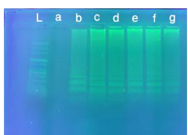
The amplification products were observed on 1.5% agarose gel as a ladder of multiple bands stained with safe stain (Fig. 6).
Fig. 5: Sensitivity of the LAMP for detection of F. hepatica under UVlight after adding SYBR Green I ,
a) negative control, b) 1 egg, c) 1-5 eggs, d) 10-15 eggs, e) 20-30 eggs, f) 50-60 eggs, g) positive
con-trol.
Fig. 6: Agarose gel electrophoresis of LAMP products using various numbers of F. hepatica egg,
L) 100 bp DNA ladder, a) negative control, b)1 egg, c)1-5 eggs, d)10-15 eggs, e)20-30 eggs, f)50-60
Discussion
Fascioliasis is one of the zoonotic diseases with worldwide distribution caused by F.
hepat-ica and F. giganthepat-ica. Fascioliasis has serious
medical and economic impact in the world (1-3). Fasciola species colonize the biliary tract of humans and ruminants (1, 19). Early diagnosis of fascioliasis by quick, precise and sensitive way is very important to avoid the severity of the disease. The most common phenotypic approaches are often laborious and prolonged (16).
Despite the clinical symptoms, researchers are still wondering why most Fasciola eggs are not seen in common parasitological methods such as formalin-ether. The intensity of infes-tation and the number of eggs per gram of feces (epg) varies greatly in human fascioliasis. In most studies, low egg outputs, e.g. 1-2 eggs per gram of feces (epg) and 1-4 cross-amplification, have been mentioned (20, 21). In some studies, especially in high prevalence areas, an output of 440 epg in humans has been reported (22). Certainly, in low-intensity contamination, parasitological methods cannot detect Fasciola in infected individuals.
Recent molecular methods, including a LAMP technique, have greatly improved the quick diagnosis of many helminths such as
Fasciola (16, 23, 24).
In a similar study to ours, the researchers added a variable number of Echinococcus
granu-losus eggs [1-2, 10, 25, 100] to the
non-contaminated dog stool. The LAMP method was capable of detecting contamination in all groups, even 1-2 eggs (17).
A study was conducted to evaluate the LAMP method and the comparison of this technique with the standard microscopic method in diagnosis of Opisthorchis viverrini in Thailand (25). All samples that were positive by the microscopic method were reported positively by the LAMP assay. Five of the 13 samples were negative by microscopy, but positive by LAMP (38.5%) (25).
A study compared the LAMP method and PCR assay in diagnosis of E. granulosus in Chi-na. In this study, the positivity rates were 12.6% and 1.6% by the LAMP and PCR assay respectively. Moreover, the LAMP method could detect the presence of E. granulsous in infected dog, four days earlier than the PCR assay (26).
LAMP method is more rapid, accurate, sen-sitive and specific in comparison to other methods, such as PCR. Additionally, fecal in-hibitors can impact the PCR results. The main problem of LAMP is that it cannot distinguish between the desired products and products of nonspecific amplification, thereby leading to false positives. There are different prevention methods of cross-contamination including, the use of UV (ultraviolet), LED (light-emitting diode) (27) and dual filter tips. Other ways to prevent contamination are closing reaction tube caps and putting them in plastic bags, cleaning environment with 75% ethyl alcohol (28) and paraffin oil added to the LAMP mix-ture(29).
Another benefit of LAMP technique is that the reactioncan be performed on a water bath and it allows easy observation of the results, using UV light. If the LAMP materials hold at ambient temperature, these materials will still retain their quality. This feature could result in the usage of this method in the field settings or in limited laboratory facilities (30).
Another benefit of this method is the early diagnosis of infection compared to other methods. LAMP was used method to identify
E. multilocularis in infected dogs. Dogs were
experimentally infected with E. multilocularis. The LAMP method was able to identify and detect E. multilocularis DNA after 12 d post-infection. While the results of the PCR meth-od were positive in 17 d after infection, and the first eggs were observed in the stool, 44 d after infection (31).
assays for IGS gene developed in this study is a flexible and quick method, which uses a wa-ter bath to maintain the temperature at 62 °C and obtains results within 60 min.
In this study, to prevent the risk of contami-nation, we autoclaved all equipment and place them under UV light for 20 min. After prepa-ration of LAMP reaction, microtubes contain-ing reaction solution were centrifuged at 12000 rpm for 3 min, and then were placed in -20 °C for 10 min. Finally, the microtubes were placed on the ice and added SYBR Green I dye. The method greatly prevented the contamination of environment and the subsequent reaction. This method of avoiding contamination was very beneficial.
Our results showed that LAMP assay has the potential to be practical in diagnosis and epidemiological researches. This method may be effective for the detection of disease in pa-tients with suspected fascioliasis with the neg-ative results of parasitological methods in en-demic countries.
Conclusion
This article was a preliminary study. In the future, the authors of this paper intend to in-vestigate and compare this method with PCR and another technique in detection of spiked
F. hepatica eggs in stool samples.
Acknowledgements
The authors would like to thank Dr. Saber Raeghi for the collection of Fasciola samples and Dr. Sahar Sajadieh for the editing of the article. This article has been extracted from the MSc thesis in the School of Medicine, Shahid Beheshti University of Medical Scienc-es, Tehran, Iran (grant No: 13.213).
Conflicts of interest
The authors declare that there is no conflict of interest.
References
1. Mas-Coma S. Epidemiology of fascioliasis in human endemic areas. J Helminthol. 2005;79(3):207-16.
2. Reaghi S, Haghighi A, Harandi MF et al. Mo-lecular characterization of Fasciola hepatica and phylogenetic analysis based on mitochondrial (nicotiamide adenine dinucleotide dehydrogen-ase subunit I and cytochrome oxiddehydrogen-ase subunit I) genes from the North-East of Iran. Vet World. 2016;9(9):1034-1038.
3. Rouhani S, Raeghi S, Spotin A. Spermatogenic and Phylo-molecular Characterizations of Iso-lated Fasciola Spp. From Cattle, North West Iran. Pak J Biol Sci. 2017;20(4):204-209. 4. Mas-Coma S, Bargues MD, Valero MA.
Fasci-oliasis and other plant-borne trematode zoono-ses. Int J Parasitol. 2005;35(11-12):1255-78. 5. Nguyen TG, Van De N, Vercruysse J, Dorny
P, Le TH. Genotypic characterization and spe-cies identification of Fasciola spp. with implica-tions regarding the isolates infecting goats in Vietnam. Exp Parasitol. 2009;123(4):354-61. 6. Le TH, De NV, Agatsuma T, et al. Human
fascioliasis and the presence of hy-brid/introgressed forms of Fasciola hepatica and Fasciola gigantica in Vietnam. Int J Parasit. 2008 May;38(6):725-30.
7. Itagaki T, Kikawa M, Terasaki K, Shibahara T, Fukuda K. Molecular characterization of par-thenogenic Fasciola sp. in Korea on the basis of DNA sequences of ribosomal ITS1 and mito-chondrial NDI gene. J Vet Med Sci. 2005;67(11):1115-8.
8. Itagaki T, Sakaguchi K, Terasaki K et al. Oc-currence of spermic diploid and aspermic trip-loid forms of Fasciola in Vietnam and their mo-lecular characterization based on nuclear and
mitochondrial DNA. Parasitol Int.
2009;58(1):81-5.
9. Rouhani S, Raeghi S, Mirahmadi H, Harandi MF, Haghighi A, Spotin A. Identification of Fasciola spp. in the East of Iran, Based on the Spermatogenesis and Nuclear Ribosomal DNA (ITS1) and Mitochondrial (ND1) Genes. Arch Clin Infect Dis. 2017;12(2):e57283. 10. Arifin MI, Höglund J, Novobilský A.
for the diagnosis of Fasciola hepatica infection in the field. Vet Parasitol. 2016;232:8-11.
11. Mezo M, González-Warleta M, Carro C, Ubei-ra FM. An ultUbei-rasensitive capture ELISA for de-tection of Fasciola hepatica coproantigens in sheep and cattle using a new monoclonal anti-body (MM3). J Parasitol. 2004;90(4):845-52. 12. Brockwell YM, Spithill TW, Anderson GR et
al. Comparative kinetics of serological and coproantigen ELISA and faecal egg count in cattle experimentally infected with Fasciola
hepat-ica and following treatment with
triclabendazole. Vet Parasitol. 2013;196(3-4):417-26.
13. Fairweather I. Reducing the future threat from (liver) fluke: realistic prospect or quixotic fanta-sy? Vet Parasitol. 2011;180(1-2):133-43. 14. Föhse L, Suffner J, Suhre K et al. High TCR
diversity ensures optimal function and homeo-stasis of Foxp3+ regulatory T cells. Eur J Im-munol. 2011 Nov;41(11):3101-13.
15. Martinez-Valladares M, Rojo-Vazquez FA. Loop-mediated isothermal amplification (LAMP) assay for the diagnosis of fasciolosis in sheep and its application under field condi-tions. Parasit Vectors. 2016 Feb 05;9:73. 16. Ai L, Li C, Elsheikha HM et al. Rapid
identifi-cation and differentiation of Fasciola hepatica and Fasciola gigantica by a loop-mediated isothermal amplification (LAMP) assay. Vet Parasitol. 2010;174(3-4):228-33.
17. Salant H, Abbasi I, Hamburger J. The devel-opment of a loop-mediated isothermal amplifi-cation method (LAMP) for Echinococcus granu-losus [corrected] coprodetection. Am J Trop Med Hyg. 2012;87(5):883-7.
18. Fernández-Soto P, Gandasegui Arahuetes J, Sánchez Hernández A et al. A loop-mediated isothermal amplification (LAMP) assay for ear-ly detection of Schistosoma mansoni in stool sam-ples: a diagnostic approach in a murine model. PLoS Negl Trop Dis. 2014;8(9):e3126. 19. Spithill TW, Dalton JP. Progress in
develop-ment of liver fluke vaccines. Parasitol Today. 1998;14(6):224-8.
20. Bendezú P, Frame A, Hillyer GV. Human fascioliasis in Corozal, Puerto Rico. J Parasitol. 1982;68(2):297-9.
21. Knobloch J, Delgado E, Alvarez A, Reymann U, Bialek R. Human fascioliasis in Ca-jamarca/Peru. I. Diagnostic methods and
treatment with praziquantel. Trop Med Parasi-tol. 1985;36(2):88-90.
22. Mas-Coma MS, Esteban JG, Bargues MD. Epidemiology of human fascioliasis: a review and proposed new classification. Bull World Health Organ. 1999;77(4):340-6.
23. Magalhães KG, Jannotti-Passos LK, Caldeira RL et al. Isolation and detection of Fasciola he-patica DNA in Lymnaea viatrix from formalin-fixed and paraffin-embedded tissues through multiplex-PCR. Vet Parasitol. 2008;152(3-4):333-8.
24. Cucher MA, Carnevale S, Prepelitchi L et al. PCR diagnosis of Fasciola hepatica in field-collected Lymnaea columella and Lymnaea viatrix snails. Vet Parasitol. 2006;137(1-2):74-82. 25. Arimatsu Y, Kaewkes S, Laha T, Sripa B.
Spe-cific diagnosis of Opisthorchis viverrini using loop-mediated isothermal amplification (LAMP) tar-geting parasite microsatellites. Acta Trop. 2015;141(Pt B):368-71.
26. Ni XW, McManus DP, Lou ZZ et al. A com-parison of loop-mediated isothermal amplifica-tion (LAMP) with other surveillance tools for Echinococcus granulosus diagnosis in canine defini-tive hosts. PLoS One. 2014;9(7):e100877. 27. Karthik K, Rathore R, Thomas P et al. New
closed tube loop mediated isothermal amplifi-cation assay for prevention of product cross-contamination. MethodsX. 2014;1:137-43. 28. Zhou D, Guo J, Xu L, Gao S, Lin Q, Wu Q, et
al. Establishment and application of a loop-mediated isothermal amplification (LAMP) sys-tem for detection of cry1Ac transgenic sugar-cane. Sci Rep. 2014 May 09;4:4912.
29. Zhang X, Liao M, Jiao P et al. Development of a loop-mediated isothermal amplification assay for rapid detection of subgroup J avian leukosis virus. J Clin Microbiol. 2010;48(6):2116-21. 30. Thekisoe OM, Bazie RS, Coronel-Servian AM
et al. Stability of Loop-Mediated Isothermal Amplification (LAMP) reagents and its amplifi-cation efficiency on crude trypanosome DNA templates. J Vet Med Sci. 2009;71(4):471-5. 31. Ni X, McManus DP, Yan H, Yang J, Lou Z, Li