Characterization of the Volatile Components and Antimicrobial
Properties of the Ethanol Leaf Extract of Uvaria chamae Grown
in Eastern Nigeria
*
1Iwu Irenus Chinonye,
2Oze Rita Nwanneamaka,
3Onu Uchenna Lynda,
4Onwumere Fidelis,
5Ukaoma Adanma Augustina
1,2,3,4Department of Chemistry, Federal University of Technology Owerri, Imo State, Nigeria
5Department of Biological Sciences, Federal University of Technology Owerri, Imo state, Nigeria
The characterization of the ethanol leaf extract of Uvaria chamae was carried out with the aim of identifying and determining the chemical compounds present in the extract. Initial phytochemical results showed the presence of flavonoids, saponins, tannins. Phenols, steroids and alkaloids. Interpreted spectrum obtained from the GC-MS revealed twelve absorption peaks. Peak 1 was identified as Benzene carboxylic acid with molecular weight of 122g and molecular formula C7H6O2. Similarly, peaks 2-12 were identified as, hexadecanoic acid methyl ester, hexadecanoic
acid, 11-octadecenoic acid methyl ester, phytol, 6-octadecanoic acid, octadecanoic acid, tetradecanamide. hexadecanoic acid-2,3-dihydroxpropyl ester, 9-octadecenamide, 9,12-octadecadienoyl chloride and 13-octadecanal with corresponding molecular formulas of C17H34O2,
C16H36O2, C19H36O2, C20H40O, C18H34O2, C18H36O2, C14H29NO, C19H38O4, C18H35NO, C18H31ClO and
C18H34O respectively. The extract inhibited the growth of some selected human pathogen; Pseudomonas aureginosa by 6mm with minimum inhibitory concentration (MIC) of 50mg/cm3.
Similar results were also obtained for Candida albicans 8mm with MIC 50mg/cm3 and Trichophyton spp 4mm with MIC OF 100mg/cm3
Keywords: (Characterization, Gas Chromatography, Phytochemicals, Mass Spectrometry, Pathogens)
INTRODUCTION
Uvaria chamae is a plant tropical to eastern Nigeria and is
commonly called finger root plant which belongs to the
family of Annonaceae. It is a small tree that grows to about
4.5m high Moses et al.,( 2013) It is commonly found in the
savannah and rain forest regions of Nigeria and other African countries. It is called “Mmimi ohia”, “Kas kaifi” and “Akisan” amongst the Ibos, Hausas and Yorubas respectively Adetunji, (1999). The fruits are yellow when ripe and have a sweet pulp which is widely eaten. The fruit carpels are in finger-like clusters. The plant has been used for treatment of fevers, tumour growth,stroke and cases of
venereal disease, Ayenusi (1978).The leaf extracts of U.
chamae has been reported to possess antibacterial
Oluremi et al., (2009), antifungal Okwuosa et al.,( 2012),
antispasmodic, anti-trypanosomal and anti-inflammatory properties. The roots of the plant has been reported to
possess antibacterial, antioxidant Kone et al., (2015),
anti-inflammatory, oxytocic. Okwu and Iroabuchi, (2009) and
anti-sickling Thierry et al., (2012) activities. Two benzyl
dihydrochalcones; chamuvaritin and chamuvarin have
been isolated from the roots of U. chamae. Uwaifo and
Bababunmi, (1984). The leaves of U. Chamae have been
used to treat wounds and sores, injuries, swellings, and to treat yellow fever. The roots, barks and leaves of U. chamae are used traditionally in the treatment of
diarrhoea, cough and urinary tract infections Chika et al.,
(2007).
The plant is rich in sesquiterpene hydrocarbons dominated
by germacrene D and γ-cadinene. (Okwwuosa et al.,
2012), Kone et al., 2015.) the root extract of U. Chamae
has been reported to contain flavonoids, alkaloids, cardiac glycosides, terpenoid and terpenes, saponin, tannin,
*Corresponding Author: Iwu Irenus Chinonye, Department of Chemistry, Federal University of Technology Owerri, Nigeria.
Tel.: +2348032444212. E-mail: [email protected].
Co-Authors Email: 2[email protected], Tel.: +2348038916979;
3[email protected], Tel.: +2348037676079;
4[email protected], Tel.: +2347038824111;
5[email protected]; Tel: +2348069380796
Research Article
proteins and sugars. The extracts and aspirin have been reported to inhibit carrageenan-induced paw oedema on albino rats and mice. Okwu and Iroabuch. (2009). The drug benzyl benzoate used in antifungal preparations has a
mutagenic compound, chamuvaritin, a benzyl
dihydrochalcone that was isolated from U. Chamae.
Therry et al., (2012). Recently, uvarinol, a novel cytotoxic
tribenzylated flavanone compound has been isolated from
U. Chamae. Uwaifo and Bababunmi, (1984). The root is
used in Nigeria as a purgative. The root bark is used for respiratory catarrh and the root extract is used in phytomedicine for the treatment of piles, menrrhegia, epiostaxis, haematuria and haemalysis. Oliver-Bever, 1986. Its root infusion is used to cure abdominal pains. The juice from the roots, stems or leaves is commonly applied to wounds and sore. Shukda and Shital. (1995.) wrote that the extracts of the roots, barks and leaves are used to treat gastroenteritis, vomiting, diarrhea, dysentery, wounds, sore throats, inflamed gums and a number of
other ailments. Irvin, (1961). Uvaria chamae methanol
extract has been shown to neutralized some biological
effects of Naja nigricollis venom. The leaf extract had
shown anti venom activity in animal models and could
potentially be used for therapeutic purpose in case of
snake bite (Omale et al.,(2012). Methanolic extracts of the
root, stem and leaf of Uvaria chamae have been evaluated
for their antibacterial activity against Methicillin-resistant
Staphylococcus aureus. The stem bark extract inhibited
the growth of all the tested organisms but the leaf extract showed the least antibacterial activity as reported by
Oluremi et al., (2009.). Uvaria chamae root contain
abundant cardiac glycosides. Similar reports in other plant
species were obtained by Okon et al., (2013). They
reported that cardiac glycosides can be used in the treatment of diseases associated with the heart and are currently used by herbalist, to treat tumour Piett, (2000).
The cardiac glycosides found in Uvaria chamae root can
similarly be used for the treatment of heart diseases.
Monosodium glutamate has detrimental effect on haematological parameters but Uvaria chamae ethanolic extract is a potent remedy against MSG-induced toxicity.
Ibukun et al., (2015) Despite its many uses for the
treatment of various diseases, its full content has not been
fully characterised.
MATERIALS AND METHOD
SAMPLE PREPARATION
The leaves of finger root (Uvaria chamae were obtained
from F.U.T.O farmland, Owerri North L.G.A. The plant was identified by Dr Ibeawuchi of Crop Science Department Federal University of Technology Owerri. The sample was cut into bits, room dried, ground into powdery form and stored in an air-tight container afterwards before the commencement of the analysis.
FROTHING TEST FOR SAPONINS
This test is based on the ability of the saponins to produce froth in aqueous solution. 5g of the plant extract was
weighed into a test tube and 50cm3 of water was added
and extracted after two hours. The water extract was shaken vigorously in a conical flask. The production of a stable froth indicates the presence of saponins in the
sample. (Iwu et al., 2018a)
TEST FOR FLAVONOIDS
5g of the sample was soaked with 20cm3 of water and left
to stand for 2 hours, it was then filtered and to the filtrate
drops of ammonia and 3cm3 of concentrated H2SO4 was
added. A yellow precipitate which disappears on storage
indicates the presence of flavonoids. Iwu et al., (2018b)
TEST FOR ALKALOIDS
5g of the sample was extracted using 20% acetic acid in
ethanol.5cm3 of the extract was treated with Wagner’s
reagent (iodine crystals and KI). A yellowish brown precipitate indicates the presence of alkaloids.
TEST FOR TANNINS
5g of the root sample was weighed into a beaker and
50cm3 of water was added and allowed to soak properly
for two hours and extracted. The extract was treated with drops of ferric chloride. A blue-black precipitate indicates
the presence of tannins. Iwu et al., (2018b)
TEST FOR STEROIDS
5cm3 of the water extract was treated with concentrated
H2SO4 in acetic anhydride. The formation of a blue-green
colour indicates the presence of steroids.
TEST FOR PHENOLS
20cm3 of the water extract was treated with 5cm3 of
concentrated sulphuric acid and drops of sodium nitrate
(NaNO3). 2cm3 of sodium hydroxide was added to the
mixture. A blue precipitate indicated the presence of
phenols. Iwu et al., (2016a)
TEST FOR GLYCOSIDES
20cm3 of the water extract was treated with Fehling
solutions of A and B in equal amount and boiled. A brownish red precipitate indicates the presence of glycoside.
PREPARATION OF SAMPLES FOR GC-MS ANALYSIS
Two hundred grams of sample was soaked in 400cm3
GC-MS EXPERIMENTAL PROCEDURES
GC-MS analysis was carried out with SHIMAZU Japan Gas Chromatography 5890-11 with a fused GC column OV 101 coated with polymethyl silicon (0.25 mm x 50 m) and the conditions are as follows: Temperature
programming from 80 – 200oC held at 80oC for 1 minute,
the rate is 5oC/min and at 200oC for 20 minutes. FID
Temperature of 300oC, injection temperature of250oC,
carrier gas is Nitrogen at a flow rate of 1 cm3/min and split
ratio of 1:75. GC-MS Gas chromatography, Mass spectrum analysis were conducted using GC-MS QP 2010
Plus Shimazu Japan with injector Temperature at 230oC
and carrier gas pressure of 100kpa. The column length was 30 m with a diameter of 0.25 mm and the flow rate of 50m/min. The eluents were automatically passed into the Mass Spectrometer with a detector voltage set at 1.5kv and sampling rate of 0.2 seconds. The Mass Spectrometer was also equipped with a computer fed Mass Spectra data bank, HERMCE Z 233 M-Z centrifuge Germany was used. Reagents and solvents such as Ethanol, Chloroform, Diethyl ether, hexane all of analytics grade was obtained
from Merck Germany. Iwu et al., (2016b, 2018c)
ANTIMICROBIAL ANALYSIS
The microorganisms; Staphylococcus aureus,
Streptococcus spp Pseudomonas aeruginosa, Aspergillus
niger, Candida albicans and Trycophton spp were used for
the analysis. They are clinical isolates of human pathogens obtained from the Federal Medical Centre Umuahia and were brought to the laboratory and resuscitated in buffered peptone broth (Secharian chemie) and thereafter into nutrient agar medium and incubated at
37oC for 24 hrs. Iwu et al., (2018b)
ANTIBACTERIAL ASSAY
The test solution of each extract was prepared by
dissolving 0.1 g of the plant extract separately in 1.0cm3 of
dimethyl sulphoxide (DMSO) to get a concentration of 100mg/cm3. The antibacterial activity was performed by filter paper disc diffusion technique. Filter paper disc (Whatman No 1.6 mm diameter) were placed in glass petri dishes and sterilized in hot air over. Iwu and Onu,
(2018).The media (10g nutrient Agar in 200cm3 distilled
water, autoclaved at115oC for 30 minutes) was cooled to
50oC. The sterile nutrient Agar media were poured into the
sterile petri dish and allowed to solidify. The bacteria were swabbed with a sterile wire loop. Each disc was
impregnated with 0.2cm3 of plant extract. .The discs were
used after drying them in an incubator at 40oC to remove
any trace of solvent. Discs were introduced into the surface
of the medium. The plates were microbated at 37oC for 24
hours to obtain zones of inhibition. The experiments were repeated three times for each extract and twice for reference antibiotics to minimize error and the average of these values were recorded. Kanayo and Ezeugo,(2006)
MINIMUM INHIBITORY CONCENTRATION (MIC).
The minimum inhibitory concentration of the extract was
determined by incorporating constant volume 0.2cm3 of
each diluents of the extract into the perforated disc on a seeded nutrient agar plate as described in the anti-microbial susceptibility test section. 0.1g of each extract
was dissolved in 1cm3 of DMSO to obtain 100mg/cm3. This
concentration of DMSO was then doubled to obtain
50mg/cm3 then doubled again to obtain 12.5mg/cm3 and
again to obtain6.25mg/cm3. Each concentration was then
used in the method earlier described to obtain zone of inhibition. The least concentration that showed inhibitory zones was taken as the MIC.
RESULTS AND DISCUSSION
The results obtained from the phytochemical screening of the leaf extract of sample are presented in table 1 below. Initial results showed the presence of flavonoids, saponins, tannins. phenols , steroids and alkaloids
Table 1. Phytochemical content of the leaf extract of uvaria chamae
Phytochemical Inference
Alkaloid ++
Flavonoid ++
Saponons ++
Steroids ++
Glycosides ++
Tannins ++
Phenols ++
Key: ++ present
Alkaloids are vast and vary a lot in their activity when ingested by man and livestock. Some alkaloids are useful and important in medicine and constitute most of the valuable drugs currently used by humans. They are reported to have marked physiological effect on animals.
Edeoga and Eriata, (2001). Iwu et al., (2018c)
Flavonoids have been shown to be highly effective scavengers of most oxidizing molecules. Tukappa and Londonkar, (2013). Flavonoids are the major nutraceutical ingredients that are in plants. The best described property of almost every group of flavonoids is their capacity to act as anti-oxidants. The flavones seem to be the most powerful flavonoid for protecting the body against reactive oxygen species (ROS). Antibacterial activity has been displayed by a number of flavonoids,. Quercetin has been
reported to completely inhibit the growth of
Staphylococcus aureus. Havesteen (1983). Flavonoids
also possess anti-inflammatory and analgesic effect as
well as anti-ulcerogenic activity. Shahid et al., (1998)
The infusions of Momordica charantia and Uvaria chamae are taken as a remedy for gonorrhea and jaundice
Draughton, (2004), Mohammed et al.,(2013) This is
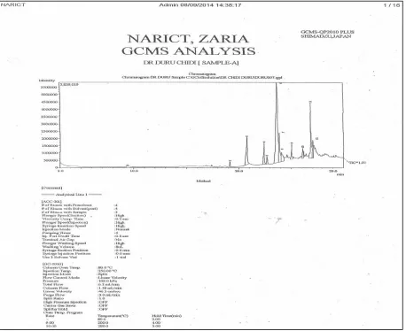
Fig 1: GC/MS GC-MS spectra of the the ethanolic leaf sample of Uvaria chamae
bioactive antibacterial agent of plant. Mandal et al., (2005.}
Saponins are also useful for utilization in foods that need sustained foam volume such as ice-creams. Plant saponins may serve as anti-feedants to protect the plant against microbes and fungi. Some plant saponins may enhance nutrient absorption and aid in food digestion. Saponins have been used as a pharmacological and/or immunological agent that modifies the effect of other agents in vaccines. Saponins from plants have been shown to significantly augment the cytotoxicity of immunotoxins and other target toxins directed against human cancer cells. Tannins are astringent, bitter plant polyphenol compounds that bind to and precipitate proteins and various other organic compounds including amino acids and alkaloids. The tannin compounds are widely distributed in many species of plants where they play a role in protection from predation and perhaps also as pesticides and in plant growth regulation. The astringency from tannin is what causes the dry puckery feeling in the mouth following the consumption of unripe fruits or red wine. Tannins are important ingredients used in process of making tannin leather. Medicinally, tannins are used as diarrhea, haemostatic and anti-hemorrhoid compounds Plant leaves with high tannin content has been used successfully as hops alternative in beer. Hutchinson and Dalziel,.(1963).
The presence of phenolic compounds in the leaf of Uvaria
chamae indicates that this plant might be an anti-microbial
agent. This is because phenols and phenolic compounds have been extensively used in disinfections and remains the standard with which other bactericides are compare. Okwu and Okwu,( 2004}. Phenolic compounds acts as electron donors and are readily oxidized to form phenolate ions. This gives rise to protonated phenol which is used as
a cleaning agent. Extracts from leaves of Uvaria chamae
therefore have potent antiseptic or bactericidal properties.
The presence of phenol further indicates that Uvaria
chamae could act as anti-inflammatory, anti-clotting,
immune enhancers and hormone modulators.
Glycosides are molecules in which a sugar is bound to another functional group via a glycosidic bond. Glycosides play numerous important roles in living organisms. Many plant store chemicals in form of inactive glycosides. Many such plant glycosides are used as medications. Some glycosides have shown some evidence of pharmacological effects in patients with hypertension or with type-2 diabetes but concluded that further study was required to determine the proper dosages
The result of the GC/MS analysis of Uvariae chamae is
The GC-MS spectrum of finger root (fig 1) has 12 absorption peaks. Peak 1 was identified as benzene carboxylic acid with percentage oil composition of 0.39 and a molecular weight of 122g, its molecular formula is
C7H6O2.. Similarly, peak 2 was identified as hexadecanoic
acid methyl ester having a % oil composition of 0.93 with a molecular weight of 270g and a molecular formula of
C17H34O2. Peak 3 was identified as hexadecanoic acid, its
% oil composition is 10.61 and its molecular weight is 256g
with a molecular formula of C16H36O2. Peak 4 was
identified as 11-octadecenoic acid methyl ester with a molecular weight of 296g and molecular formula of
C19H36O2 and its % composition was 4.75. Peak 5 was
identified as Phytol with molecular weight of 296g and
molecular formula C20H4OO. Its % oil composition is 1.92.
Peak 6 was identified as 6-octadecanoic acid with
molecular formula C18H34O2 and molecular weight 282g
and % oil composition of 45.04. Peak 7 was identified as
Octadecanoic acid with molecular formula of C18H36O2 and
molecular weight 284g with % oil composition of 9.49. Peak 8 was identified as Tetradecanamide. Its molecular
weight and formula are 227g and C14H29NO respectively.
Peak 9 was identified as Hexadecanoic acid, 2,3-dihydroxypropyl ester with a molecular weight and formula
of 330g and C19H38O4 and % oil composition of 2.99. Peak
10 was identified as 9-octadecenamide with molecular
weight and formula of 281g and C18H35NO. Its % oil
composition is 2.32. Peak 11 was identified as 9,12-octadecadienoyl chloride with molecular weight and
formula of 298g and C18H31ClO. Its % oil composition is
12.55. Peak 12 was identified as 13-octadecanal with
molecular weight and formula of 266g and C18H34O
respectively. Its % oil composition is 4.46.
Table 2. Interpreted values from the GC/MS spectrum of Uvaria chamae leaf extract Chromatographic
peak
% oil composition
Chemical name Molecular
formula
Molecular weight
1 0.39 Benzene carboxylic acid C7H6O2 122
2 0.93 Hexadecanoic acid methyl ester C17H34O2 270
3 10.61 Haxadecanoic acid C16H32O2 256
4 4.75 11-octadecenoic acid methyl ester C19H36O2 296
5 1.92 Phytol C20H40O 296
6 45.04 6-octadecenoic acid C18H34O2 282
7 9.49 Octadecanoic acid C18H36O2 284
8 4.55 Tetradecanamide C14H29NO 227
9 2.99 Hexadecanoic acid,2,3 dihydroxypropyl ester C19H38O4 330
10 2.32 9-Octadecenamide C18H35NO 281
11 12.55 9,12-octadecadienoyl chloride C18H31ClO 298
12 4.46 13-octadecanal C18H34O 266
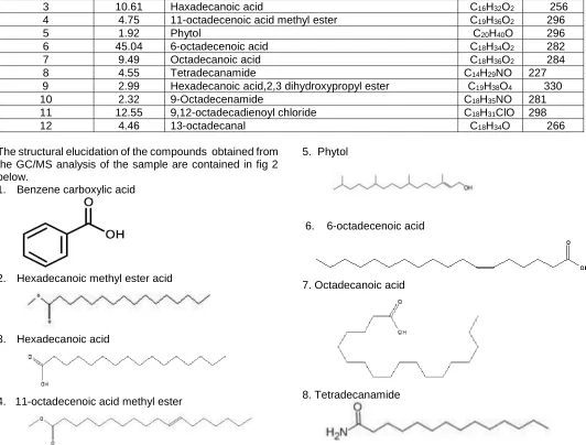
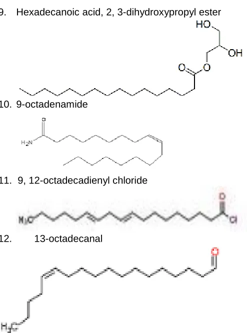
The structural elucidation of the compounds obtained from the GC/MS analysis of the sample are contained in fig 2 below.
1. Benzene carboxylic acid
2. Hexadecanoic methyl ester acid
3. Hexadecanoic acid
4. 11-octadecenoic acid methyl ester
5. Phytol
6. 6-octadecenoic acid
7. Octadecanoic acid
8. Tetradecanamide
9. Hexadecanoic acid, 2, 3-dihydroxypropyl ester
10. 9-octadenamide
11. 9, 12-octadecadienyl chloride
12. 13-octadecanal
Fig 2: Structures of the compounds obtained from the
GC-MS spectrum OF Uvaria chamae leaf extract
Antimicrobial analysis of Uvaria chamae
The antimicrobial analysis result obtained from the leaf
extract of Uvaria chamae are shown in table 3 below.
Table 3: Result of the antimicrobial analysis of Uvariae chamae leaf
Extract concen-tration (mg/ml)
Diameter of zone of inhibition (mm)
Asper-gillus Spp
Candida albicans
Tricho-phyton Spp
Strepto-cocus spp
Pseudo-monas aeruginosa
Staphylo-coccus spp
200 ---- 8 4 ---- 6 ----
100 ---- 4 2 ---- 4 ----
50 ---- 2 ---- ---- 2 ----
25 ---- ---- ---- ---- ---- ----
M.I.C 50
mg/ml 100 mg/ml
50 mg/ml
The extract inhibited the growth of Pseudomonas
aureginosa by 6mm with MIC of 50mg/cm3.table 3. This
organism is agram negative gamma-proteobacteria which
belong to the family Pseudomonaceae. It causes
bacteremia, pneumonia, folliculitis, swimmer ear which is an ear infection accompanied with swelling, ear pus. Itching, discharge and difficulty in hearing, eye
inflammation with associated pains, pus, swelling redness
and impaired vision Pseudominas spp causes bacterial
blight in guinea corn. Similarly the extract showed activity against Candida albican with 8mm diameter of inhibition.
Candida albicans is a fungus which causes yeast infection.
Candida yeast normally live in the skin and mucous
membranes without causing infection; however,
overgrowth of these organisms can cause symptoms to develop. They cause diseases based on the area of the body they affect; candidiasis that develop in the mouth or throat is called thrush while in the vagina is called yeast infection. Invasive candidiasis occurs when candida species enter the bloodstream and spread throughout the body. The extract also showed activity against
Trichophyton spp 4mm. This pathogen is a fungus that
cause tinea, athletes’ foot, ring worm, jock itch, nail, beard and skin infections. The extracts exhibited some level of inhibitory effects against some of the studied pathogens which have been implicated in one bacterial infection to the
other in human and plant. Okwuosa et al, (2012), Kone et
al., (2015)
CONCLUSION
The work so far carried out has shown that the leaf extract
of Uvaria chamae is rich in phytochemicals and other
volatile compounds. The compounds have far reaching medicinal applications. The extract may be applied in
cases involving bacteremia, pneumonia, folliculitis,
swimmer ear which is an ear infection accompanied with swelling, ear pus. Itching, discharge and difficulty in hearing, eye inflammation with associated pains, pus, swelling redness and impaired vision, candidiasis, tinea, athletes foot, ring worm, jock itch , nail , beard and skin infections.
REFERRENCES
Adelodun VO, Elusiyan CA, OlorunmolanFO, Adewoyin FB, Omisore NO, Adepiti AO, Agbedahunsi JM, Adewunmi CO, 2013. Evaluation of antitrypanosomal and anti-inflammatory activities of selected Nigerian medicinal plants in mice. Afr. J. Tradit.Complement.
Altern. Med. 10(6), pp 469-476
Adetunji, LK. (1999). Great Secret of Nature. Natural Links Centre, Lagos.
.Ayedoun MA, Moudachirou M, Adeoti BS, Menut C, Lamaty G, Bessiére JM. 1999 Aromatic plants of tropical West Africa. VII. Essential oil of leaf and root bark of UvariachamaeP. Beauv. from Benin. J. Essent. Oil Res; 11: pp23-26
Ayensu ES. 1978 Medicinal Plants of West Africa. Reference Publications, Inc., Algonac, Michigan 41 Chika CO, Jude NO, Beatrice NA,(2007} The Effects Of
And Leaves Of UvariaChamaeOn Some Hospital Isolates. Journal of American Science, 3(3)pp 68-73 Draughon FA.2004. Use of botanicals as bio-preservatives
in foods. Food Technol, ; 58(2): pp20-30
Edeoga HO, Eriata DO. 2001 Alkaloid, tannin and saponin content of some Nigeria medicinal plants. J Med Aromatic Plant Sci;25 pp344-349.
Ekundayo EO, Ezeogu LI. (,2006). Evaluation of antimicrobiabial activities of Extracts of five plant used
in Traditional Medicine in Nigeria. International
Journal of Tropical Medicine 3; pp93-96
Havsteen B.1983. Flavonoids a class of Natural products of high pharmacological potency. Biochempharmacol ; 32(7) pp1141-1148
Hutchinson J, Dalziel J. 1963. Flora of West Tropical Africa. 2nd ed. Vol. 11. Crown Agents, London pp1-544.
Irvin FR. 1961 Woody Plants of Ghana with Special Reference to their Uses. Oxford
University Press, London, pp 695.
Ibukun OO, Monday T, Abiola SO , Ololade SO. 2015 Haematological effect of ethanolic extract of Uvaria chamae on monosodium glutamate (MSG)-induced toxicity in sprague-dawley rats Annals of Biological Research 6 (7) pp17-22
Iwu IC, Ogukwe CE, Akah CM. Chijioke-Okere M, Onu UL, Iwu JO. 2016a Phytochemical and Antimicrobial Properties of the Root and leaf Extract of Carica papaya. International Journal of Innovative Research and Development 5(8) pp 173-179
Iwu IC, Onu UL, Chijioke-okere M, Ukaoma AA, Azorji JN. 2016b GC-MS, Phytochemical and Antibacterial Analysis of Pentaclethra macrophylla Leaf. The International Journal of Science and Technology 4 (7) pp 151-159
Iwu IC, Chijioke-okere M, Onu UL, Uchegbu R. 2018a. GC-MS, Phytochemical and Antimicrobial Analysis of the Leaf of Newboudia laevis P. Benth. International Journal of Innovative Research and Development 7 (7) pp 242- 250
Iwu IC, Onu UL, Ukaoma AA, Oze RN. 2018b Phytochemical, Antimicrobial and Gc/Ms Analysis of the Root of Stachytarpheta Cayennensis (L .Vahl) Grown in Eastern Nigeria. International Research
Journal of Natural Sciences.6 (2), pp.1-14.
Iwu IC, Onu UL. 2018. GC-MS, Phytochemical and Antimicrobial of Pentaclethra macrophylla Bark P.
Benth. International Journal of Science and
Technolodge 6 (7) pp96-103
Iwu IC, Oze RN, Onu UL, Amarachi, N and Ukaoma AA. 2018c Phytochemical and GC/MS Analysis of the Rhizome of Zingiber officinale plant grown in Eastern part of Nigeria. African Journal of Biology and Medical Research Volume 1, Issue 1, (pp. 43-54)
Kone M, Toure A, Ouattara K, Coulibaly A. 2015.
Phytochemical Composition, Antioxidant and
Antibacterial Activities of Root of Uvaria chamae P. Beauv. (Annonaceae) used in Treatment of Dysentery
in North of Côte d’Ivoire.Int..J.Pharmacog. Phytochem. Res., 7(6) pp 1047-1053
Mandal P, Babu SP, Mandal NC.2005. Antimicrobial activity of saponins from Acacia auriculiformis. Fitoterapia;76(5) pp462-465.
Mohammed M, Musa AM, Adeiza AA, Musa SH, Lande L.
2013. Bioactive caffeic glycoside ester and
antimicrobial activity of various extracts from the leaf of Stachytarpheta angustifolia Mill Vahl (Verbenaceae). J Pharmacognosy and Phytochem;2(3) pp77-85.
Moses OS, Olowu RA, Noura SD, William NS. 2013.
1-Nitro-2-phenylethane dominates the chemical
composition of the leaf essential oil of Uvaria chamae from Badagry, Nigeria. Am. J. Essential Oils Nat. Prod., 1(1), pp48-50
Oguntimein B, Ekundayo O, Laakso I, Hiltunen I. 1989 Volatile constituents of Uvaria chamae leaves and root bark. Planta Med. 55 pp312-313
Okogun JI. 1985 Drug Production Efforts in Nigeria. Medicinal Chemistry Research and
Missing Link. Being a Text of a Lecture given to the Nigeria Acad Sci.pp29-52
Okon JE, Udosen IR, Mbong EO. 2013 . Phytochemical screening and effect of ethanolic root extract of Uvaria chamae haematological parameters on albino rats in Akwa Ibom State, Nigeria Merit Research Journal of Environmental Science and Toxicology Vol. 1(2) pp. pp016-020
Okwu DE, Iroabuchi F. 2009. Phytochemical Composition and Biological Activities of Uvaria chamae and Clerodendoron splendens E-Journal of Chemistry, 6(2), pp553-560
Okwu DE. and Okwu ME .2004. Chemical composition of Spondias mombialinn plants parts, J. sustain, Agric environment 6 pp140-147
Okwuosa OM, Chukwura EI, Chukwuma GO, Okwuosa CN, Enweani IB, Agbakoba NR, Chukwuma CM, Manafa PO and Umedum CU . 2012. Phytochemical and antifungal activities of Uvaria. Chamae leaves and roots, Spondias mombin leaves and bark and Combretum racemosum leaves. Afr. J. Med. Sci., 41, pp99-103
Oliver-Bever B. 1986 Medicinal Plants in Tropical West Africa. Cambridge University Press, UK , pp134-135 Oluremi BB, Osungunna MO, Omafuma OO. 2010
Comparative assessment of antibacterial activity of uvariachamae parts African Journal of Microbiology Research 4 (13), pp. 1391-1394
Omale J, Ebiloma UG, Idoko GO. 2013 Uvaria chamae
(Annonaceae) Plant Extract Neutralizes Some
Biological Effects of Naja nigricollis Snake Venom in Rats British Journal of Pharmacology and Toxicology 4(2) pp41-50
Piett PG . 2000. Flavonoids as antioxidant. Journal of Natural Products, 63(7) pp1035 – 1042.
Shukda P, Shital PM,1994 An Introduction to the Taxonomy of Angiosperms, Vikas Publishing House, New Delhi, pp, 204
Thierry AC, Joachim DG, Kpoviessi SDS, Accrombessi GC, Moudachirou M, Gbeassor M. 2012.Antihemolytic Properties of Extracts of Six Plants Used in the Traditional Treatment of Sickle Cell Disease in Benin. J. Appl. Pharm. Sci., 2(3) pp8-13
Tukappa A, Londonkar RL. 2013 Evaluation of antimicrobial and antioxidant activities of different methanol extracts of Rumex vesicarius L. Am J Drug Discovery Dev; 3 pp72-83.
Uwaifo AO, Bababunmi EA. 1984. Liver carcinogenesis in tropical Africa. IARC Sci Publ. 63, pp59-88
Accepted 16 May 2019
Citation: Iwu IC, Oze RN, Onu UL, Onwumere F, Ukaoma AA (2019). Characterization of the Volatile Components and Antimicrobial Properties of the Ethanol Leaf Extract of
Uvaria chamae Grown in Eastern Nigeria. International
Journal of Herbs, Spices and Medicinal Plants. 4(2): 050-057.
Copyright: © 2019. Iwu et al. This is an open-access article