ISSN 0015–5659 www.fm.viamedica.pl
Address for correspondence: N. Boyaci, MD, Department of Radiology, Medical School, Harran University, 63300, Sanliurfa, Turkey, tel: +90 (414) 318 22 88, fax: +90 (414) 318 31 92, e-mail: [email protected]
Evaluation of left renal vein and inferior
vena cava variations through routine
abdominal multi-slice computed tomography
N. Boyaci, E. Karakas, D.S. Dokumacı, S. Yildiz, H. Cece
Department of Radiology, Medical School, Harran University, Sanliurfa, Turkey
[Received 24 August 2013; Accepted 17 December 2013]
Background: The aim of this study was to evaluate the frequency of left renal vein (LRV) and inferior vena cava (IVC) variations and the effect of gender on this frequency, as well as the presence of associated abdominal pathologies. Materials and methods: Multi-slice computed tomography (MSCT) images from 746 patients were evaluated retrospectively.
Results: Left renal vein variations were identified in 9.8% of cases, while re-troaortic LRV (RLRV) and circumaortic LRV (CLRV) were found in 7.4% and 2.4% of cases, respectively. No significant correlation was found between gender and LRV variations (p = 0.797). Of the cases with LRV and IVC variations, 73% had abdominal pathologies, the most common of which were nephrolithiasis, which appeared in 18 (32%) cases, and renal cysts, which appeared in 14 (25.4%) cases. Conclusions: MSCT is a rapid and reliable method of identifying LRV and IVC va-riations and associated abdominal pathologies. (Folia Morphol 2014; 73, 2: 159–163) Key words: inferior vena cava, left renal veins, multi-slice computed tomography
INTRODUCTION
Left renal vein (LRV) and inferior vena cava (IVC)
variations are caused by variations in the anastomoses of the posterior cardinal, subcardinal, and supracar-dinal veins during embryonic development and can
take a number of forms [2]. LRV variations include retroaortic LRV (RLRV) and circumaortic LRV (CLRV),
which can be found in 2.1% and 8.7% of cases, re-spectively [10]. Inferior vena cava variations include
double IVC, left IVC, and azygos continuation of the IVC, which occur in 0.2–3%, 0.2–0.5%, and 0.6% of
cases, respectively [6, 10]. It is crucial to determine the
presence of LRV and IVC variations before beginning
retroperitoneal surgical interventions, interventio-nal vascular procedures, or donor nephrectomies for transplantation, and the vascular structures and
associated abdominal pathologies can be evaluated quickly and reliably by means of multi-slice computed
tomography (MSCT) [8]. The purpose of this study is to evaluate the frequency of LRV and IVC variations
and the effect of gender on this frequency, as well as the presence of associated abdominal pathologies.
MaTERIals aND METhODs
Patients and study design
The study included 395 male and 351 female
patients with a mean age ± standard deviation of 40.69 ± 23 (range 1–95) years. Of the 746 total
cases, 570 were 18 years old or older. The presence of
patients that were carried out because of abdominal problems between January and July 2012. Patients with nephrectomy and some of the paediatric pa-tients were excluded from the study because their vascular structures could not be evaluated, due to
motion artefact and poor opacification. Approval was obtained from the local ethics committee prior to the study. The LRV was evaluated via axial
com-puterised tomography (CT) images from the renal hilum to the IVC and was considered structurally normal when it drained into the IVC by crossing
anterior to the aorta. The LRV was considered RLRV
when it drained into the IVC by passing posterior to the aorta and was considered CLRV when one LRV
crossed anterior to the aorta and another crossed
posterior to the aorta to join the IVC. The presence and localisation of the IVC were evaluated via axial CT images and were considered structurally normal when on the right side of the aorta. When the IVC was on the left side of the aorta, this was defined as a transposed left IVC, and when IVCs were found
on both sides of the aorta, this was reported as
a double IVC. A prerenal IVC was defined as an azygos continuation of the IVC that passes posterior
to the diaphragmatic crura to enter the thorax as
the azygos vein.
Imaging method
Abdominal CT examinations were performed using a multi-detector row CT (16-slice, Siemens,
Somatom Emotion, Germany) and following stand-ard protocols. Slices were obtained from the region extending from the dome of the diaphragm to the end of the pelvis. Examinations were performed us-ing intravenous contrast medium durus-ing the portal
venous phase. A non-ionic contrast medium with an
iodine concentration of 300 mg/cc was administered intravenously into the antecubital vein with an
au-tomatic injector pump at a dose of 1–2 mg/kg after a 70 s delay time. For the examinations requiring intestinal opacification, a non-ionic contrast medium
with iodine diluted in water was orally administered to the patients before the examination, in addition to intravenous contrast medium.
The obtained axial images were uploaded onto
a Picture Archiving Communication System (PACS)
workstation, and the examination results were evalu-ated together with the axial images and reformatted multi-planar reconstruction (MPR) images as nec-essary. Image analysis was performed by the same radiologist in all cases.
Statistical analysis
The data obtained were processed using the Sta-tistical Package for the Social Sciences (SPSS) Version
15.0 (SPSS Inc, Chicago, IL, USA), and the statistical analysis was evaluated at a p < 0.05 significance
level. An independent t-test was used to compare the data for males and females. The relationships between the categorical variables were determined
using a c2 test. The percentages of detectable LRV
variations and IVC variations were calculated.
REsUlTs
A total of 746 patients were enrolled in the stu-dy, 395 (52.9%) males and 351 (47.1%) females
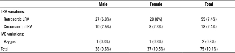
(Table 1). LRV and IVC variations were found in 75 (10.1%) cases, while LRV variations were identified
in 73 (9.8%) cases, of which 55 (7.4%) were RLRV
(Fig. 1) and 18 (2.4%) were CLRV (Fig. 2). Among
the female patients, total LRV variations, RLRVs, and
CLRVs numbered 36 (10.3%), 28 (8%), and 8 (2.3%),
respectively, and the corresponding values among men were 37 (9.3%), 27 (6.8%), and 10 (2.5%). No
statistically significant correlation was found between gender and overall LRV variations, RLRV, or CLRV (p = 0.801, p = 0.551, p = 0.823, respectively). Only 2 (0.3%) patients had azygos continuations of the IVC (Fig. 3). No other variations of the IVC were observed.
Overall, 1 male and 1 female patient had IVC variations, with no statistically significant correlation
being found between gender and the presence of variations (p = 0.933). The numbers and percentages
of LRV and IVC variations among the patients are
presented in Table 2.
During the CT, 55 (73.3%) cases with vascular
variations were found to have various abdominal pathologies, of which 18 (32%) had nephrolithiasis, Table 1. Distribution of patients by gender
Female Male P
Frequency 351 (47.1%) 395 (52.9%)
14 (25.4%) had renal cysts, and 8 (14.5%) had
hepa-tic hydatid cysts. Other CT findings included hepahepa-tic
and splenic lacerations, ascites, ovarian cysts, uterine myomas, perforated appendicitis, and ileus.
DIsCUssION
In a retrospective study of 746 patients, LRV and IVC variations were found in a total of 75 (10.1%)
cases: 73 (9.7%) had LRV variations and 2 (0.3%) had
IVC variations. No significant correlation was found between gender and variations in the LRV or IVC. A further 73.3% of cases with LRV variations were found to have abdominal pathologies on the CT, the
most common of which were nephrolithiasis, in 18 (32%) cases, and renal cysts, in 14 (25.4%) cases.
There have been several studies investigating the
frequency of LRV and IVC variations using enhance
-ment helical CT, MSCT angiography, conventional
angiography, and magnetic resonance angiography.
These studies have reported a variation frequency ranging from 3% to 10% for LRV [1, 3–5, 12, 15]. According to our study, variations of the LRV or IVC exist in 10.1% of society. Therefore, LRV or IVC va -scular variations exist in one out of every 10 people. Imaging the renal vascular anatomy preoperatively is essential in invasive therapy planning. Vascular variations should be known before surgery because this will prevent vascular complications. The
presen-ce of LRV and IVC variations should be investigated
prior to retroperitoneal surgical interventions, partial nephrectomies, laparoscopic procedures, and donor nephrectomies for transplantation [13]. In the present Figure 1. Axial computed tomography image of a 55-year-old male patient demonstrates retroaortic left renal vein (arrows).
Figure 2. Axial computed tomography images of a 55-year-old male patient demonstrate circumaortic left renal vein with both (A) retroaortic (arrowheads) and (B) preaortic (arrows) components.
Figure 3. Axial computed tomography images of a 73-year-old female patient demonstrate azygos continuation of the inferior vena cava (arrow).
study, the RLRV rate was 6.9%, which is quite high when compared with those of other studies [1, 3,
4, 12, 15]. As in many other studies, no significant
correlation was found between LRV variations and gender in our study [5, 7, 15].
The development of LRV and IVC occurs between
the 4th and 8th gestational weeks as a result of a num
-ber of anastomoses of the posterior cardinal, subcar-dinal, and supracardinal veins [12]. Many variations may develop during this complex formation process due to changes that may occur during various stages
[2]. A transposed left IVC runs parallel to the aorta on its left side, and this condition has a prevalence of 0.2–0.5% [10]. A double IVC refers to the presence of the IVC on the left side as well, and the prevalence of this condition has been reported to be 0.2–3.0% [10]. In an azygos continuation of the IVC, the IVC merges with the azygos vein by passing posterior to
the diaphragmatic crura in the prerenal segment. Its prevalence was reported to be 0.6% [6]. In our study, this variation was found in 2 (0.3%) cases. In 1 case of
azygos continuation of the IVC, the patient also had
situs ambiguous-heterotaxy syndrome [9]. It is vital to
be aware of the presence of a LRV and IVC variation
prior to surgical interventions or the implantation of
vena cava filters because this may prevent possible
haemorrhages and potentially fatal complications.
Further more, the recognition of such vascular struc -tures, which may be confused with lymphadenopa-thies, is crucial for disease staging and management planning.
IVC and LRV variations can be evaluated via veno -graphy, which is an invasive interventional method [11] or evaluated through colour Doppler
ultraso-nography (CDUS), which is a non-invasive imaging technique. However, CDUS is operator-dependent, and it can be difficult to evaluate obese or immobi -le patients. Venous structures can be evaluated via
a magnetic resonance imaging, without the use of
contrast media [5], although magnetic resonance imaging is both time-consuming and expensive when
compared with CT. The disadvantages of CT are its use of ionizing radiation and the possible nephrotoxicity
of the contrast media.
In our study, the most common abdominal patho-logies in cases with vascular variation were nephro-lithiasis, in 18 (32%) cases, and renal cysts, in 14 (25.4%) cases. This may be due to the negative effects
of these variations on renal function. In a previous
study, an increase in the intrarenal venous impedance index was found to result from the compression of the retroaortic LRV behind the aorta [14]. Such variations may have negative effects on renal haemodynamics.
Our study has several limitations. Principally, all
cases included in the study were patients who had
undergone abdominal CT examinations for various
reasons, and because healthy people cannot be given
CT scans due to the radiation risk, the present study is unable to make a comparison with the variation rate
in healthy people. The main strength of the present study is that it includes the large study population
examined via MSCT.
CONClUsIONs
In conclusion, MSCT is a rapid and reliable method of identifying LRV and IVC variations and associated
abdominal pathologies. The presence of the LRV and
IVC variations is not related to gender. Due to the con
-tribution of the MPR-reformatted images, a reliable assessment can be made via MSCT. Incidentally, de -tected vascular variations should always be reported
when found during routine CT scans. Renal patholo -gies are the most common patholo-gies seen in cases
with renal vein variations. Further studies of larger
series are needed to investigate the effects of these variations on renal haemodynamics and function. Table 2. Distribution of left renal vein (LRV) and inferior vena cava (IVC) variations in patients
Male Female Total
LRV variations:
Retroaortic LRV 27 (6.8%) 28 (8%) 55 (7.4%)
Circumaortic LRV 10 (2.5%) 8 (2.3%) 18 (2.4%)
IVC variations:
Azygos 1 (0.3%) 1 (0.3%) 2 (0.3%)
REfERENCEs
1. Atalar MH, Kosar MI, Salk I, Isleyen M (2012) Left renal vein abnormalities detected during routine abdominal computed tomography imaging: clinico-radiological
significance. Folia Morphol, 71: 168–172.
2. Bass JE, Redwine MD, Kramer LA, Huynh PT, Harris JH, Jr. (2000) Spectrum of congenital anomalies of the inferior vena cava: cross-sectional imaging findings. Radio-
graphics, 20: 639–652.
3. Bouali O, Labarre D, Molinier F, Lopez R, Benouaich V, Lauwers F, Moscovici J (2012) Anatomic variations of the
renal vessels: focus on the precaval right renal artery. Surg
Radiol Anat, 34: 441–446.
4. Dilli A, Ayaz UY, Kaplanoglu H, Saltas H, Hekimoglu B
(2013) Evaluation of the left renal vein variations and inferior vena cava variations by means of helical computed
tomography. Clin Imag, 37: 530–535.
5. Dilli A, Ayaz UY, Karabacak OR, Tatar IG, Hekimoglu B
(2012) Study of the left renal variations by means of magnetic resonance imaging. Surg Radiol Anat, 34:
267–270.
6. Ginaldi S, Chuang VP, Wallace S (1980) Absence of hepatic segment of the inferior vena cava with azygous continu
-ation. J Comput Assist Tomogr, 4: 112–114.
7. Koc Z, Ulusan S, Oguzkurt L, Tokmak N (2007) Venous va -riants and anomalies on routine abdominal multi-detector
row CT. Eur J Radiol, 61: 267–278.
8. Kumar S, Neyaz Z, Gupta A (2010) The utility of 64 channel multidetector CT angiography for evaluating
the renal vascular anatomy and possible variations:
a pictorial essay. Korean J Radiol, 11: 346–354.
9. Minniti S, Visentini S, Procacci C (2002) Congenital ano -malies of the venae cavae: embryological origin, imaging features and report of three new variants. Eur Radiol, 12:
2040–2055.
10. Phillips E (1969) Embryology and anomalies. In: Ferris EJ, Hipona FA, Kahn PC, Phillips E, Shapiro JH eds. Venograp -hy of the inferior vena cava and its branches. Williams &
Wilkins, Baltimore, pp. 1–32.
11. Satyapal KS, Kalideen JM, Haffejee AA, Singh B, Robbs JV (1999) Left renal vein variations. Surg Radiol Anat, 21:
77–81.
12. Trigaux JP, Vandroogenbroek S, De Wispelaere JF, Lacrosse M, Jamart J (1998) Congenital anomalies of the inferior vena cava and left renal vein: evaluation with spiral CT. J Vasc Interv Radiol, 9: 339–345.
13. Turkvatan A, Ozdemir M, Cumhur T, Olcer T (2009) Mul
-tidetector CT angiography of renal vasculature: normal anatomy and variants. Eur Radiol, 19: 236–244.
14. Yagci B, Tavasli B, Karabulut N, Kiroglu Y (2008) Clinical significance and renal haemodynamics of incidentally de -tected retroaortic left renal vein: assessment with venous
Doppler sonography. Br J Radiol, 81: 187–191.
15. Yesildag A, Adanir E, Koroglu M, Baykal B, Oyar O, Gul -soy UK (2004) [Incidence of left renal vein anomalies in